About gutsandgrowth
I am a pediatric gastroenterologist at GI Care for Kids (previously called CCDHC) in Atlanta, Georgia. The goal of my blog is to share some of my reading in my field more broadly. In addition, I wanted to provide my voice to a wide range of topics that often have inaccurate or incomplete information.
Before starting this blog in 2011, I would tear out articles from journals and/or keep notes in a palm pilot. This blog helps provide an updated source of information that is easy to access and search, along with links to useful multimedia sources.
I was born and raised in Chattanooga. After graduating from the University of Virginia, I attended Baylor College of Medicine. I completed residency and fellowship training at the University of Cincinnati at the Children’s Hospital Medical Center. I received funding from the National Institutes of Health for molecular biology research of the gastrointestinal tract.
During my fellowship, I had the opportunity to work with some of the most amazing pediatric gastroenterologists and mentors. Some of these individuals included Mitchell Cohen, William Balistreri, James Heubi, Jorge Bezerra, Colin Rudolph, John Bucuvalas, and Michael Farrell. I am grateful for their teaching and their friendship. During my training with their help, I received a nationwide award for the best research by a GI fellow.
I have authored numerous publications/presentations including original research, case reports, review articles, and textbook chapters on various pediatric gastrointestinal problems. In addition, I have been recognized by Atlanta Magazine as a "Top Doctor" in my field multiple times.
Currently, I am the vice chair of the section of nutrition for the Georgia Chapter of the American Academy of Pediatrics. In addition, I am an adjunct Associate Clinical Professor of Pediatrics at Emory University School of Medicine. Other society memberships have included the North American Society for Pediatric Gastroenterology Hepatology and Nutrition (NASPGHAN), American Academy of Pediatrics, the Food Allergy Network, the American Gastroenterology Association, the American Association for the Study of Liver Diseases, and the Crohn’s and Colitis Foundation.
As part of a national pediatric GI organization called NASPGHAN (and its affiliated website GIKids), I have helped develop educational materials on a wide-range of gastrointestinal and liver diseases which are used across the country. Also, I have been an invited speaker for national campaigns to improve the evaluation and treatment of gastroesophageal reflux disease, celiac disease, eosinophilic esophagitis, hepatitis C, and inflammatory bowel disease (IBD). Some information on these topics has been posted at my work website, www.gicareforkids.com, which has links to multiple other useful resources.
I am fortunate to work at GI Care For Kids. Our group has 17 terrific physicians with a wide range of subspecialization, including liver diseases, feeding disorders, eosinophilic diseases, inflammatory bowel disease, cystic fibrosis, DiGeorge/22q, celiac disease, and motility disorders. Many of our physicians are recognized nationally for their achievements. Our group of physicians have worked closely together for many years. None of the physicians in our group have ever left to join other groups. I have also worked with the same nurse (Bernadette) since I moved to Atlanta in 1997.
For many families, more practical matters about our office include the following:
– 14 office/satellite locations
– physicians who speak Spanish
– cutting edge research
– on-site nutritionists
– on-site psychology support for abdominal pain and feeding disorders
– participation in ImproveCareNow to better the outcomes for children with inflammatory bowel disease
– office endoscopy suite (lower costs and easier scheduling)
– office infusion center (lower costs and easier for families)
– easy access to nursing advice (each physician has at least one nurse)
I am married and have two sons (both adults). I like to read, walk/hike, bike, swim, and play tennis with my free time.
I do not have any financial relationships with pharmaceutical companies or other financial relationships to disclose. I have helped enroll patients in industry-sponsored research studies.
“Common sense is not so common.” Voltaire,, Dictionnaire Philosophique 1764
A website that I learned about recently from the Journal of Pediatrics article, “The Elephant in the Examination Room: Addressing Parent and Child Mobile Device Use as a Teachable Moment:” commonsensemedia.org
“Common Sense is the nation’s leading nonprofit organization dedicated to improving the lives of kids and families by providing the trustworthy information, education, and independent voice they need to thrive in the 21st century.”
This website has extensive resources for families regarding all forms of media. This includes advice on apps, age for using smartphones, encouragement for device-free dinners, movie/TV reviews and more.
M Barroso et al. Gastroenterol 2018; 154: 2087-96.
Background: “Western-like diets –mainly characterized by high intake of red and processed meats, refined grains, simple sugars, and saturated fats and low intake of fruits, vegetables, and whole grains– have been associated with low-grade chronic inflammation, which is involved in the etiology of inflammatory conditions.” Ref: Br J Nutr 2015; 114: 999-1012.
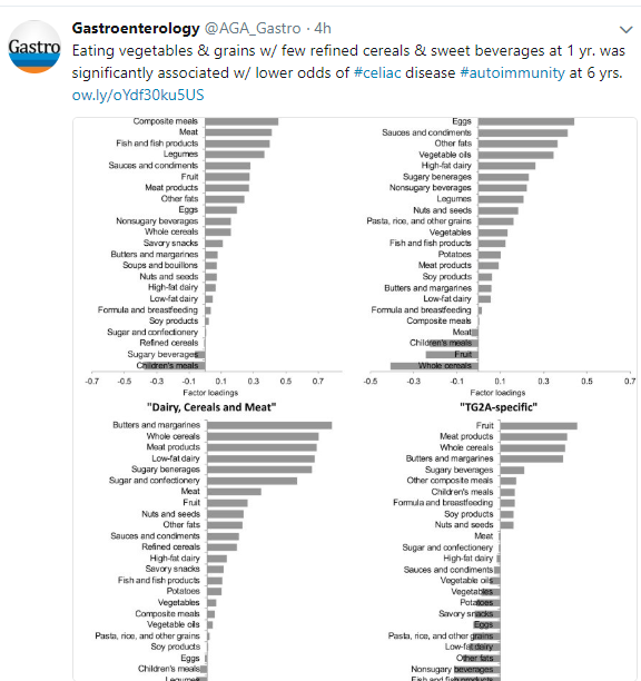
To examine how diet may influence the development of celiac autoimmunity, defined by TG2A positivity, the authors examined a subset of patients (n=1997) from the prospective Generation R study (Netherlands); 27 in this cohort developed celiac autoimmunity (1.4%).
Key finding:
- Higher adherence to a “prudent” diet which had a higher intake of vegetables, vegetable oils, pasta, and grains and low consumption of refined cereals and sweet beverages at 1 year of age was associated with a lower odds of celiac autoimmunity at 6 years of age with an odds ratio of 0.67.
This study is limited by the relatively low number who had celiac autoimmunity and by its use of a food questionnaire.
My take: This study indicates that diet plays a role in the development of celiac along with other disease, but this likely involves a complex mix of components rather than a single toxic agent.

Related blog posts:
A recent study (Z Kiss et al. JPGN 2018; 67: 6-12) provides more data on normative values for eosinophil counts in the GI tract. For their report, the authors reviewed 3 databases for a systematic search of the literature. They screened 1316 abstracts but found only 8 articles with complete/relevant data. Among these 8 articles, data regarding each segment of the GI tract was present in as few as 3 articles and as many as 6 articles. The authors provide confidence intervals (CIs) and prediction intervals (PIs); the latter account for the wider uncertainty due to insufficient data.
Key points:
Normal eosinophil cell number per high-power field (HPF area = 0.2 mm squared):
- Duodenum 8.26 with CI 4.71-11.8 and PI of 0 to 20.57
- Terminal ileum 11.52 with CI 7.21-15.83 and PI of 0 to 60.64
- Cecum 14.12 with CI 9.05-19.19 and PI of 0 to 38.64
- Ascending colon 13.25 with CI 8.65-17.86 and PI of 0 to 35.42
- Transverse colon 11.52 with CI 7.80-15.23 and PI of 0 to 25.85
- Descending colon 10.32 with CI 7.22-13.42 and PI of 0 to 49.10
- Sigmoid colon 8.80 with CI 6.82-10.77 and PI of 0 to 32.49
- Rectum 7.39 with CI 4.20-10.59 and PI of 0 to 22.33
Other points:
- The authors note that eos/HPFis a flawed measurement due to technical parameters of the microscope. Some HPFs are bigger than others –this could affect eosinophil count up to 5-fold. The authors specify an HPF to be =0.2 mm squared.
- Obtaining appropriate mucosal samples for normal number of eosinophil counts can be difficult. Even patients with functional disorders like irritable bowel syndrome and nonulcer dyspepsia could have abnormal numbers of eosinophils.
My take: These numbers of expected eosinophil counts for pediatric histology are a good starting point. The prediction intervals remain large due to insufficient data.
Related blog posts:

Gibbs Gardens
A recent study (P Kumar et al. JPGN 2018; 67: 75-9) examined suspected NAFLD in 12 to 18 year olds using data from NHANES. In the analysed cohort, there were 124 suspected NAFLD and 1385 without suspicion of NAFLD. This subset was weight to represent a U.S. population of over 18 million.
Key definitions:
- Suspected NAFLD was defined by abnormal ALT (>25.8 U/L for boys and >22.1 U/L for girls) who did not have another explanation (eg. viral hepatitis, medication)
- Lean BMI was defined by BMI less than 85th% for age
- Hypertriglyceridemia ≥ 150
- Low HDL ≤ 40 mg/dL
- HOMA-IR =fasting glucose x insulin (microU/mL) divided by 405. Insulin resistance was defined as HOMA-IR ≥ 3
Key findings:
- Suspected NAFLD affects ~8% of lean adolescents in the U.S.
- Hypertriglyceridemia was noted in 10 of 124 suspected NAFLD and was a risk factor (P=0.028) as was Low HDL which occurred in 15 (P=0.016) and IR which occurred in 43 (P=0.053)
My take: Elevated ALT, a marker for fatty liver disease, is common even in adolescents without obesity. Elevated triglycerides, low HDL, and insulin resistance are all risk factors for suspected NAFLD in non-overweight/non-obese teens.
Related blog posts:

Cumberland Island 2018

A recent study (PC Church et al. JPGN 2018; 67: 53-8) examined how well EGD findings were detected by MRE in 188 children (mean age 14 years).
Key findings:
- EGD was macroscopically abnormal in 93 (49%) with ulcerations being the most common abnormality in 66 (35%).
- In contrast, the local radiologist identified UGI inflammation in 7 (4%) and the central radiologists identied UGI inflammation in 20 (22%). “There was no agreement between local and central radiologists when examining the UGI as a whole (κ=-0.02, P-0.59)”
- The local radiologists “correctly identified only 5 of 93 (8%) patients with UGI findings on EGD.” The central radiologists “correctly identified 9 of 45 (30%) patients with UGI findings on EGD.”
The authors state that “the Porto criteria mandate the performance of EGD for all pediatric patients suspected of having IBD. Our study has demonstrated that MRE cannot be relied upon as the sole method of evaluating the UGI.”
My take: For those who take care of children with IBD, this study will not come as a surprise as many of the UGI findings (found at endoscopy) are subtle. This study does quantify the much higher sensitivity of endoscopic evaluation and is similar to studies that have compared capsule endoscopy to MRE.
Related blog posts:

Cumberland Island 2018

Abstract Link: Nutritional Considerations in Pediatric Pancreatitis: A Position Paper from the NASPHAN Pancreas Committee and ESPHAN Cystic Fibrosis/Pancreas Working Group.
M Abu-El-Haija et al. JPGN 2018; 67: 131-43. This working group made ~27 recommendations (summarized in Table 1) and indicated the quality of evidence supporting the recommendation as well as the agreement among team members –virtually all received at least 12 of 13 votes.
Here are the ones that grabbed my attention:
For Acute Pancreatitis (AP):
- 1a & 1aa. Children with mild AP should be started on a regular diet –preferably via mouth as compared to nasogastric route
- 1b. Enteral nutrition (EN) should be attempted in children with severe AP within 72 hours from presentation, once deemed hemodynamically stable.
- 1.4 Even in severe AP, jejunal tube feeding should be reserved for those unable to tolerate oral or NG tube feeding
For Acute Recurrent Pancreatitis (ARP):
- 2.1a & 2.1b. Children should receive a regular-fat diet in between bouts of ARP and a regular-fat diet can safely be started within 1 week after the onset of a bout of AP (except in those with very elevated triglycerids (>1000 mg/dL)
- 2.2a & 2.3a. PERT is NOT recommended in children with ARP without eocrine pancreatic insufficiency (EPI). Antioxidants are NOT recommended (insufficient supporting evidence)
For Chronic Pancreatitis (CP):
- 3.1b & 3.12a. Recommends routine followup every 3-6 months and a regular diet
- 3.3a, 3.4a, & 3.5a Monitoring: recommends checking fat-soluble vitamin levels every 6 to 12 months, checking for EPI with elastase (or 72 hr fecal fat) every 6-12 months, and BMD (bone mineral density) if CP and malnutrition (especially if Vit D deficiency or hx/o fractures)
My take: This report provides a methodical approach for the care of children with these pancreatic disorders.
Related blog posts:

Tide pools and wide beach at Cumberland Island 2018



Briefly noted: P Singh et al. Clin Gastroenterol Hepatol 2018; 16: 823-36. After a systemic review which selected 96 articles from a pool of 3843 published between 1991 through 2016, the authors determined a pooled global prevalence of 1.4% in 275,818 individuals based on seroprevalence (positive TTG or EMA). Biopsy-confirmed celiac disease was noted in 0.7% in 138,792 individuals.
In their study, biopsy-proven disease was most prevalent in Argentina, Egypt, Hungary, Finland, Sweden, New Zealand, and India.

Related blog posts:
Briefly noted: A recent study (EA Spencer et al.Inflamm Bowel Dis 2018; 24: 1335-42) examined phenotype and serology in 399 children with newly diagnosed ulcerative colitis (PROTECT study).
Key findings:
- 65% had positive serology for pANCA; 62% in those <12 and 66% in those ≥12 years
- 19% had positive serology for anti-CBir1; 32% in those <12 and 14% in those ≥12 years
- High titer (≥ 100)) pANCA positivity was associated with more extensive disease but not with PUCAI values or Mayo endoscopic subscores.
My take: The serology titers for IBD, in my view, have academic interest but do not routinely enhance patient care.
Related blog post:

Amelia Island
Briefly noted: A small pilot study (n=9) (SS Sundaram et al. J Pediatr 2018; 198: 67-75) showed that treatment (with home CPAP) of obstructive sleep apnea (OSA) was associated with improved alanine aminotransferase levels, reduced metabolic syndrome markers and lower F(2)-isoprostanes (a marker of oxidative stress) in pediatric patients with nonalcoholic fatty liver disease (NAFLD). All nine of the participants were Hispanic males with a median age of 11.5 years; they had a median BMI of 29.5 and had biopsy-proven NAFLD. The improvement in NAFLD parameters occurred despite an increase in BMI. The authors note that studies in adults have shown contradictory findings with regard to whether treatment of OSA helps NAFLD.
My take: This study suggests potential beneficial liver effects of treating OSA. Regardless, treatment of OSA could be considered a quality metric in the care of children with NAFLD as better sleep at night has additional clear benefits.
Related blog posts:

Outside Mercedes-Benz Stadium (Atlanta)