Unknown's avatar

About gutsandgrowth

I am a pediatric gastroenterologist at GI Care for Kids (previously called CCDHC) in Atlanta, Georgia. The goal of my blog is to share some of my reading in my field more broadly. In addition, I wanted to provide my voice to a wide range of topics that often have inaccurate or incomplete information. Before starting this blog in 2011, I would tear out articles from journals and/or keep notes in a palm pilot. This blog helps provide an updated source of information that is easy to access and search, along with links to useful multimedia sources. I was born and raised in Chattanooga. After graduating from the University of Virginia, I attended Baylor College of Medicine. I completed residency and fellowship training at the University of Cincinnati at the Children’s Hospital Medical Center. I received funding from the National Institutes of Health for molecular biology research of the gastrointestinal tract. During my fellowship, I had the opportunity to work with some of the most amazing pediatric gastroenterologists and mentors. Some of these individuals included Mitchell Cohen, William Balistreri, James Heubi, Jorge Bezerra, Colin Rudolph, John Bucuvalas, and Michael Farrell. I am grateful for their teaching and their friendship. During my training with their help, I received a nationwide award for the best research by a GI fellow. I have authored numerous publications/presentations including original research, case reports, review articles, and textbook chapters on various pediatric gastrointestinal problems. In addition, I have been recognized by Atlanta Magazine as a "Top Doctor" in my field multiple times. Currently, I am the vice chair of the section of nutrition for the Georgia Chapter of the American Academy of Pediatrics. In addition, I am an adjunct Associate Clinical Professor of Pediatrics at Emory University School of Medicine. Other society memberships have included the North American Society for Pediatric Gastroenterology Hepatology and Nutrition (NASPGHAN), American Academy of Pediatrics, the Food Allergy Network, the American Gastroenterology Association, the American Association for the Study of Liver Diseases, and the Crohn’s and Colitis Foundation. As part of a national pediatric GI organization called NASPGHAN (and its affiliated website GIKids), I have helped develop educational materials on a wide-range of gastrointestinal and liver diseases which are used across the country. Also, I have been an invited speaker for national campaigns to improve the evaluation and treatment of gastroesophageal reflux disease, celiac disease, eosinophilic esophagitis, hepatitis C, and inflammatory bowel disease (IBD). Some information on these topics has been posted at my work website, www.gicareforkids.com, which has links to multiple other useful resources. I am fortunate to work at GI Care For Kids. Our group has 17 terrific physicians with a wide range of subspecialization, including liver diseases, feeding disorders, eosinophilic diseases, inflammatory bowel disease, cystic fibrosis, DiGeorge/22q, celiac disease, and motility disorders. Many of our physicians are recognized nationally for their achievements. Our group of physicians have worked closely together for many years. None of the physicians in our group have ever left to join other groups. I have also worked with the same nurse (Bernadette) since I moved to Atlanta in 1997. For many families, more practical matters about our office include the following: – 14 office/satellite locations – physicians who speak Spanish – cutting edge research – on-site nutritionists – on-site psychology support for abdominal pain and feeding disorders – participation in ImproveCareNow to better the outcomes for children with inflammatory bowel disease – office endoscopy suite (lower costs and easier scheduling) – office infusion center (lower costs and easier for families) – easy access to nursing advice (each physician has at least one nurse) I am married and have two sons (both adults). I like to read, walk/hike, bike, swim, and play tennis with my free time. I do not have any financial relationships with pharmaceutical companies or other financial relationships to disclose. I have helped enroll patients in industry-sponsored research studies.

Hepatitis C -New Studies & New Recommendations (2017)

Briefly noted:

EJ Gane et al. Gastroenterol 2017; 152: 1366-71.  This phase 2, open-label study (n=53) examined the efficacy of ledipasvir plus sofusbuvir for 8 or 12 weeks for Hepatitis C virus (HCV) genotype 2.  An SVR was noted in 96% with 12 weeks of treatment and 74% with 8 weeks of treatment.  The only patient in the 12 week group without an SVR did not complete treatment.  Overall, this study stands in contrast with the ION-3 study which showed that 8 weeks of therapy led to an SVR of 94% among genotype 1.  Thus, this study is consistent with ledpasvir having more potency against genotye 1 and the need for a 12 week course with genotype 2 HCV.

IM Jacobson et al. Gastroenterol 2017; 152: 1372-82. This retrospective study examined 402 patients with HCV genotypes 1, 4 or 6 with Child-Pugh A compensated cirrhosis who were treated with Elbasvir/GrazoprevirKey finding: SVR12 was 98% and 89% for treatment-naive and treatment-experienced patients after 12 weeks of therapy. The authors noted that baseline tests were done to look for resistance-associated substitutions (RASs).  They recommend: “GT1a patients with RASs require extension of therapy to 16 weeks and addition of ribavirin.”

IM Jacobson et al. Gastroenterol 2017; 152: 1378-87. This AGA clinical practice update makes recommendations for patients have achieved an SVR after HCV therapy. These recommendations are mainly expert opinion given the recent advent of newer treatments for HCV and lack of data regarding long-term outcomes after these treatments. Recommendations from authors:

  • SVR should be confirmed by undetectable HCV RNA at 12 weeks after treatment regimen
  • Reconfirmation of SVR at 48 weeks after treatment is recommended.
  • Surveillance for HCC with liver imaging ± AFP should be pursued twice annually for an indefinite duration in all patients with stage 3 fibrosis or liver cirrhosis post-SVR.  Surveillance is “not recommended for patients with stages 0-2 fibrosis post SVR.”
  • Endoscopic screening for varices is recommended for all patients with cirrhosis, independent of SVR. Repeat screening should be considered if no varices or small varices are noted 2-3 years later.  If there are still no varices at 2-3 years, no further endoscopic screening is recommended.

Related blog posts:

If this picture and reports are accurate, this man’s family urged him to come inside. He said he was keeping an eye on it.


Defining the Role for Elastography

The ability to determine if a patient has cirrhosis/severe fibrosis with a noninvasive test can help determine appropriate monitoring and treatment for many liver conditions. As such the AGA has provided recommendations for the use of vibration-controlled transient elastography (VCTE).

  • JK Lim et al. Gastroenterol 2017; 152: 1536-43.
  • S Singh et al. Gastroenterol 2017; 152: 1544-77.

Many recommendations are based on the specific unit of measurement, kilopascals (kPa)

Specific recommendations (most with low  or very low quality evidence):

  • “In adults with chronic HCV, we can accurately diagnosis cirrhosis …with VCTE-defined liver stiffness of ≥12.5 (±1) kPa.”  The AGA suggests using VCTE rather than MRE for detection of cirrhosis.
  • “In adults with chronic HCV who have achieved SVR…we can accurately rule out advanced fibrosis (F3 and F4) with post-treatment VCTE-..of ≤9.5 (±1) kPa.” . Even in patients who have had HCV eradicated, if cirrhosis has been identified, careful followup is recommended.
  • “In adults with chronic HBV, we can accurately diagnosis cirrhosis…with VCTE…of ≥11.0 (±1) kPa.”
  • “The AGA makes no recommendation regarding the role of VCTE in the diagnosis of cirrhosis in adults with NAFLD.” For NAFLD, VCTE is not as helpful as with chronic HCV and HBV.  Currently, liver biopsy remains the “gold standard.” However, for noninvasive imaging, “the AGA suggest using MRE, rather than VCTE, for detection of cirrhosis.
  • For adults with suspected compensated cirrhosis, a VCTE of 19.5 or greater can be used “to assess the need for esophagogastroduodenoscopy to identify high risk esophageal varices.”

My take: These elastography recommendations are applicable for adults.  For pediatric patients, these reports suggest that elastography may be helpful in specific circumstances as well.

Related posts:

Omaha Beach

Low Rate of Ocular Disease in Pediatric Crohn’s Disease

A recent study (S Naviglio et al. Inflamm Bowel Dis 2017; 23: 986-90) confirms that there is a low rate of ocular disease in pediatric inflammatory bowel disease (IBD); in this cohort, half had Crohn’s disease (CD) and half had ulcerative colitis.

In this single center study, 94 children with a median age of 13.4 yrs were offered ophthalmologic examination (2014-2016).  None of these patients reported ocular symptoms.  The authors assert that 70% had intestinal remission, though 64% had elevated fecal calprotectin levels (>100 mg/kg). Key finding: One patient (1.06%) had ocular finding of uveitis (previously diagnosed prior to study)

The authors indicate that hepatobiliary manifestations, present in 9, were the most common extraintestinal IBD manifestation (EIM). Arthropathy occurred in 8, cutaneous manifestations occurred in 6 and ‘metastatic’ CD occurred in 4.

My take:  Ocular disease is an infrequent EIM in pediatric patients with IBD.

Related articleK Hata et al. Inflamm Bowel Dis 2017; 23: 1019-24. This article found that patients with EIMs were more likely to have chronic pouchitis after colectomy for ulcerative colitis. Overall, chronic pouchitis developed in 3.3%, 7.6% and 16.6% at 2, 5, and 10 years respectively. Key finding: preoperative EIM yielded a HR of 4.52.

Hepatitis B Reactivation Due to Immunosuppressive Therapies

The topic of Hepatitis B virus (HBV) reactivation has been discussed on this blog before (see link below).  Another excellent review on this topic (R Lomba, TJ Liang. Gastroenterol 2017; 152: 1297-1309) has been published.  The authors examine the course and mechanisms of HBV reactivation.  They divide the risk of reactivation into three groups: high, moderate and low risk and proposed management.

High risk groups, which have >10% risk of reactivation) include the following

  • B-cell-depleting agents including rituximab, ofatumumab, alemtuumab, and ibritumumab
  • High-dose corticosteroids (>20 mg/day in adults)
  • Antracyclines including doxorubicin
  • Potent TNF-α inhibitors: infliximab, adalimummab, certolizumab, and golimumab
  • Local therapy ofr HCC including TACE (transarterial chemoembolization)

Moderate groups (1-10% reactivation) include cytokine-based Rx (eg. abatacept, ustekinumab, natalizumab, vedolizumab), cyclosporine, systemic chemotherapy, moderate corticosteroid dosing

Low risk groups (<1% reactivation) include thiopurines (azathioprine, 6-mercaptopurine), and methotrexate as well as short-term low-dose corticosteroids.

Management:

  • For HBV screening, the authors recommend HBsAg and anti-HBc testing
  • Prophylactic therapy with potent oral anti-HBV therapies are recommended for those at moderate or high risk of reactivation.  In those at low risk, the options include prophylactic treatment or watchful monitoring.
  • A more detailed algorithm is provided in Figure 3.  In those with HBsAg positivity, if HBV DNA is less than 2000 U/mL, this algorithm suggests monitoring labs (HBsAg, ALT, HBV DNA every 3 months)

Mechanisms of HBV reactivation are discussed.  For example, with TNF-α inhibitors “can activate a unique host antiviral pathway, the APOBEC (apolipoprotein B mRNA editing enzyme, catlytic polypeptide-like) proteins, that cause the degradation of cccDNA in HBV-infected cells. Thus, blocking this endogenous antiviral pathway may lead to a higher HBV replication state and HBV reactivation.”

My take: In pediatric gastroenterology, we do not see a lot of HBV reactivation. Nevertheless, we do use many of the medications which can trigger HBV reactivation and need to keep these recommendations in mind.

Related blog post: What HBV Testing is Needed Before TNF Inhibitor Therapy

Suntrust Park

Dreaded Nausea (2017)

This post provides followup to a previous post: Dreaded Nausea.

A recent study (AC Russell, AL Stone, LS Walker, Clin Gastroenterol Hepatol 2017; 15: 706-11) provides even more reasons to dread nausea.

This prospective study of 871 children with functional abdominal pain examined the comorbidity of nausea.  Followup data were collected from 392 patients at median of 8.7 years later.

Key findings:

  • At baseline, 44.8% of patients reported nausea. This group reported worse abdominal pain, somatic symptoms and depression than those without nausea.
  • At followup, “those with nausea in childhood continue to have more severe GI (P<.001) and somatic symptoms (P=.003)…as well as higher levels of anxiety (P=.02) and depression (P=.02).”  Anxiety and depression remained significant after controlling for baseline abdominal pain severity.
  • At the followup evaluation, the prevalence of any functional GI disorder (FGID) was 85 (48%) of those who had nausea at baseline compared with 77 (36%) for those without nausea at baseline.

In their discussion, the authors reiterate findings from previous work on this patient sample: “current and lifetime diagnoses of anxiety disorders are substantially higher in adolescents with a history of FAP [functional abdominal pain] compared with healthy controls (lifetime, 51% vs. 20%; current 30% vs 12%). The lifetime risk of depressive disorder is also significantly higher in those with FAP (40% vs. 16%).”  They also note some limitations in their work, including the absence of formal screening for postural orthostatic tachycardia syndrome (POTS).

My take (borrowed from authors): This study “suggests that nausea is more than just a comorbid symptom of FAP and may have a different underlying etiology” and increases likelihood of persistent symptoms as well as anxiety and depression.

Briefly noted: RJ Shulman et al. Clin Gastroenterol Hepatol 2017; 15: 712-9. This randomized, double-blind study showed that added psyllium reduced frequency (but not severity) of abdominal pain in children (n=103) with irritable bowel syndrome. Psyllium was dosed at 6 g/day for 7-11 year olds, and 12 g for 12-18 year olds. Interestingly, this study did not show that psyllium caused a difference in normal stools or other mechanistic reasons for improvement, like breath hydrogen, breath methane, intestinal permeability or microbiome composition.

Related posts:

Atlanta Zoo, Bald Eagle


More Data for Ustekinumab in Crohn’s Disease

Briefly noted: C Ma et al. Inflamm Bowel Dis 2017; 23: 833-9.  This retrospective study examined the response to ustekinumab in 104 patients with Crohn’s disease.  All patients had achieved a steroid-free ustekinumab induction.  92.3% had failed anti-TNFα therapy.

Key findings:

  • 71.8% maintained a response at 52 weeks
  • 64.4% maintained an endoscopic or radiographic response

POWER — Practice Guide on Obesity and Weight Management, Education, and Resources

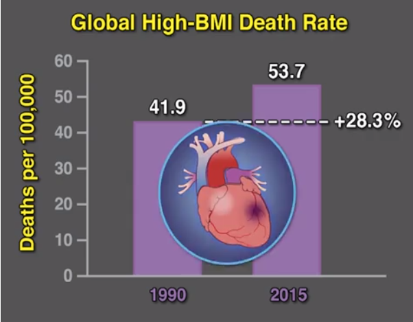
Recently, the American Gastroenterological Association (AGA) has published a large amount of information regarding obesity and the potential role for gastroenterologists.  In addition to publishing an entire Special Issue supplement of Gastroenterology (152: (7): 1635-1801, the AGA has published a “white paper” (Clin Gastroenterol Hepatol 2017; 15: 631-49).  The AGA has also addressed coding issues and episodic care issues: Clin Gastroenterol Hepatol 2017; 15: 650-64.

Some useful points from these articles:

  • “Severe obesity [as classified by] the American Heart Association…BMI>120% of the 95% for age and sex or a BMI ≥35” (“class 2 obesity in adults”) Class 3 obesity is BMI >140% of 95% for age and sex or a BMI ≥40.
  • Intensive lifestyle interventions ‘average weight losses of up to 8 kg in 6 months’ but maintaining weight loss has been a challenge. “However, both the DPP and Look AHEAD have shown that weight loss, followed by substantial weight regain, was associated with greater improvements in health than not having lost weight at all.”
  • Good idea to review medications that affect weight.  Medications associated with weight gain include antidiabetics, some antihypertensives (eg. nadolol, propranolol), antidepressants (eg. lithium, mirtazapine, SSRIs, tricyclic antidepressants), antipsychotics (clozapine, olanzapine, quetiapine, risperdione), some antieleptics (carbamazepine, gabapentin, pregabalin, valproic acid), 1st generation antihistamines and glucocorticoids.
  • Is there a best diet? On this topic, the authors (pg 1749 of supplement): “there appears to be little weight loss advantage or difference in metabolic health outcomes between dietary approaches and improvements in health are relative to degrees of weight loss.  Caloric restriction is the fundamental premise of every successful weight loss strategy, whether that is achieved by lowering fat or carbohydrate, fasting, or using meal replacements...the best diet ultimately is the one you can stick to long enough

The information available in these publications are overlapping and cannot be summarized adequately in a short post.  The white paper, in particular, does an excellent job of summarizing the reasons for obesity, the steps a clinician should take, identification of comorbidities, management (diet, exercise, pharmacologic agents, endoscopic therapies, and surgery), and outcomes.

My take (borrowed from the authors):  “obesity is possibly the greatest health care issue of our day…Although lifestyle changes, including an individualized reduced-calorie diet and physical activity, are the cornerstones of treatment, new medications and bariatric endoscopic therapies and surgery can be effective tools.”

Related blog posts:

Mural Near Sullivan’s Island


IBD Shorts and Postop Crohn’s Management

C Ma et al. Inflamm Bowel Dis 2017; 23: 833-9.  This retrospective study examined the ongoing response to ustekinumab in 104 patients with Crohn’s disease.  All patients had achieved a steroid-free ustekinumab induction.  92.3% had failed anti-TNFα therapy.Key findings:

  • 71.8% maintained a response at 52 weeks
  • 64.4% maintained an endoscopic or radiographic response

Related blog post: Closer Look at Ustekinumab Data

O Truffinet et al JPGN 2017; 64: 721-25. This small study with 8 children with Crohn’s disease examined the use of tacrolimus.  Six of eight showed a response to tacrolimus (target 8-15) with a clinical response at 2 months and 4 of 8 in clinical remission.  Adverse effects were common, occurring in 6 of 8.  These included renal dysfunction, diabetes, paresthesia and tremor.

J Adler et al.  JPGN 2017; 64: e117-e124. Using ImproveCareNow registry, the authors identified perianal disease (PD) in 1399 of 6679 cases (21%).  PD was more common in blacks than whites: 26% vs. 20%.  Overall, this study showed a higher rate of PD than previously recognized.

J Amil-Dias et al JPGN 2017; 64: 818-35.  This is an ESPGHAN IBD Porto Group guideline for surgical Crohn’s disease management in children.  There are 25 graded statements.  Here are a few:

  • #7 & #8. If needing surgery for CD pancolitis, the authors recommend subtotal colectomy and ileostomy.  Possible reanastomosis at later date if no significant rectal and/or perianal disease.  Ileal pouch-anal anastomosis is NOT recommended.
  • #13. Monitor Vitamin B12 if >20 cm resection of terminal ileum
  • #16. Postoperative management “should be based on ileocolonoscopy.” Figure 1 details recommendations, including need for assessment postoperatively.
  • In patients with high-risk factors, anti-TNF therapy is recommended postoperatively.  In those without high-risk factors, the authors indicate that thiopurines are reasonable with and advancing to anti-TNF if Rutgeerts i2 or greater at followup assessment.  High-risk factors include growth failure, short duration from diagnosis to surgery, extensive resection (>40 cm), and penetrating disease.

Related blog post:

Musee d’Orsay


One Way Fecal Microbiota Transplant May Work: Changing Bile Acids

Breifly Noted:

From MedPage Today: Fecal transplant success may depend on bile acid metabolism

An excerpt:

the transplants change patterns of bile acid metabolism in the gut, making the environment inhospitable to C. diff colonization.

In three studies reported at Digestive Disease Week (DDW) 2017, it was demonstrated that individuals with C. diff who respond to fecal transplant showed a different pattern of microbiota species composition compared with baseline and/or with those who fail to respond. But that’s not all: the responders also showed distinct, altered profiles of those elements involved in bile acid metabolism.

Vincent Van Gogh; Hopital Saint-Paul (1889)