Unknown's avatar

About gutsandgrowth

I am a pediatric gastroenterologist at GI Care for Kids (previously called CCDHC) in Atlanta, Georgia. The goal of my blog is to share some of my reading in my field more broadly. In addition, I wanted to provide my voice to a wide range of topics that often have inaccurate or incomplete information. Before starting this blog in 2011, I would tear out articles from journals and/or keep notes in a palm pilot. This blog helps provide an updated source of information that is easy to access and search, along with links to useful multimedia sources. I was born and raised in Chattanooga. After graduating from the University of Virginia, I attended Baylor College of Medicine. I completed residency and fellowship training at the University of Cincinnati at the Children’s Hospital Medical Center. I received funding from the National Institutes of Health for molecular biology research of the gastrointestinal tract. During my fellowship, I had the opportunity to work with some of the most amazing pediatric gastroenterologists and mentors. Some of these individuals included Mitchell Cohen, William Balistreri, James Heubi, Jorge Bezerra, Colin Rudolph, John Bucuvalas, and Michael Farrell. I am grateful for their teaching and their friendship. During my training with their help, I received a nationwide award for the best research by a GI fellow. I have authored numerous publications/presentations including original research, case reports, review articles, and textbook chapters on various pediatric gastrointestinal problems. In addition, I have been recognized by Atlanta Magazine as a "Top Doctor" in my field multiple times. Currently, I am the vice chair of the section of nutrition for the Georgia Chapter of the American Academy of Pediatrics. In addition, I am an adjunct Associate Clinical Professor of Pediatrics at Emory University School of Medicine. Other society memberships have included the North American Society for Pediatric Gastroenterology Hepatology and Nutrition (NASPGHAN), American Academy of Pediatrics, the Food Allergy Network, the American Gastroenterology Association, the American Association for the Study of Liver Diseases, and the Crohn’s and Colitis Foundation. As part of a national pediatric GI organization called NASPGHAN (and its affiliated website GIKids), I have helped develop educational materials on a wide-range of gastrointestinal and liver diseases which are used across the country. Also, I have been an invited speaker for national campaigns to improve the evaluation and treatment of gastroesophageal reflux disease, celiac disease, eosinophilic esophagitis, hepatitis C, and inflammatory bowel disease (IBD). Some information on these topics has been posted at my work website, www.gicareforkids.com, which has links to multiple other useful resources. I am fortunate to work at GI Care For Kids. Our group has 17 terrific physicians with a wide range of subspecialization, including liver diseases, feeding disorders, eosinophilic diseases, inflammatory bowel disease, cystic fibrosis, DiGeorge/22q, celiac disease, and motility disorders. Many of our physicians are recognized nationally for their achievements. Our group of physicians have worked closely together for many years. None of the physicians in our group have ever left to join other groups. I have also worked with the same nurse (Bernadette) since I moved to Atlanta in 1997. For many families, more practical matters about our office include the following: – 14 office/satellite locations – physicians who speak Spanish – cutting edge research – on-site nutritionists – on-site psychology support for abdominal pain and feeding disorders – participation in ImproveCareNow to better the outcomes for children with inflammatory bowel disease – office endoscopy suite (lower costs and easier scheduling) – office infusion center (lower costs and easier for families) – easy access to nursing advice (each physician has at least one nurse) I am married and have two sons (both adults). I like to read, walk/hike, bike, swim, and play tennis with my free time. I do not have any financial relationships with pharmaceutical companies or other financial relationships to disclose. I have helped enroll patients in industry-sponsored research studies.

In NASH, is ALT Wrongly Used as a Marker of Liver Injury?

According to a recent report (Hepatology 2015; 61: 153-60), elevation of alanine aminotransferase (ALT) which is frequently used as an indicator to select patients for further investigations (eg. liver biopsy) is NOT a good indicator of liver parenchymal injury in patients with nonalcoholic fatty liver disease (NAFLD).

The researchers enrolled 440 patients and divided them into three groups: no NAFLD (n=60), NALFLD with normal ALT (n=165), and NAFLD with elevated ALT (n=215). The patients were overweight/obese patients prospectively recruited from newspaper ads, general medicine clinics and hepatology clinics at several VA hospitals. Those with history of alcohol abuse were excluded.

Numerous investigations were performed including liver fat by proton magnetic resonance spectroscopy (H-MRS), liver biopsy (n=293), and insulin resistance measurements.

Key findings:

  • NAFLD & NASH patients with elevated ALT had higher liver triglyceride content (P<0.0001), worse adipose tissue insulin resistance (ATIR) (P<0.0001), and lower plasma adiponectin (P<0.05).
  • Steatosis was worse on liver biopsy in those with NASH and elevated ALT (P<0.0001).
  • There were no differences in liver inflammation (P=0.62), ballooning (P=0.13), or fibrosis (P=0.12). Thus, patients with normal versus elevated ALT had similar severity of NASH liver histology.

Take-home message:  In adults, ALT values are “poor surrogate markers of disease activity” in NAFLD.  ALT values, in these patients, are indicative of metabolic disease.  Given the staggering numbers of individuals, adults and children, with fatty liver disease, the lack of simple screen tool is quite problematic.  Equally problematic is a lack of a simple treatment approach.

Related blog posts:

Refractory Constipation -Terrific Update

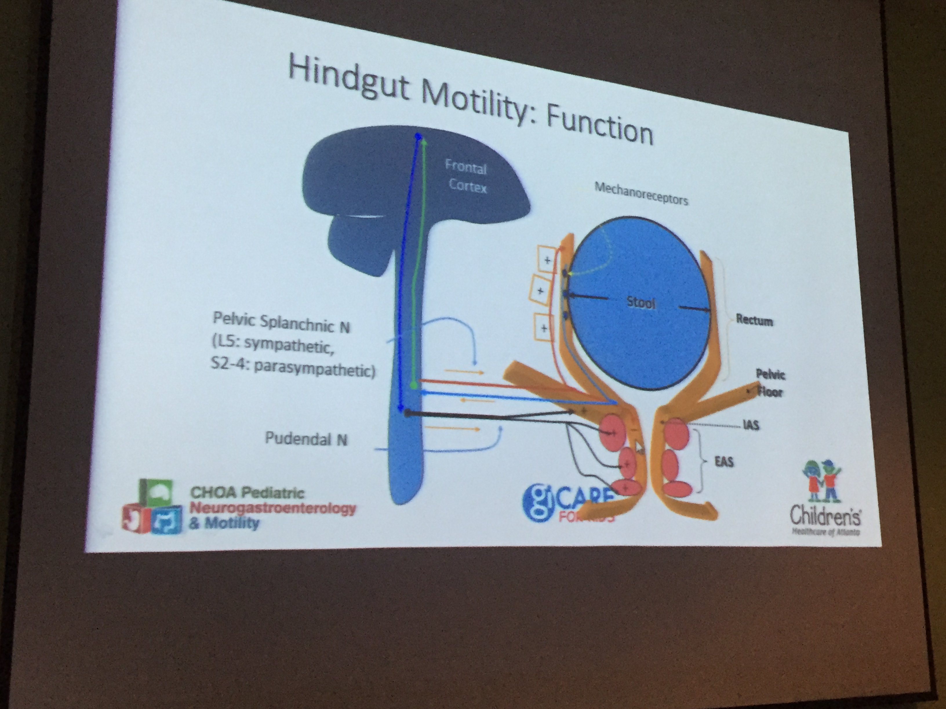
Recently I attended a terrific talk by my partner, Jose Garza.  This lecture provided a great deal of information on refractory constipation for both pediatricians and pediatric gastroenterologists alike.

Elements of the talk included diagnosis, pathophysiology and differential diagnosis.

Rome III Criteria -Helpful in Diagnosis of Constipation

Rome III Criteria -Helpful in Diagnosis of Constipation

JG1 pathophys

Is it Hirschsprung's Disease?

Is it Hirschsprung’s Disease?

Some of the more useful points.

  1. AXR should not be used to make diagnosis of constipation.
  2. Many refractory constipation patients are stooling fine and actually labeled as constipation instead of a functional abdominal pain disorder.  That is, they are complaining of stomach pain and have been erroneously told they are constipated (see point #1).
  3. Miralax remains a 1st line agent for constipation. In individuals with fecal soiling, if miralax is not working and they have had appropriate cleanout, then senna laxative may be helpful.
  4. Sitz markers are particularly helpful in proving stooling when teenager claims to not be stooling for a month and in proving functional fecal retention rather than nonretentive soiling.
  5. If good treatment is not working, then refer to neurogastroenterology.

Related blog posts:

JG3 -Help


FMT for Crohn’s Disease -Small Study

A recent prospective open-label study (Suskind DL, et al. Inflamm Bowel Dis 2015; 21: 556-63) adds a bit more information on fecal microbial transplantation (FMT) for individuals with Crohn’s disease (CD). This study included 9 patients, ages 12-19, and 11 authors.

Study details:

  • Pretreated with rifaximin 200 mg TID x 3 days and Miralax 17 gm TID x 2 days prior, and omeprazole day before and morning of procedure
  • Patients continued on prior treatment: 4 on methotrexate, 2 on thiopurine, and 3 on mesalamine (1 mesalamine patient was receiving methotrexate also)
  • Stool donor was a screened parent
  • Stool administered via NG
  • Stool DNA studied –>metagenomic sequencing

Key findings:

  • FMT engraftment was demonstrated in 7 of 9 patients
  • Mean PCDAI improved –baseline 19.7 –>6.4 at 2 weeks–>8.6 at 6 weeks
  • 5 of 9 patients who did not receive additional medical therapy were in remission at 6 and 12 weeks.

One particular advance with this study was the correlation between microbial species before and after FMT.  One speculation from the authors was that “the more divergent a Crohn’s patient is from his donor the more the potential benefit of transplantation.”  Thus, this same study with unrelated donor stool (eg. OpenBiome) would be of interest as well.

My Take: While this study offers encouragement for bigger studies, we will not know if FMT is effective (& how to administer optimally) until we have studies with more patients than authors.

Related blog posts:


Can Apple Make Research Cool?

For anyone who has looked at Apple’s March presentation, there is big news with regard to research (thanks to Seth for this information).  Here’s a link to the March announcement –around minute 16 there is the research presentation: Apple March Event

Screenshot: Rationale for Apple iPhone for Research -Large Research Pool

Screenshot: Rationale for Apple iPhone for Research -Large Research Pool

The presentation makes it clear that Apple wants to dramatically increase the participation in research studies by leveraging 700 million iPhone users.  Using an app called, “ResearchKit”, Apple has partnered with leading academic centers to help study Parkinson’s, Diabetes, Asthma, Cardiovascular disease, and Breast Cancer.  For the GI community, I hope that someone will work collaboratively to add inflammatory bowel disease to the list.

Besides increased participation, iPhone-based research has the ability to lower research costs, collect data at frequent intervals, and allow a wider demographic representation.

A shorter ~4 minute video on a separate area of the website explains ResearchKit: ResearchKit video

 Screenshot: Research Kit


Screenshot: ResearchKit

NBC News provides a condensed summary along with the caveat that there will be concerns about accuracy of data collected with ResearchKit.  That being said, most critics have not always appreciated the impact of previous Apple innovations.

Has someone from our national organization (NASPGHAN) or from ImproveCareNow started working with Apple? If not, this looks like a great opportunity.

Transoral Fundoplication for Refractory Gastroesophageal Reflux

A new endoscopic technique’s efficacy has recently been reported (Gastroenterol 2015; 148: 324-33).  Since this technique is not likely to be broadly applicable to the pediatric population for some time, I will not delve into all of the details.

In essence, a carefully selected group (n=129 from a screened group of 696) of adult patients with persistent regurgitation underwent transoral fundoplication; this eliminated troublesome regurgitation in 67% compared to 45% who were randomized to sham/PPI.  Severe complications were rare.

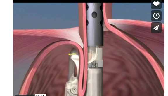
Here is a picture of the technique:

Transoral Fundoplication

Transoral Fundoplication

Link: Description and a video animation of the procedure

Bottomline: This endoscopic procedure along with the Stretta procedure and the LINX device (using magnets) offer alternatives to surgical fundoplication in carefully-selected patients with refractory gastroesophageal reflux symptoms.

New drugs approved by FDA:

Ceftolozane (Zerbaxa) -combines a cephalosporin with a beta-lactamase inhibitor (tazobactam).  Indications: complicated intra-abdominal infections (in combination with metronidazole), and complicated urinary tract infections. From FDA: FDA approves new antibacterial drug Zerbaxa

Viekira Pak -combination of 3 new drugs: ombitasvir, paritaprevir, and dasabuvir along with  older drug: ritonavir.  Indications: Hepatitis C genotype 1. From FDA: FDA approves Viekira Pak to treat hepatitis C

Related blog posts to fundoplication:

Improving MRI for NAFLD

From MedicalNewsToday – a summary of a recent Hepatology study by Jeffrey B. Schwimmer, MD and colleagues.

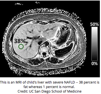
Excerpts from http://www.medicalnewstoday.com/releases/289088.php along with image.

In this study, the researchers compared a new MRI technique to the standard liver biopsy method of assessing fat in the liver. To do this, the team enrolled 174 children who were having liver biopsies for clinical care. For each patient, the team performed both MRI-estimated PDFF and compared the results to the standard pathology method of measuring fat on a liver biopsy.

Screenshot from MedicalNews

Screenshot from MedicalNewsToday

The team found a strong correlation between the amount of liver fat as measured by the new MRI technique and the grade of liver fat determined by pathology. This is an important step towards being able to use this technology for patients. Notably, the correlation was influenced by both the patient’s gender and the amount of scar tissue in the liver. The correlation between the two techniques was strongest in females and in children with minimal scar tissue.

Depending on how the new MRI technology is used, it could correctly classify between 65 and 90 percent of children as having or not having fatty liver tissue.”

“… However, further refinements will be needed before this or any other MRI technique can be used to diagnose NAFLD in an individual child.

Infantile Hemangioma -Propranolol Effective

While infantile hemangiomas are not much of a GI disorder, I still thought a recent article (NEJM 2015; 372; 735-46) merited a brief mention. This trial included 456 patients who received treatment in a randomized, double-blind trial.

Key finding: “this trial shows that oral propranolol at a dose of 3 mg per kilogram per day for 6 months is effective in the treatment of infantile hemangioma.”

  • Successful treatment noted in 60% compared with 4% of placebo patients.
  • 88% of propranolol patients showed improvement by week 5 compared with 5% in placebo group.
  • Eligible patients between 35-150 days had a proliferating hemangioma requiring systemic therapy with a minimal diameter of 1.5 cm.

Short Takes on IBD Articles

Singh S, et al. Gastroenterol 2015; 148: 64-76.  In this study, the authors identified 21 trials with 2006 participants to examine the comparative efficacy of pharmacologic interventions to prevent relapse of Crohn’s disease (CD) after surgery.  Conclusion: “anti-TNF monotherapy appears to be the most effective strategy for postoperative prophylaxis for CD.” The relative risk of clinical relapse and endoscopic relapse with anti-TNF monotherapy was estimated to be between 0.02-0.20 and 0.005-0.04, respectively. Thus, those at highest risk for recurrence, including younger individuals, smokers, penetrating CD, perianal CD, and recurrent surgeries) are most likely to benefit.(Related blog post: More Lessons in TNF Therapy (Part 1) | gutsandgrowth)

Pariente B, et al. Gastroenterol 2015; 148: 52-63. The researchers in this cross-sectional study developed the Lémann Index which measures cumulative structural bowel damage in patients with CD.  My only complaint with this study was the associated editorial on pages 8-10, titled “The Holy Grail, or Only Half Way There?”  There are too many medical advances compared to ‘the holy grail’ and, in my opinion, this shouldn’t be one of them.

Zitomersky NL et al. Inflamm Bowel Dis 2015; 21: 307-14.  In this study the authors examine the relationship between the development of antibodies to infliximab (ATI) and the risk of surgery in a cross-sectional cohort of pediatric and young adult patients.  Not surprisingly, development of ATI, which was noted in 20% of cohort, correlated with reductions in infliximab levels and higher risk of surgery.  Interestingly, prior (but not current) immunomodulator therapy was associated with lower antibody levels (P=0.007).  Perhaps, “step-up” therapy may lower the risk of ATI. (This was a point noted by James Markowitz in a previous post: More NASPGHAN Meeting Notes: IBD Hot Topics | gutsandgrowth)

Rogler G, Vavricka S. Inflamm Bowel Dis 2015; 21: 400-08. This review article discusses the exposome in IBD.  Exposures include air pollution, diet, drugs, infections, water pollution, food additives, and smoking.  These exposures influence the gut microbiome and genetic susceptibility. “Only environmental influences…explain the rising incidence in IBD worldwide. The investigation of the exposome…is an enormous challenge…[but] of crucial importance.” (Related blog post: What do you know about the “exposome”? | gutsandgrowth)

Kalmon RS. Inflamm Bowel Dis 2015; 21: 428-35. Review article provides information when there is a prior personal or family history of malignancy (=avoid thiopurines).  Figure 2 is a suggested algorithm for those with IBD and a previous diagnosis of cancer.

  • In those in which the cancer is adequately controlled, the recommendations indicate that if it has been more than 2 years since completion of therapy to use a ‘step-up’ management and favor methotrexate over thiopurines
  • In those with less than 2 years since completion of cancer treatment and not responsive to 5-ASAs/antibiotics, then “consider monotherapy with biologic agents.”
  • In those still receiving chemotherapy, the authors suggest “hold immunosuppression and follow course of IBD.  If IBD not well controlled despite chemotherapy, 5-ASAs and antibiotics, treat flares with steroids, then consider biologic agents.”

More Evidence of Anesthetic Neurotoxicity

One of the more troubling commentaries that I read recently (Rappaport BA et al NEJM 2015; 372: 796-97) provides additional insight into the issue of anesthetic neurotoxicity.

The possibility that anesthetic agents could result in learning disabilities and other neurologic impairments is not new (Pediatrics 2011); however, the data has become more concerning.

Key points:

  • “Compelling evidence from animal models is supported by a small number of observational studies in children who underwent anesthesia early in life.” Exposure to multiple (but not single) episodes of anesthesia and surgery were associated with increased risk of learning disabilities.
  • Anesthetics which have been implicated include propofol, ketamine, sevoflurane, etomidate, desflurane, and isoflurane.  Histologic changes, in animal models, have included apoptosis and cell death, changes in neuronal morphology, and decreased number of synapses.
  • “In June 2014, SmartTots convened a meeting…the participants concluded that the current data from animal studies are now sufficiently convincing that large-scale clinical studies are warranted.” SmartTots Consensus Statement
  • “Care providers should be made aware of the potential risks that anesthetics pose to the developing brain…and parents should consider how urgently surgery is needed, particularly in children under 3 years of age.”

Take-home message:  While recognizing that confounding variables make it difficult to be certain, it appears that anesthetics (particularly prolonged or repeated courses) can result in neurologic changes.  There is enough information available to recommend avoiding truly elective procedures which require anesthetics in young children.


Dropping Weight by Adding Fiber in Diet

A recent study showed that increasing fiber in the diet helped participants lose weight.  The details are noted in this LA Times story: To lose weight, experts suggest a focus on fiber

Here’s an excerpt:

If you’re trying to lose weight, you could count your calories, keep track of precisely how much salt and sugar your eat, and make sure you hit certain targets for protein, carbohydrates, cholesterol and the various types of fat. Or you could set all of that aside and concentrate on just one thing: Eating at least 30 grams of fiber each day.

In a yearlong clinical trial involving 240 obese people who had metabolic syndrome, those who focused on fiber lost almost as much weight as those who followed the American Heart Assn.’s extremely detailed dietary recommendations.

Related blog posts:

Some funny headlines form Freakomomics website –here’s one:

Screen Shot 2015-02-22 at 9.35.46 AM