Alpha-1-Antitrypsin Deficiency

A recent terrific review on Alpha-1-Antitrypsin (A1AT) Deficiency: P Strnad et al. NEJM 2020; 382: 1443-55

Background:

  • 95% of A1AT deficiency is due to homozygosity for the Z allele; prevalence of 1 in 2000 in those of European descent.
  • A1AT protects the lung tissue against attack by the enzyme neutrophil elastase.
  • The presence of A1AT genetic variants suggests that these mutations may confer a selective advantage, perhaps by amplifying the inflammatory response to invasive respiratory/gastrointestinal infections.

Pathophysiology/Clinical Features:

  • Table 1 lists the key alleles/mutations associated with A1AT deficiency -17 deficiency/null listed including: F, I, Iners, King’s, M-malton, M-procida, Pittsburgh, Queen’s, S, S-iyama, Z, QO-bellingham, QO-granitefalls, QO-hongkong
  • S allele deficiency often results in disease (emphysema, cirrhosis) in the setting of SZ heterozygotes.  The disease is typically less severe than in ZZ disease.  This allele is the most common deficiency variant (1 in 5 in Southern Europe, 1 in 30 in U.S.)
  • Z allele deficiency is the most common severe deficiency variant.  Carrier frequency: 1 in 27 persons in Northern Europe, 1 in 83 in the U.S. It is NOT seen in China, Japan, Korea, Malaysia, or Northern and Western Africa.

PI ZZ Genotype:

  • 73% of infants with PI ZZ genotype had elevated ALT level in the 1st 12 months of life
  • Cholestatic jaundice noted in 10% of infant; 15% of these infants progress to juvenile cirrhosis
  • Only 15% with abnormal ALT values by 12 years of age
  • 35% of adults with ZZ genotype show clinically-significant liver fibrosis. Risk factors for advanced fibrosis: male gender, metabolic syndrome/obesity, and alcohol consumption.

Lung Disease Due to A1AT Deficiency:

  • The clinical features of lung disease due to A1AT deficiency are “mainly indistinguishable from those of nonhereditary emphysema…this is partly why severe A1AT deficiency remains undiagnosed in approximately 90% of case, with an interval of 5 to 7 years from the onset of symptoms to diagnosis.”  When the diagnosis is late, lung disease has become irreversible.
  • Early diagnosis allows lifestyle changes (eg. smoking cessation), reduction in occupational risks, and access to therapies.

MZ Phenotype:

PI MZ genotype is more susceptible to multiple disorders, including a predisposition to COPD (at least among smokers) with odds ratio of 1.4.  Other conditions with increased risk: NAFLD-related cirrhosis (OR 3-7), Alcoholic cirrhosis, and CF-associated liver disease

Treatment:

  • Smoking cessation
  • Plasma-purified A1AT infusions.  “Randomized, controlled trials have focused on decreased loss of lung density as the primary efficacy outcome;” however, augmentation therapy has not been to shown to effect other measures, “such as FEV1, quality of life, or exacerbation of COPD.”

Related blog posts:

Also, this study was previously alluded to by this blog, but now is in print:

Briefly noted: X Lu et al. SARS-CoV-2 Infection in Children (NEJM 2020; 382: 1663-5). In 171 Chinese children with confirmed SARS-CoV-2 infection, 41.5% had fever during illness; 27 (15.8%) had no symptoms of infection or radiographic findings. Three required ICU/ventilator support; all had coexisting conditions.  One 10 month old child with intussusception died.
Stratifying Cystic Fibrosis Liver Disease with Ultrasonography

A recent multicenter case-controlled study (MJ Siegel, J Freeman, W Ye et al. J Pediatr 2020; 219: 62-9) show that ultrasonography can identify children with cystic fibrosis (CF) at increased risk for developing advanced CF liver disease; it is well-recognized that currently advanced CF liver disease occurs predominantly in children.

Among 722 participants who underwent ultrasonography as part of a planned 9 year research study, 65 had heterogeneous liver pattern and 592 had a normal liver pattern at baseline.

Key findings at planned 4-year interim analysis in a subgroup of 55 with (baseline) heterogeneous liver pattern and 116 with (baseline) normal liver pattern:

  • Those with heterogeneous liver pattern had higher median biochemical markers of liver disease: ALT 42 vs 32, GGT 36 vs 15, AST to platelet ratio index: 0.7 vs 0.4
  • Those with heterogeneous liver pattern were at ~9-fold increased risk of developing a nodular liver pattern (23% vs. 2.6%).
  • The authors did not identify specific clinical risk factors that predict the development of nodular liver ultrasound pattern

My take: Even among those with a heterogeneous pattern, the majority will not develop a ultrasonography (nodular) pattern of advanced liver disease.  Much more insight in this area is needed with the advancement in medical treatments and with the more availability of elastography.

Related blog posts:

Island Ford Nat’l Recreation Area/Chattahoochee River

Elevated Bilirubin in Newborns with Down Syndrome

Elevated bilirubin in newborns with Down syndrome has been previously reported but the frequency has not been well-described.  A recent retrospective report (TM Bahr J Pediatr 2020; 219; 140-5) compared 357 neonates with Down syndrome to 377,368 controls.

Key findings:

  • Compared with control subjects, neonates with Down syndrome had 4.7 times the risk of having an initial total serum bilirubin exceeding the 95th percentile (23.5% vs 5.0%), 8.9 times the need for phototherapy (62.2% vs 7.0%) and 3.6 times the readmission rate for jaundice (17.4 vs 4.8 per 1000 live births).

The authors note that the basis for the increased risk of hyperbilirubinemia may be early hemolysis related to “neocytolysis” which is due to destruction of RBCs following a change from low to high oxygen exposure. Other factors could include slower bilirubin conjugation/elimination and poor feeding.

My take: This study indicates that infants with Down syndrome have a substantial risk of hyperbilirubinemia.  And, while you are checking a bilirubin, it is worthwhile to obtain a direct bilirubin as cholestasis is increased in infants with Down syndrome too; the latter is often transient and/or associated with other organ involvement.

Related blog post: Neonatal cholestasis and Down syndrome

Island Ford Nat’l Recreation Area/Chattahoochee River, Sandy Springs

Autoimmune Hepatitis Outcomes, Grand Rounds on Splenomegaly, Hydroxychloroquine for SARS-CoV-2 & Zantac Warning

Here’s a commentary explaining why hydroxychloroquine is NOT proven effective:

Annals of Internal Medicine -Link: A Rush to Judgment? Rapid Reporting and Dissemination of Results and Its Consequences Regarding the Use of Hydroxychloroquine for COVID-19

Some of the key points:

  • While the study suggested more rapid clearance of SARS-CoV-2 virus at day 6 in those treated with hydroxychloroquine/azathioprine (n=20), the authors excluded 6 from the treatment group including one patient who died and three who were transferrred to the ICU.  In addition, the treatment group had a lower viral load at the start of treatment.
  • Other viral infections, including influenza, have also had in vitro data suggesting efficacy with hydroxychloroquine but this did not translate into clinical efficacy in clinical trials.
  • “The hydroxychloroquine shortage not only will limit availability to patients with COVID-19 if efficacy is truly established but also represents a real risk to patients with rheumatic diseases who depend on HCQ for their survival.”

————————–

A Di Giorgio et al (J Pediatr 2020; 218: 121-9) provide long-term data (median f/u of 14.5 years) from a retrospective review on 83 children with autoimmune hepatitis (AIH, n=54)/autoimmune sclerosing cholangitis (ASC, n=29). Median age at presentation, between 2000-2004 was 12.1 years

Key findings:

  • 29% had histologic evidence of cirrhosis at diagnosis
  • At a median followup of 14.5 years, 99% were alive, 11 underwent transplantation.  In those who underwent transplantation, 5-year and 10-year survival was 95% and 88% respectively.
  • ASC was associated with IBD in 73% of cases, compared to 33% of AIH patients.
  • Treatment: 95% of all patients had normalization with transaminases with immunosuppressive treatment (most commonly azathioprine with prednisone 2.5-5 mg/day). ASC patients also received ursodeoxycholic acid 15-20 mg/kg/day.
  • Immunologic remission: 47% achieved immunologic remission which required normal IgG levels and negative/low ANA/SMA <1:20 in addition to normal transaminases.
  • Liver transplantation was needed in 28% of ASC compared to 9% of AIH patients; overall, 83% experienced 15-year transplant-free survival. Median age of those needing a liver transplant was 19.3 years.
  • Immunosuppression withdrawal was attempted in 12 patients after a median of 4.5 years of treatment.  9 were able to stay off immunosuppression.
  • An increase in case frequency was noted during the last 4 decades at this center, from 3.6 cases/year to 5.4 case/year.
  • Four patients had isolated infrequent autoantibodies of anti-SLA (n=3) nad antiLC-1 (n=1). SLA =liver soluble antigen, LC-1 =liver cytosol antibody type 1.  Thus, in those with suspected AIH/ASC, testing for these autoantibodies is important in ~5%.
  • Pathology: 18% did not have classical features of interface hepatitis.  Instead, some had lymphocytic/lymphoplasmocytic infiltrate without spillover into the parenchyma.
  • Progression from AIH to ASC occurred in 3 patients on followup cholangiography.
  • ASC would have been overlooked in 41% if one relied on pathology alone -reaffirming need for biliary imaging.

My take: This article has a number of useful points and with an overarching message that long-term outcomes are good for children with AIH/ASC.

Related blog posts:

B Freiberg et al. 2020; 218: 221-31. This grand rounds describes the extensive workup of a 12 year old with splenomegaly ultimately due to splenic vein stenosis.  The report provides a nice review of hepatologic, hematologic, infectious, and other causes of splenomegaly as well as a work-up algorithm. (look for everything).

Initial evaluation per algorithm should start with CBC/d, retic, blood smear, liver biochemistries, GGT, coags, EBV VCA IgM, CMV IgM, Parvovirus IgM, and complete abdominal ultrasound with doppler.

Hepatologic causes of splenomegaly include the following:

  • cirrhosis with portal hypertension
  • autoimmune hepatitis/autoimmune sclerosing cholangitis
  • congenital hepatic fibrosis
  • hepatoportal sclerosis
  • nodular regenerative hyperplasia
  • storage disease and inborn errors of metabolism which includes lipidosis (Gaucher, Niemann-Pick), mucopolysaccharidoses, defects in carbohydrate metabolism (galactosemis, hereditary fructose intolerance), sea-blue histiocyte syndrome
  • anatomic disorders: portal/splenic thrombosis, Budd-Chiari, cysts, hamartomas, hemangiomas, hematoma, peliosis

Other causes of splenomegaly: infecions, hematologic-oncologic, and rheumatic disorders

Related blog posts:

The U.S. Food and Drug Administration today announced it is requesting manufacturers withdraw all prescription and over-the-counter (OTC) ranitidine drugs from the market immediately. This is the latest step in an ongoing investigation of a contaminant known as N-Nitrosodimethylamine (NDMA) in ranitidine medications (commonly known by the brand name Zantac). The agency has determined that the impurity in some ranitidine products increases over time and when stored at higher than room temperatures and may result in consumer exposure to unacceptable levels of this impurity. As a result of this immediate market withdrawal request, ranitidine products will not be available for new or existing prescriptions or OTC use in the U.S.

New FDA testing and evaluation prompted by information from third-party laboratories confirmed that NDMA levels increase in ranitidine even under normal storage conditions, and NDMA has been found to increase significantly in samples stored at higher temperatures, including temperatures the product may be exposed to during distribution and handling by consumers. The testing also showed that the older a ranitidine product is, or the longer the length of time since it was manufactured, the greater the level of NDMA. These conditions may raise the level of NDMA in the ranitidine product above the acceptable daily intake limit.

Liver Shorts March 2020 & COVID-19 Screenshots

Sofusbuvir and Ribavirin for children with hepatitis C infection (3-12 yrs, genotype 2 or 3) P Rosenthal et al. Hepatology 2020; 71: 31-43. n=54.  SVR12 was 98% (one patient did not complete treatment).

Alpha-one antitrypsin heterozygositiy contributes to cirrhosis in fatty liver disease. Liver Transplantation 2020; 26: 17-24. From the discussion: “unexpected PASD+ globules, in the context of advanced liver disease, are a specific finding that indicates the presence of a mutant A1AT allele.”  Of 196 explanted livers from NASH patients, 21 (11%) has PASD+ globules; however, among NASH patients the frequency was 47%.  Also, the Z allele was present in 10% of all tested liver explants, this exceeds the 2% rate in the general population.  Thus, in agreement with other studies, A1AT heterozygosity contributes to chronic liver failure, but may affect fatty liver disease more than other chronic liver diseases.

Durability of HBsAg Loss in Hepatitis B AS Alawad et al. Clin Gastroenterol Hepatol 2020;18: 700-09.  In this retorspective study form NIH, 89/787 HBsAg-positive patients cleared HBsA; 65 had confirmed clearance. (spontaneous in 19, post-interferon in 22, and post-NA treatment in 24). 62 of 65 remained negative after a mean time of 9.6 years. 3 patients had seroreversion at a mean of 20 months after stopping therapy, though this was transient in 2 of 3 and may have been a false-positive.

Are Medications Contributing to Obesity and Fatty Liver Disease? ~25% of U.S. adults take a prescription medication  that often produces obesity as an adverse effect. (Hales CM et al. Obesity Week 2019, Link to Abstract T-OR-2037). PRESCRIPTION MEDICATIONS THAT PROMOTE WEIGHT GAIN: Prevalence of Use Among U.S. Adults, 2013-2016 Common obesogenic medications in this cohort, (n=11,055), included all glucocorticoids, beta-blockers, and antihistamines and some agents among antidepressants, antipsychotics, antidiabetics and progestin-only contraceptives.  Medications were defined as promoting weight gain according to the Endocrine Society Clinical Practice Guideline for the Pharmacological Management of Obesity (J Clin Endocrinol Metab, 2015).

———————————————

If you have not seen this on YouTube, highly recommend this virtual choir link: Rodean School -Hallelujah


More fallout from Coronavirus: NY Times: Coronavirus May Add Billions to Nation’s Health Care Bill Insurance premiums could spike as much as 40 percent next year, a new analysis warns, as employers and insurers confront the projected tens of billions of dollars in additional costs of treating coronavirus patients.

Topical (& Tasty) Tweets:

Projected 20-Year and 30-Year Survival Rates for Pediatric Liver Transplant Recipients (U.S.)

A recent study (MG Bowring et al. JPGN 2020; 70: 356-63) provides data on pediatric liver transplantation (LT) survival rates and projected survival rates.

This retrospective cohort study included 13,442 first-time pediatric (<18) LT recipients from 1987-2018.

Key findings:

  • Projected 20-year survival rate for pediatric LT from 2007-18: 84.0%
  • Prior 20-year survival rates: 72.8% (1997-2006 cohort) and 63.6% (1987-1996 cohort)
  • Projected 30-year survival rates for pediatric LT from 2007-18: 80.1%
  • Prior 30-year survival rates: 68.6% (1997-2006 cohort) and 57.5% (1987-1996 cohort)
  • Projected outcomes with split LT (28% of 2007-2018 cohort) are similar to outcomes with whole LT

My take: While projections can overestimate and underestimate survival rates, the clear trend has been a remarkable improvement in long-term outcomes.  This published data can provide current expectations when counseling families, though with ongoing improvements in management/development of tolerance, the hope is for even better outcomes.

Related blog posts:

View from the top of Blood Mountain, Ga

“Crushing it:” Practice Guidance for Hepatitis C

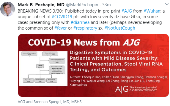
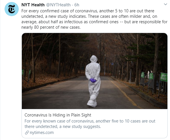
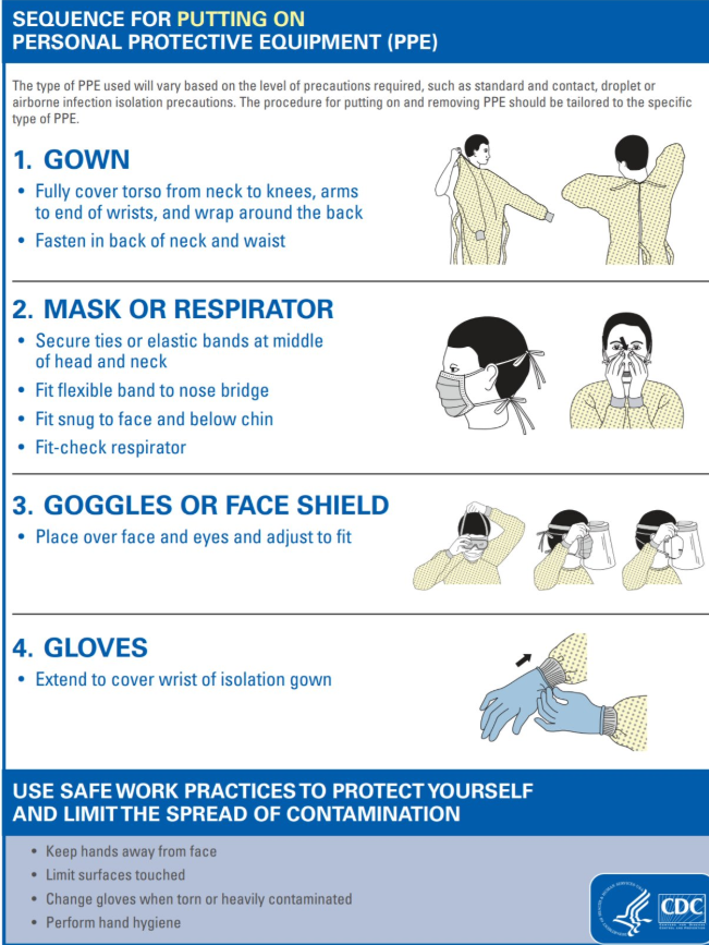
Today’s post on Hepatitis C follows a few screenshots from twitter regarding the coronavirus epidemic.

Pediatric report of coronavirus in children: NEJM Full link: SARS-CoV-2 Infection in Children A recent review of 72,314 cases by the Chinese Center for Disease Control and Prevention showed that less than 1% of the cases were in children younger than 10 years of age (n=171)…3 patients required intensive care support and invasive mechanical ventilation; all had coexisting conditions. There was one death in a 10-month-old child with intussusception had multiorgan failure and died 4 weeks after admission.

——-

As noted yesterday, this post will review a recent practice guidance for hepatitis C

Some specific recommendations for children:

Testing:

  • “All children born to HCV-infected women should be tested for HCV infection. Testing is recommended using an antibody-based test at or after 18 months of age.”
  • “Testing with an HCV-RNA assay can be considered in the first year of life, but the optimal timing of such testing is unknown” (but can be done as early as 2 months of life).
  • “The siblings of children with vertically-acquired chronic HCV should be tested for HCV infection, if born from the same mother.”

Counseling for parents:

  • “Parents should be informed that hepatitis C is not transmitted by casual contact and, as such, children with HCV infection do not pose a risk to other children and can participate in school, sports, and athletic activities, and engage in all other regular childhood activities without restrictions.”
  • “Parents should be informed that universal precautions should be followed at school and in the home of children with HCV infection. Educate families and children about the risk and routes of HCV transmission, and the techniques for avoiding blood exposure, such as avoiding the sharing of toothbrushes, razors, and nail clippers, and the use of gloves and dilute bleach to clean up blood.”

Treatment:

  • “Direct-acting antiviral (DAA) treatment with an approved regimen is recommended for all children and adolescents with HCV infection aged ≥3 years as they will benefit from antiviral therapy, regardless of disease severity.”
  • Early treatment in childhood is expected to be cost-effective compared to treatment at later ages based on previous studies

This chart provides recommendations for pediatric patients who have not received prior direct-acting antivirals. More information at HCVguidelines.org

“Crushing it:” Two More Pediatric Hepatitis C Trials

Before today’s planned blog post, I wanted to mention a good NY Times article which highlights how long the virus which causes COVID-19 can be present on surfaces:

Full link from NY Times: How Long Will Coronavirus Live on Surfaces or in the Air Around You?

An excerpt:

The virus lives longest on plastic and steel, surviving for up to 72 hours. But the amount of viable virus decreases sharply over this time. It also does poorly on copper and cardboard, surviving four to eight hours; the latter finding suggests packages that arrive in the mail should be safe — unless the delivery person has coughed or sneezed on it or has handled it with contaminated hands.

That the virus can survive and stay infectious in aerosols is also important for health care workers.

For weeks experts have maintained that the virus is not airborne. But in fact, it can travel through the air and stay suspended for that period of about a half-hour.

The virus does not linger in the air at high enough levels to be a risk to most people who are not physically near an infected person. But the procedures health care workers use to care for infected patients are likely to generate aerosols.

The original article from NEJM:  Aerosol and Surface Stability of SARS-CoV-2 as Compared with SARS-CoV-1

——–

This “C” virus was hard to cure until recently.  More good news from recently published studies for pediatric hepatitis c virus (HCV) treatment:

  • KB Schwarz et al. Hepatology 2020; 71: 422-30. 
  • MM Jonas et al. Hepatology 2020; 71: 456-62.
  • AASLD-IDSA Practice Guidance Panel. Hepatology 2020; 71: 686-721

In the first study of an all oral regimen of ledipasvir-sofosbuvir, sustained virological response at 12 weeks after dosing (SVR12) was achieved in 33 of 34 (97%) of children 3-<6 yrs of age with genotypes 1 or 4 (only 1 with type 4). No serious adverse effects were reported. Dosing: 33.75 mg/150 mg if <17 kg or 45 mg/200 mg if ≥17 kg. The one non-responder discontinued treatment due to drug taste.  Pharmokinetic studies in 13 patients confirmed appropriate medication dosing.

In the second study of glecaprevir/pibrentasvir (G/P), as part of the DORA phase 2/3 nonrandomized, open-label trial, adolescents 12-17 received the ‘adult’ regimen of 300 mg/120 mg daily for 8-12 weeks in accordance with indication duration based on adult data.  Among the 47 patients (genotypes 1, 2, 3, 4), 100% achieved SVR12. Safety profile was consistent with prior studies in adults.

The third publication, which is quite lengthy, highlights updated recommendations for HCV in adults and children (this will be reviewed in tomorrow’s post).

Related blog posts:

Biliary Atresia Biomarkers 2020

Two recent studies provide more information on biliary atresia (BA) biomarkers.

  • OG Behairy et al. JPGN 2020; 70: 344-9.
  • S Shamkar et al. JPGN 2020; 70: 350-5.

Behairy et al report on the use of serum IL-33 in a cohort of 90 infants, 30 with BA, 30 with cholestasis due to other causes, and 30 healthy infants.

  • Using a cut-off of 20.8 pg/mL, IL-33 had a specificity of 95% and sensitivity of 96.7% for identifying BA.
  • Interestingly, the test performed better in those with advanced fibrosis.  The mean value of IL-33 in those with grade 3/6 was 88.2 compared to 37.2 for 1/6 and 70.9 for 2/6. In comparison, the children with cholestasis due to other liver disease had a level of 18.5 for those with 3/6 fibrosis

The authors note in a prior study that IL-33 was higher in BA infants than those with a choledochal cyst.

While this is a small study, I disagree with the editorial (pg 278-9) which largely discounted the potential role of IL-33.  “IL-33 is elevated with many other diseases (bronchopulmonary dysplasia, asthma, allergy, and more) It, therefore, cannot easily be used as a highly specific marker for fibrosis. Furthermore, the use of IL-33 as a prognostic marker, is from a clinical point of view not of great importance, as follow-up clinical decisions are generally made based on patients’ clinical course.”

Shankar et al provide data on GGT values in BA (n=113 infants).

These infants underwent Kasai procedure at a median of 61 days

  • 12.3% had normal (<200) GGT values.
  • Those with normal GGT had worse outcomes: earlier need for liver transplantation (14 vs 20 months) and poorer transplant survival.
  • 9/14 (64%) with normal GGT and 53/99 (53.5%) of elevated GGT underwent liver transplantation

The authors note that decreased levels of GGT has been associated with reduced glutathione metabolism which could impari adaptive response to oxidative stress, leading to further hepatocyte injury.

My take: In my experience, I have had very few BA patients with GGT values <200 (lower than 10%).  The development of other biomarkers like MMP-7 and IL-33 increase the likelihood that BA will be recognized sooner and if elevated, could obviate the need for a liver biopsy prior to operative cholangiogram.  Nevertheless, practitioners cannot wholly rely on any the current biomarkers.

Related blog posts:

Fall on UNC Campus, Chapel Hill

What to Expect After Pediatric Liver Transplantation: Cognitive Function and Quality of Life

A recent study (D Ohnemus et al. Liver Transplantation 2020; 26: 45-56, editorial 9-11) examined health-related quality of life (HRQOL) and cognitive functioning approximately 15 years after liver transplantation (LT).

Study details:

Median age 16 years.  Original group was a SPLIT research cohort recruited from 20 centers and then tested at multiple time points; for this study, 8 sites of the original 20 were included.  It is noted that patients with serious neurologic injury were excluded. Among an initial group of 108, there were 79 available for potential enrollment.  In this group, 65 parent surveys were completed and 61 child surveys.

Key findings:

  • For cognitive and school functioning, 60% and 51% of parents reported “poor” functioning, respectively (>1 SD below the health mean).  41% of children rated their cognitive function as poor.
  • Adolescents’ self-reported overall HRQOL was similar to that of healthy children; in contrast, parents rated their teenage children as having significantly worse HRQOL than healthy children in all domains.
  • The cognitive score in the poor functioning group at the latest time point was lower than at first time point measurement (ages 5-6 years and at least 2 years after LT), “suggesting that difficulties intensified in adolescence for those who have problems in early childhood.”
  • Almost half had received special educational services.

The editorial notes that the PedsQL Cognitive Functioning Scale scores used by the investigators were considered subjective.  “The more objective PedsPCF scores fell within the normal range.”

My take: This report indicates that a majority of children are likely to have some cognitive deficits and many are likely to have reduced HRQOL following liver transplantation; in addition, if these problems are detected at a younger age, they are likely to persist.

Related blog posts:


Mural on Atlanta’s Beltway