Neonatal Cholestasis for Neonatologists

I recently had the opportunity to review the topic of neonatal cholestasis with my neonatal colleagues.  I reviewed two related conditions: parenteral nutrition associated liver disease (PNALD) and neonatal acute liver failure (NALF).  Some of the material incorporates recommendations from NASPGHAN cholestasis guidelines and from NASPGHAN cholestasis slidesets. Much of the slideset information is publicly available on a YouTube lecture by Dr. Linda Book (link at bottom).

Full lecture: Neonatal Cholestasis for Neonatologists
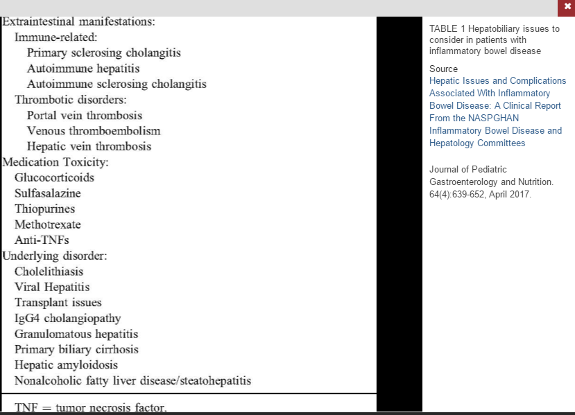
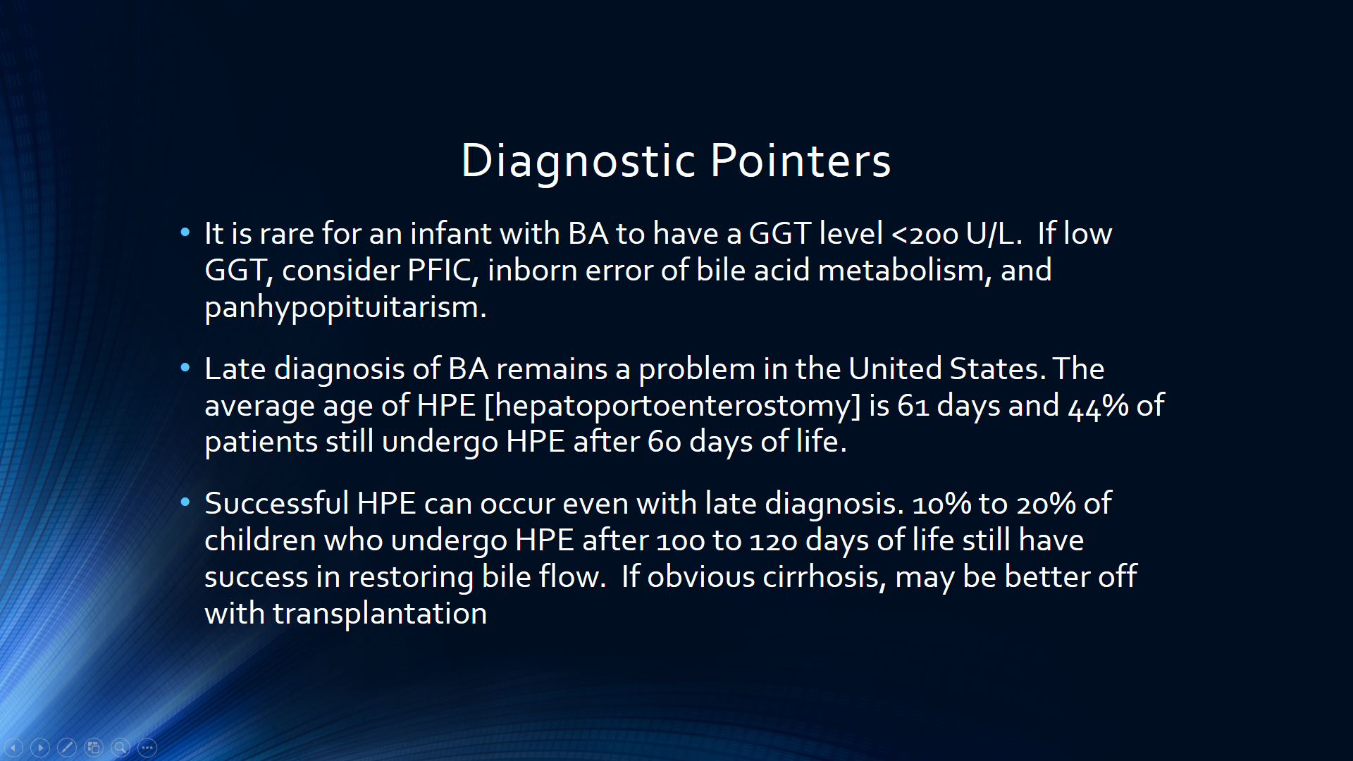
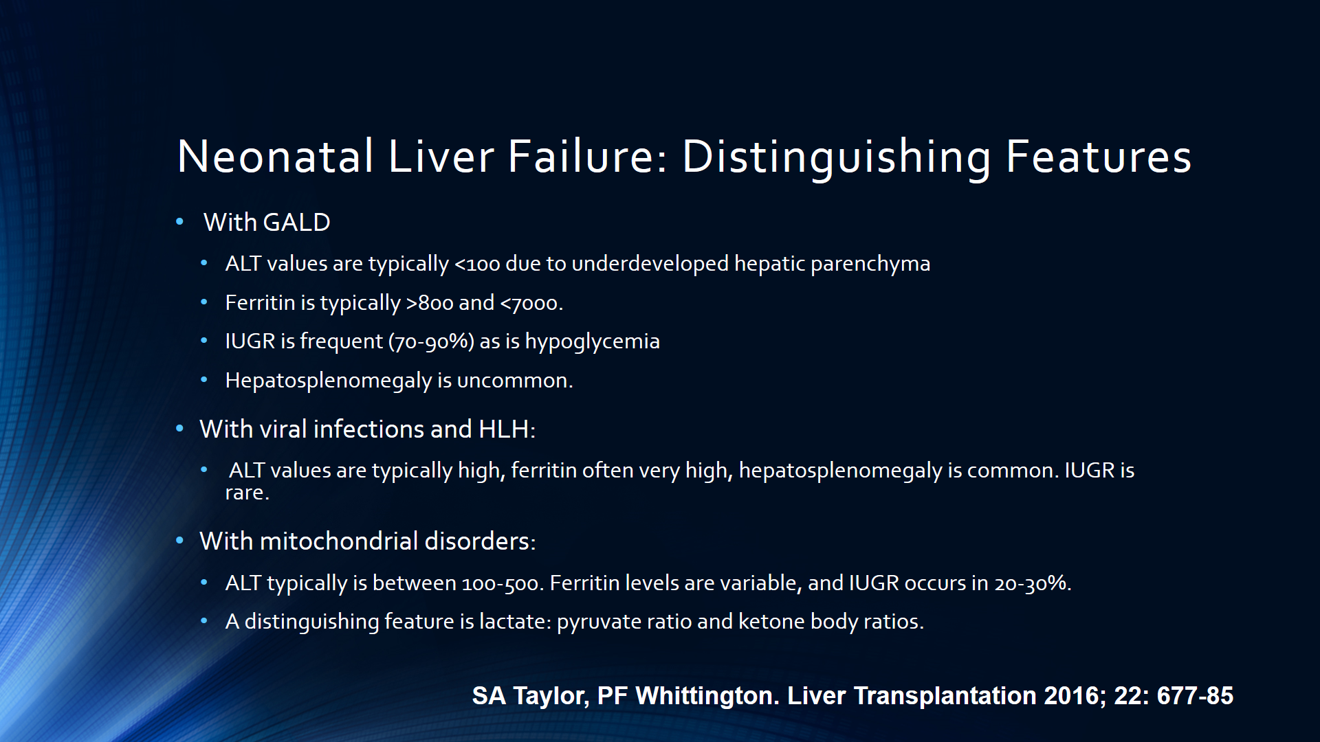
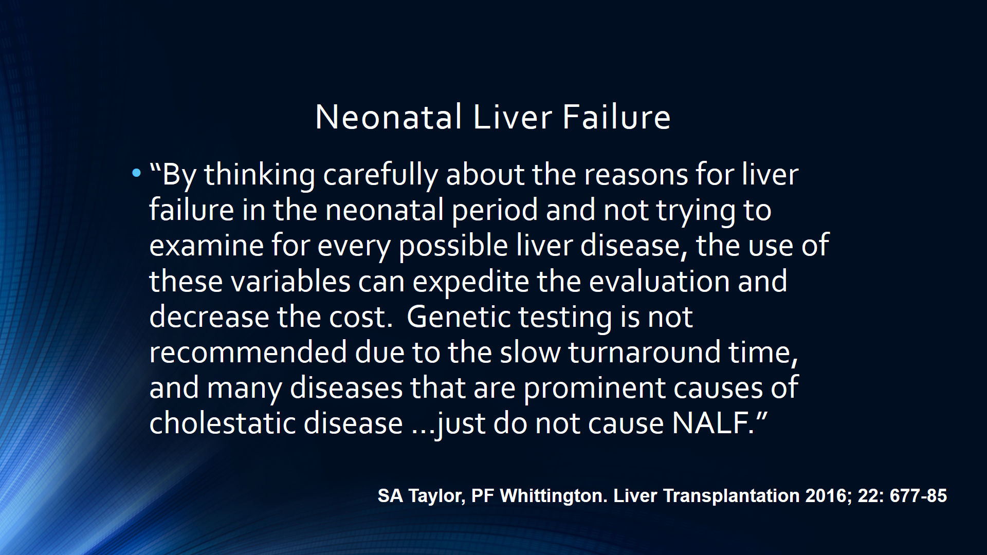
Some screenshots:

Related blog posts:

Disclaimer: These blog posts are for educational purposes only. Specific dosing of medications (along with potential adverse effects) should be confirmed by prescribing physician.  This content is not a substitute for medical advice, diagnosis or treatment provided by a qualified healthcare provider. Always seek the advice of your physician or other qualified health provider with any questions you may have regarding a condition.


Predicting Future Liver Disease with GGT Levels in Biliary Atresia Patients

A recent study (AJ Freeman, VL Ng, S Harpavat, A Hrycko, Z Apted, P Bulut, T Leong, SJ Karpen. Clin Gastroenterol Hepatol 2017; 15: 1133-35) describes the predictive value of γ-glutamyltransferase (GGT) in predicting thrombocytopenia/portal hypertension among biliary atresia patients.

In this retrospective study from three centers who had followup for at least 4 years, GGT values at 2 years of age were examined among biliary atresia patients (n=46) who continued with their native liver.

Key findings:

  • GGT ≥100 U/L had a predictive positive relationship with thrombocytopenia at 4, 5, and 6 years of age.  Patients with elevated GGT had lower platelet count (160 vs. 211) and their values continued to decline. GGT ≥100 U/L at 2 yrs predicted thrombocytopenia (<150) at age 4 with a sensitivity of 0.88, specificity of 0.57.
  • Patients with normal GGT values had “essentially stable platelet counts over the next 4 years.” GGT <100 U/L at 2 yrs predicted a low risk of thrombocytopenia with negative predictive value of 0.89, 0.92, and 0.93 at age 4, 5, and 6 respectively.

My take: This study quantitates a useful point –patients with biliary atresia and elevated GGT values are likely to develop evidence of portal hypertension.

Brevard, NC

Briefly Noted: Outpatient Liver Biopsy

A small retrospective study (R Bolia et al. JPGN 2017; 65: 86-88) with 497 patients (626 biopsies) found that all complications were identified within 8 hours.  Thirty (48%) had complications, with a subcapsular hematoma being most common (n=14).  Less common adverse events included fever (n=5), skin site ooze (n=3), intraperitoneal bleeding (n=3), hemobilia (n=2), anaphylaxis to gelfoam (n=2), and sepsis (n=1). In this study, the majority of biopsies were performed by interventional radiology (n=492); though, the complication rate was similar in both groups.

The authors conclude that their data support the outpatient liver biopsies in children.

My take: I disagree with the authors’ conclusion to some extent.  Their population is too small to detect rare but severe complications.  Our empiric practice is watch children older than 6 years of age for 6 hours and watch younger children (or others deemed at increased risk) for 24 hours.

Related blog posts:

Prague

Slim Pickings: Data for 2nd-Line Autoimmune Hepatitis Pediatric Therapy

A recent study (AN Zizzo et al. JPGN 2017; 65: 6-15) performed a systematic review and meta-analysis of pediatric autoimmune hepatitis (AIH) studies.

The most remarkable finding was that there were only 76 patients from 15 qualifying studies.

Other findings:

  • Response to mycophenolate mofetil (MMF) with 34 patients was 36% (according to abstract) at 6 months  (discrepancy in article –results state 38% response)
  • Response to cyclosporine with 15 patients was 83% (discrepancy in article –results state 86% response)
  • Response to tacrolimus with 4 patients was 50%
  • Adverse effects were very common, particularly with cyclosporine (64% noted at least 1 adverse effect)

The article has an associated editorial (N Kerkar, pg 2-3).  “The adverse event profile of cyclosporine with gingival hyperplasia, hypertrichosis, nephrotoxicity, and neurotoxicity made it challenging for long-term use in children.”  Besides the small number of patients, “the studies that were included were largely “observational”‘ which limits their findings as well.  The study authors recommend MMF as the preferred option for 2nd-line therapy.

My take: Fortunately, most patients with autoimmune hepatitis respond to first line therapy with azathioprine/steroids.  It is unclear what is the optimal 2nd-line treatment for refractory patients.

Related blog entries:

Egret, Shem Creek

Hepatitis B Reactivation Due to Immunosuppressive Therapies

The topic of Hepatitis B virus (HBV) reactivation has been discussed on this blog before (see link below).  Another excellent review on this topic (R Lomba, TJ Liang. Gastroenterol 2017; 152: 1297-1309) has been published.  The authors examine the course and mechanisms of HBV reactivation.  They divide the risk of reactivation into three groups: high, moderate and low risk and proposed management.

High risk groups, which have >10% risk of reactivation) include the following

  • B-cell-depleting agents including rituximab, ofatumumab, alemtuumab, and ibritumumab
  • High-dose corticosteroids (>20 mg/day in adults)
  • Antracyclines including doxorubicin
  • Potent TNF-α inhibitors: infliximab, adalimummab, certolizumab, and golimumab
  • Local therapy ofr HCC including TACE (transarterial chemoembolization)

Moderate groups (1-10% reactivation) include cytokine-based Rx (eg. abatacept, ustekinumab, natalizumab, vedolizumab), cyclosporine, systemic chemotherapy, moderate corticosteroid dosing

Low risk groups (<1% reactivation) include thiopurines (azathioprine, 6-mercaptopurine), and methotrexate as well as short-term low-dose corticosteroids.

Management:

  • For HBV screening, the authors recommend HBsAg and anti-HBc testing
  • Prophylactic therapy with potent oral anti-HBV therapies are recommended for those at moderate or high risk of reactivation.  In those at low risk, the options include prophylactic treatment or watchful monitoring.
  • A more detailed algorithm is provided in Figure 3.  In those with HBsAg positivity, if HBV DNA is less than 2000 U/mL, this algorithm suggests monitoring labs (HBsAg, ALT, HBV DNA every 3 months)

Mechanisms of HBV reactivation are discussed.  For example, with TNF-α inhibitors “can activate a unique host antiviral pathway, the APOBEC (apolipoprotein B mRNA editing enzyme, catlytic polypeptide-like) proteins, that cause the degradation of cccDNA in HBV-infected cells. Thus, blocking this endogenous antiviral pathway may lead to a higher HBV replication state and HBV reactivation.”

My take: In pediatric gastroenterology, we do not see a lot of HBV reactivation. Nevertheless, we do use many of the medications which can trigger HBV reactivation and need to keep these recommendations in mind.

Related blog post: What HBV Testing is Needed Before TNF Inhibitor Therapy

Suntrust Park

Acute Liver Failure -Pediatric ICU Management

Full Text Link: Intensive Care Management of Acute Liver Failure

This article provides a very good overview of this topic starting withe diagnosis, epidemiology and proceeding to specific management issues/outcomes.

Table 1 reviews etiologies –indeterminant is most common. Table 2 shown below reviews management principles and Table 3 reviews specific treatments based on etiology. Table 4 reviews grades of encephalopathy.

My take (from authors): “Despite recent advances in supportive care and the improvements in outcomes observed…the practical intensive care management of PALF remains poorly defined…Current treatment options are merely supportive and based on incomplete adult data and local institutional experience.”

Related blog posts:

Liver Briefs May 2017

Briefly noted:

O Jeanniard-Malet et al. JPGN 2017; 64: 524-7. This survey of 28 centers in France assessed clinical practice with regard to primary prophylaxis in portal hypertension. More than 75% use endoscopy to screen for varices in patients with chronic liver conditions. “In cases of grade 2 varices with red marks and grade 3 varices >90% of centres perform sclerotherapy or endoscopic variceal ligation.”

Y-D Ren et al. Hepatology 2017; 65: 1765-8. FMT for chronic HBV? This small study with 5 patients who received fecal microbiota transplantation in an effort to clear HBeAg.  There were 13 controls.  Patients in both group received either ongoing entecavir or tenofovir antiviral therapy (& had received for at least 3 years). FMT was given every 4 weeks (1 to 7 treatments). HBeAg declined gradually after each round.  Three patients in the FMT arm cleared HBeAg compared with none in the control arm.  Two of the three cleared HBeAg after on FMT and the third after two rounds of FMT.

Y Sun et al. Hepatology 2017; 65: 1438-50.  In this report, the authors propose to augment the liver biopsy classification in patients with Hepatitis B.  Their goal is to provide more information about dynamic changes regarding fibrosis using three terms:

  • Predominantly progressive: thick/broad/loose/pale septa with inflammation
  • Predominantly regressive: delicate/thin/dense/splitting septa
  • Indeteminate

Using this new designation, they characterized 71 paired liver biopsies before and after entecavir for 78 weeks.  Before treatment: 58%, 29%, and 13% for progressive, regressive and indeterminate; after treatment: 11%, 11%, and 78% respectively.

Rodin Museum, Gates of Hell


Mauriac Syndrome (Glycogenic Hepatopathy)

A case report (T Malikowski et al. Gastroenterol 2017; 152: 947-49) provides some insight into a fairly common problem –elevated liver tests in the setting of poorly controlled type 1 diabetes mellitus.  This 18-year-old had presented with a glucose of 497 mg/dL, elevated lactate, aspartate aminotransferase 257 U/L, and alanine aminotransferase 178 U/L.

The authors note that Mauriac syndrome “occurs in young patients as a result of poorly controlled type 1 diabetes mellitus.”  It may result in growth retardation, pubertal delay, and cushingoid features.

“Glycogenic hepatopathy is a underrecognized complication of Mauriac syndrome that presents with abdominal pain, nausea, vomiting, elevated serum transaminases, elevated plasma lactate levels, and hepatomegaly  The pathogenesis stems from an accumulation of glycogen in the liver…The diagnosis…is made …when all other causes of liver disease have been excluded…When glucose control is achieved, prognosis is excellent.”

My take: There are many potential reasons for elevated liver enzymes associated with type 1 diabetes mellitus, including celiac disease, and autoimmune hepatitis.  However, familiarity with glycogenic hepatopathy helps with pattern recognition and helps explain the frequent concurrence of liver disease with poorly controlled type 1 diabetes mellitus.

Liver Problems with Inflammatory Bowel Disease

A recent review (Full text: LJ Saubermann et al. JPGN 2017; 64: 639-52)  discusses the hepatic issues and complications associated with inflammatory bowel disease.

Key topics:

  • Primary Sclerosing Cholangitis (PSC)
  • Autoimmune Hepatitis (AIH)
  • Autoimmune Sclerosing Cholangitis (ASC)
  • Portal Venous Thrombosis/hypercoagulability
  • Cholelithiasis (more common in Crohn’s disease if diseased terminal ileum)
  • Viral hepatitis
  • Drug-Induced Liver Disease
  • Fatty Liver disease

Many of these topics have been discussed previously on this blog.  A couple of pointers in this review:

PSC:

  • Greater risk of colorectal carcinoma
  • IBD-PSC patients are at higher risk for pouchitis
  • GGT of >252 U/L “was highly sensitive (99%) and had good specificity (71%) for PSC” [or ASC]
  • The authors recommend “screening all newly diagnosed patients with IBD with ALT and GGT
  • Immunosuppressive therapy is NOT effective
  • Vancomycin therapy is currently being tested (clinical trials: NCT02137668 & NCT01802073)

AIH:

  • Less frequent in IBD patients than PSC
  • Most common treatment is prednisone/azathioprine
  • 40-80% of children have cirrhosis at AIH diagnosis, but “progression to end-stage liver disease is rare and …with appropriate treatment, 80% of patients achieve remission.”

ASC:

  • ASC is an overlap syndrome between AIH and PSC
  • “It is important that children with IBD and apparent AIH are routinely investigated for evidence of biliary disease with MRCP”
  • “ASC responds to the same immunosuppressive combination therapy used for AIH”

HAV/HBV Immunization:

  • HAV vaccination is effective in patients with IBD…although the rate [seroconversion] was significantly lower” in patients receiving anti-TNF therapy (92.4% vs 99.1% in one study).
  • In those needing HBV immunization: “One strategy evaluated to improve HBV immunity in adults with IBD is an accelerated course with double vaccine doses at 0, 1, and 2 months.”

Methotrexate (MTX):

  • “The extent of histological features of hepatotoxicity secondary to long-term MTX use in IBD has been infrequently described; however, the inicdence of significant abnormal histological findings appears to be rather low.”

My take: This article is a good starting point for liver-related issues in IBD.  For concerns regarding medications, the NIH livertox website is more useful and much more comprehensive.

Related blog entries:

DILI:

PSC:

AIH:


Proof That Diet Changes Can Improve a Fatty Liver

A recent prospective study (M Markova, O Pivovarova, et al. Gastroenterol 2017; 152: 571-85) showed that among individuals with nonalcoholic fatty liver disease (NAFLD) and type 2 diabetes that a diet high in protein (animal or plant) reduced liver fat over a 6 week period.

Among 37 participants, body fat and intrahepatic fat were detected with MRI and spectroscopy, respectively. Protein was increased to 30% of the diet. Fat was reduced to 30% and carbohydrates to 40% of diet composition. .

Key findings:

  • With a high animal protein diet, liver fat was reduced by 36%.  In the high plant protein diet group, liver fat was reduced by 48%.
  • Theses changes were unrelated to change in body weight.  However, these changes were correlated with down-regulation of lipolysis and lipogenic indices.

Some of the findings may be limited to older patients as this cohort was older than 60 years of age.  In the pediatric population, the dietary factor that has been linked most closely to NAFLD has been fructose, mainly in sugar-sweetened beverages (R Patusco et al. Top Clin Nutr 2017; 32: 27-46 -thanks to Ben Gold for this reference).

My take: This study shows improvement in liver fat with increased protein/reduced dietary fat.  While this study indicates that dietary modification is important in treating NAFLD, the optimal dietary intervention (eg. higher protein, lower sugar, lower fat) remains uncertain.

Related posts: