Opiates, Inflammatory Bowel Disease and Mortality

A recent retrospective study (NE Burr et al. Clin Gastroenterol Hepatol 2018; 16: 534-41) with 3517 patient’s with Crohn’s disease (CD) and 5349 with ulcerative colitis (UC) examined the frequency of opioid prescriptions and the relationship to fatal outcomes.

Key findings:

  • Compared to 1990-93, the period of 2010-13 saw a sharp rise in the use of opiods in England: 10% compared to 30%.
  • Prescription of strong opioids (>3 prescriptions per calendar year) was associated with premature mortality: Hazard ratio 2.18 for CD and 3.3 for UC.

This study is in agreement with other data showing increasing use of opiate prescriptions worldwide for chronic noncancer pain (although there has been a drop in the past year).  As with other studies of patients with inflammatory bowel disease, this study shows an association between opioid use and mortality.

My take: Needing an opioid may be a marker for more severe disease. Whether the opioid use directly contributes to mortality remains unclear.


113 Recommendations for Crohn’s Disease Management from ACG

Full Text Link: ACG Clinical Guideline: Management of Crohn’s Disease. GR Lichtenstein et al. Am J Gastroenterol 2018; 113:481–517

A few of the recommendations from Table 1:

  • (Insurance companies –please read this one): #1 Fecal calprotectin is a helpful test that should be considered to help differentiate the presence of IBD from irritable bowel syndrome (IBS) (strong recommendation, moderate level of evidence).
  • #9 Perceived stress, depression, and anxiety, which are common in IBD, are factors that lead to decreased health-related quality of life in patients with
    Crohn’s disease, and lead to lower adherence to provider recommendations. Assessment and management of stress, depression, and anxiety should be
    included as part of the comprehensive care of the Crohn’s disease patient (strong recommendation, very low level of evidence)
  • #24, 25 Anti-TNF agents (infliximab, adalimumab, certolizumab pegol) should be used to treat Crohn’s disease that is resistant to treatment with corticosteroids (strong recommendation, moderate level of evidence). Anti-TNF agents should be given for Crohn’s disease refractory to thiopurines or methotrexate (strong recommendation, moderate level of evidence).
  • #26 Combination therapy of infliximab with immunomodulators (thiopurines) is more effective than treatment with either immunomodulators alone or
    inflximab alone in patients who are naive to those agents (strong recommendation, high level of evidence).
  • #27 For patients with moderately to severely active Crohn’s disease and objective evidence of active disease, anti-integrin therapy (with vedolizumab) with
    or without an immunomodulator is more effective than placebo and should be considered to be used for induction of symptomatic remission in patients with
    Crohn’s disease (strong recommendation, high level of evidence).
  • #30 Ustekinumab should be given for moderate-to-severe Crohn’s disease patients who failed previous treatment with corticosteroids, thiopurines, methotrexate, or anti-TNF inhibitors or who have had no prior exposure to anti-TNF inhibitors (strong recommendation, high level of evidence).
  • #46 Oral 5-aminosalicylic acid has not been demonstrated to be effective for maintenance of medically induced remission in patients with Crohn’s disease,
    and is not recommended for long-term treatment (strong recommendation, moderate level of evidence).
  • # 58 In high-risk patients, anti-TNF agents should be started within 4 weeks of surgery in order to prevent postoperative Crohn’s disease recurrence
    (conditional recommendation, low level of evidence).

From Table 2:

  • #9 Symptoms of Crohn’s disease do not correlate well with the presence of active inflammation, and therefore should not be the sole guide for therapy. Objective evaluation by endoscopic or cross-sectional imaging should be undertaken periodically to avoid errors of under– or over treatment.
  • #23 Routine use of serologic markers of IBD to establish the diagnosis of Crohn’s disease is not indicated.
  • #30 Small bowel imaging should be performed as part of the initial diagnostic workup for patients with suspected Crohn’s disease.
  • #44 Insufficient data exist to support the safety and efficacy of switching patients in stable disease maintenance from one biosimilar to another of the same biosimilar molecule.

Disclaimer: These blog posts are for educational purposes only. Specific dosing of medications/diets (along with potential adverse effects) should be confirmed by prescribing physician/nutritionist.  This content is not a substitute for medical advice, diagnosis or treatment provided by a qualified healthcare provider. Always seek the advice of your physician or other qualified health provider with any questions you may have regarding a condition.

Eliminating Gluten Challenge for the Diagnosis of Celiac Disease

Many patients receive a gluten-free diet (GFD) prior to a definitive diagnosis of celiac disease.  The diagnostic yield of serology can significantly decrease within a month after institution of a GFD.  A recent study (VK Sarna et al. Gastroenterol 2018; 154:886-96) has identified an HLA-DQ-Gluten Tetra

mer Blood test which can accurately identify celiac disease despite the implementation of a GFD.  This test quantifies HLA-DQ-gluten tetramer binding to T cells with flow cytometry. Key findings:

  • For patients receiving a GFD, the sensitivity was 97% and the specificity was 95% for the diagnosis of celiac disease
  • For patients not receiving a GFD, the sensitivity was 100% and the specificity was 90% for the diagnosis of celiac disease

My take: An accurate test to determine if celiac disease is present for those who have started a GFD would be quite helpful.  This HLA-DQ-Gluten Tetramer blood test still needs further validation in more patient populations. This test is NOT commerically-available at this time.

Morgan Falls

Related blog posts:

Disclaimer: These blog posts are for educational purposes only. Specific dosing of medications (along with potential adverse effects) should be confirmed by prescribing physician.  This content is not a substitute for medical advice, diagnosis or treatment provided by a qualified healthcare provider. Always seek the advice of your physician or other qualified health provider with any questions you may have regarding a condition.

Chattahoochee river -Morgan Falls

POEM for Achalasia in Children

Briefly noted: S Miao et al. JPGN 2018; 66: 257-62.  In this retrospective study, the authors examined the use of peroral endoscopic myotomy (POEM) in children.  They , describe a successful outcome in all 21 patients (range 11 months to 18 years).  Complications included subcutaneous emphysema (n=4), pneumoperitoneum (n=1), mediastinal emphysema ((n=4), pneumonia (n=1) and mucosal injury (n=1). The authors: “although Heller myotomy is still widely accepted as the standard treatment for achalasia in children, POEM …may provide a better treatment …due to less hospitalization, less trauma, …and long-term efficacy.”

Related blog posts:

Amber Cove, Dominican Republic

Capsule Endoscopy More Sensitive than MRE for Crohn’s Disease

Briefly noted: B Gonzalez-Suarez et al. IBD 24: 775-80.

In 47 patients with established (n=32) or suspected Crohn’s disease (n=15), MRE was first performed to exclude strictures and then subsequently capsule endoscopy (CE) (with patency capsule in 10 patients). Key finding: Small bowel lesions were found in 36 of 47 with CE compared with 21 of 47 with MRE (76.6% vs 44.7%, P=0.001)

Related blog post: Head-to-Head: Capsule endoscopy compared to colonoscopy

Time Will Tell: Granulomatous Upper GI Inflammation

A recent retrospective study (K Queliza et al. JPGN 2018; 66: 620-23) describes seven patients with granulomatous disease in the upper GI tract who were diagnosed with ulcerative colitis.

This study examined patients at a single center between 2007-2016 with ages ranging from 2 years to 17 years.  Median time of followup is not provided.  Two patients required colectomy.  All patients had non-casseating granulomas identified in either the stomach or duodenum (or both) along with moderate to severe pancolitis.  All of the patients had extensive investigations, generally cross-sectional imaging (MRE or CT) or capsule endoscopy

Key point::

  • “The final classification of IBD was based on expert opinion from gastroenterologists, radiologists, and pathologists upon thorough review of the medical records.”

My take: This study highlights the confusion of the essentially binary classification of IBD into either Crohn’s disease or ulcerative colitis, when in fact there are hundreds of genetic mutations which give rise to inflammatory bowel disease.  Given that granulomas are a hallmark of Crohn’s disease and there are no pathognomic features of ulcerative colitis, only time will tell if these patients have an ulcerative colitis phenotype.  I wonder how many centers would take exception to this classification and describe these patients as ‘indeterminate’ colitis/IBDU (IBD unclassified).

Related blog posts:

Lactobacillus rhamnosus GG Associated with Increased Necrotizing Enterocolitis in Observational Study

A recent retrospective study (AF Kane et al. J Pediatr 2018; 195: 73-9) with 640 VLBW infants found that the probiobiotic, Lactobacillus rhamnosus GG (LGG), was associated with an increased risk of necrotizing enterocolitis (NEC).

LGG supplementation was started at a median age of 6 days at a dose of 2.5 to 5 x 10 to the 9th CFU/day.

Key finding:

  • LGG group had an aOR of 2.10 for developing NEC.  LGG group NEC incidence was 16.8% whereas NEC incidence was 10.2% prior to institution of LGG.

The authors note their findings are in contrast to findings from 38 randomized trials (10,520) which have found that probiotics lowered the risk of NEC.

My take: This study reinforces the need for further studies to identify which factors and probiotic strains are likely to lead to reduced rates of NEC.

Related blog posts:

Joint Mobility –Not Associated with Increased Functional GI Disorders

According to a recent study (M Saps et al. JPGN 2018; 66: 387-90), joint hypermobility is not associated with an increased risk of functional gastrointestinal disorders (FGIDs).

From a school-based study of 654 children from a public school in Cali, Columbia, 148 (22.6%) were identified as having an FGID. Among this group, 136 children participated in the study along with 136 age/sex-matched healthy controls. Joint laxity was assessed to establish a Beighton score.

Key finding:

  • There was no significant difference in joint laxity between the FGID group and the control group, with OR of 1.03.

The implication of this study is that previous associations between joint hypermobility (JH) and FGIDs could be due to selection bias at tertiary care centers.  Alternatively, “it is possible that the association between FGIDs and JH exists, but it is only limited to a subset of patients that consult at specialized clinics.”

My take: This article challenges the idea that JH increases the risk of FGID.  Based on this study, if JH is a risk factor, it is hard to detect in a general population.

Related blog post:


Amber Cove, Dominican Republic

Vedozlizumab -Detectable in Breastmilk

A recent correspondence (M Julsgaard et al.Gastroenterol 2018; 154: 752-65) shows that vedolizumab is detectable in varying concentrations in breastmilk.  The authors collected samples from 5 mothers who were receiving vedolizumab (VDZ) for inflammatory bowel disease.

Key findings:

  • Peak VDZ concentration in breastmilk was 0.318 mcg/mL which was 1/179th of the corresponding concentration of the maternal serum levels.
  • The authors calculated a maximum oral dose of 0.048 mg/kg/day for breastfed infants based on this peak level. “This minute quantity is furthermore anticipated to undergo proteolysis in the stomach” and be bound/excreted in GI tract.
  • VDZ was detectable in all samples for 30 minutes prior to infusion (trough) through 14 days.

This study is in agreement with another study showing that levels in the breastmilk were minute (~1/100) of serum levels (A Lahat et al. J Crohns Colitis. 2018 Jan 5;12(1):120-123).

My take: These are low levels of VDZ –nevertheless further monitoring of infants to determine conclusively whether VDZ enterally causes any adverse effects is warranted.

Views from Mike O’Callahan -Pat Tillman Bridge


Pediatric Home and Office Biologic Infusions -What is Needed

A recent clinical report (E Barfield et al. JPGN 2018; 66: 680-86) will be influential.  This guideline is from the North American Society for Pediatric Gastroenterology, Hepatology, and Nutrition.  Congratulations to my partner, Chelly Dykes, who is one of the coauthors.

Full textAssuring Quality for Non-Hospital Based Biologic Infusions in Pediatric Inflammatory Bowel Disease: A Clinical Report from the North American Society for Pediatric Gastroenterology, Hepatology and Nutrition

For many years, our office has had an office-based infusion center which has provided infusions in a safe and cost-effective manner.  Recently, there have been some situations in which home-based infusions have been proposed either to lower costs and/or for convenience.  This report succinctly describes the hurdles that need to be addressed before recommending this treatment pathway. As noted below, patient safety encompasses a great deal more than infusion reactions. Delays in infusions (which can increase risk of loss of response) due to reactions and lapses in communication are additional issues.

Recommendation 1: Home- or office-based infusions should ensure safe administration of the biologic infusion, provide reliable execution of infusion-related orders (eg, laboratories for therapeutic drug monitoring, dose optimization protocols, etc), and be equipped to recognize and respond to potential complications.

  • Infusion reactions:  ” Infusion reactions associated with infliximab and vedolizumab can range from mild reactions such as fever and chills, dyspnea, pruritus, or urticaria (in approximately 5%–10%), to severe reactions including anaphylaxis, convulsions, and hypotension (<1%)”
  • Emergencies: “In the event of an urgent or emergent reaction during home- or office-based infusions, the in-home services agency (IHSA) nurse needs to be able to contact the appropriate ordering medical team member expeditiously by phone or pager to review/clarify specific concerns or needs to have an established clear policy on how to proceed with managing the reaction.” 
  • Communication: “We identified the lack or inconsistency of on-call coverage by the primary medical team when home- or office-based infusions occur as a significant barrier to safely initiating or continuing home- or office-based infusion programs. Difficulty in reaching a knowledgeable team member is a breach in reliable care and represents serious patient risk.”
  • Related work: “In addition to administering the biologic infusion, executing all other infusion-related orders is an important safety consideration. Implementing unique home infusion protocols is linked to treatment efficacy.”  

Recommendation 2: Pediatric home- or office-based infusions, particularly for patients 12 years and younger, should be staffed by a pediatric nurse professional with Pediatric Advanced Life Support (PALS) certification and clinical experience with pediatric patients.

Recommendation 3: Evidence-based standard of care for biologic therapy maximizing effectiveness and treatment sustainability should be established before initiating home or office-based infusions.

Recommendation 4: Home- or office-based infusion pathways that decrease opportunity loss for patients and families and deliver high-quality, patient-centered care should be supported and reproduced.

Recommendation 5: Pediatric gastroenterologists should ensure appropriate shared liability with IHSAs to deliver high-quality care in home-based infusions for children by executing pragmatic steps as outlined below:

  1. “Document discussion with the patient and family about the indication, risks, and adverse event management …
  2. Refer the patient to an accredited, licensed IHSA based on patient’s insurance coverage. If no accredited, licensed IHSA for the pediatric patient exists, this is grounds for not initiating home- or office-based infusions…
  3. & 4. Use an infusion protocol… with clear directives on recognition of signs/symptoms of reactions and administration of reaction medications and use of EMS or parent transport to an emergency room.
  4. Maintain accurate documentation and communication of therapy type, dose, and frequency.
  5. Provide a reliable communication mechanism for the IHSA to notify provider of changes or infusion-related events
  6. Regularly reviewing ongoing IHSA performance with regard to delivery of services, accurate laboratory ordering and turnaround time, safety and quality concerns and timely redressal of these issues.
  7. Switch to another IHSA if the performance reliability is unsatisfactory. …we acknowledge that changing IHSAs may be difficult.”

Recommendation 6: A more equitable division of labor should be established to offset increased administrative burden placed on the pediatric gastroenterologist and medical team to effectively facilitate and maintain home- or office-based infusions, especially when driven by payer-mandated policies.

Recommendation 7: …Among patients receiving home- or office-based infusions, unreliable follow-up care with the provider as scheduled is grounds for discontinuation of home- or office-based biologic therapy.

Recommendation 8: A proper appeals process should be in place to prevent cost transference from payer to patient in payer-mandated decisions for home- or office-based infusions.

Our office practice:

  • Emergencies: In our office, there is always one physician dedicated to being available to assess patients who are receiving infusions.  This helps insure safety and in addition, helps to make sure that minor medical problems do not needlessly postpone important treatment.
  • Documentation: With our office-based infusions, each infusion is documented by the administering nurse.  This documentation along with labs are embedded in the medical record (EPIC) to help modify treatment.
  • Communication: In our office, prior to each infusion, each patient’s chart is reviewed and specific orders are given.  This assures that needed blood tests/imaging, additional treatments (eg. iron infusion), insurance authorizations, necessary followup, and personalized adjustments are made.  This type of communication needs to be replicated for home-based infusions; hence, the use of home-based infusions could result in a huge increase in uncompensated work for the treating physician.

My take: In my experience, office-based infusions can be provided safely and in a cost-effective manner.  While the convenience of home-based infusion is desirable, before implementing broadly, issues regarding communication, safety protocols, and documentation to allow modifications in therapy need to be proactively addressed. Families may not realize some of the complexities involved in managing infusions and how these issues could affect their child’s long-term response to biologic therapy.

Related blog posts:

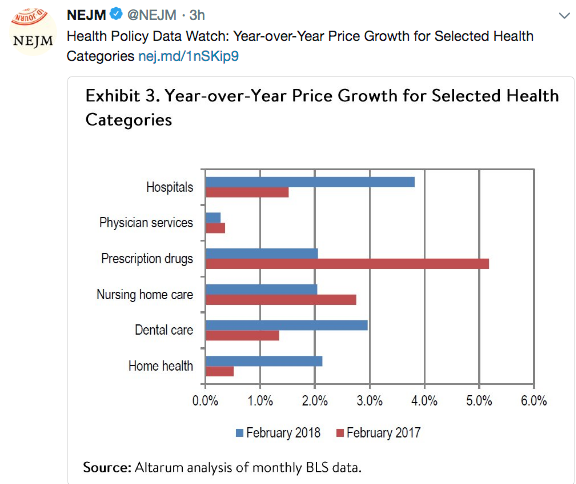
The following image relates to another convenience-related health trend:

Disclaimer: These blog posts are for educational purposes only. Specific dosing of medications/diets (along with potential adverse effects) should be confirmed by prescribing physician/nutritionist.  This content is not a substitute for medical advice, diagnosis or treatment provided by a qualified healthcare provider. Always seek the advice of your physician or other qualified health provider with any questions you may have regarding a condition.