PEG 3350 is Not Associated with Elevated Glycol Levels

Everyday parents ask me if Miralax (polyethylene glycol) is safe; this has been driven by social media claims of neurotoxicity and by articles in the NY Times (see prior blog references) indicating that more testing is needed.

A recent study (KC Williams et al. J Pediatr 2018; 195: 148-53) examines one of the areas of concern, whether miralax could result in toxic levels of glycols.  In this study with 9 treated children (ages 6-12 years) and 18 controls, careful study of potentially toxic agents, ethylene glycol (EG), diethylene glycol (DEG), and triethylene glycol (TEG), were measured every 30 minutes for 3 hours after receiving 17 g of PEG 3350.

Key findings:

  • Baseline blood levels of EG (390.51 ng/mmL) and TEG (2.21 ng/mL) did not differ between control and treated groups
  • Baseline DEG levels were lower in the PEG 3350 group (40.12 ng/mL vs 92.83 ng/mL, P=.008)
  • After PEG 3350 dose, EG and TEG levels remained well below toxic levels; DEG levels did not change.  The increases in EG and TEG, which peaked at 90 minutes, were not sustained at levels different from controls.
  • EG peaked at 1032.8 ng/mL. TEG peaked at 35.17 ng/mL
  • The highest levels of EG and DEG were actually identified in control patients. Thus, “all children are exposed routinely and have measureable amounts in the blood.”

With regard to TEG toxicity, in the discussion, the authors note that, based on animal studies, “very large doses of TEG are needed to cause side effects.” Even doses of 4000 mg/kg of TEG daily for 90 days did not result in local or systemic toxicity.  The authors note that TEG concentration in PEG 3350 is “approximately 22.1-30.6 mcg per 17 gram dose of PEG 3350.”

With regard to EG and DEG, “the average EG and DEG content of the PEG samples in this study were a 100 and 800 times less, respectively, than this required 0.2% cutoff” [FDA limit].  The agency of Toxic Substances and Disease Registry profile for EG, has indicated that “EG blood levels greater tan 0.2 mg/mL are needed for acute toxic poisoning.  The average level of EG at the 90-minute peak of 1100 +/- 350 ng/mL was 182 times lower than this level.”  For chronic exposure EG toxicity, the authors estimate that one would need to take “40 capfuls [17 gram each] of PEG 3350 per day for up to a year.”  The EPA also has advisories with regard to EG.  To achieve toxic levels for a 10-kg child, this would necessitate that the child “would have to drink 1 L of water with 50 capfuls (858 g) in 1 day or 15 capfuls (258 g) per day for 10 days.”

An important limitation of this study is that there may be other metabolites that are not measured that could cause neurotoxicity.

My take: This study shows that the theoretical risk of glycol toxicity is highly unlikely.  My advice for miralax usage: (borrowed from expert review): “Generally speaking, if your child has been prescribed PEG 3350 as part of his/her treatment plan, and you feel this medicine provides benefit, you should feel safe continuing PEG 3350. At this time, PEG 3350 appears to be safe based on current medical literature. We recommend discussing any concerns you have about the safety of PEG 3350 with your child’s health care provider. If you would prefer for your child to stop taking PEG 3350, discuss other treatments options with your child’s health care team before stopping PEG 3350 therapy. Although abruptly stopping PEG 3350 is not considered dangerous, it could lead to a relapse/worsening of constipation.”

Disclaimer: These blog posts are for educational purposes only. Specific dosing of medications/diets (along with potential adverse effects) should be confirmed by prescribing physician/nutritionist.  This content is not a substitute for medical advice, diagnosis or treatment provided by a qualified healthcare provider. Always seek the advice of your physician or other qualified health provider with any questions you may have regarding a condition.

Related blog posts:

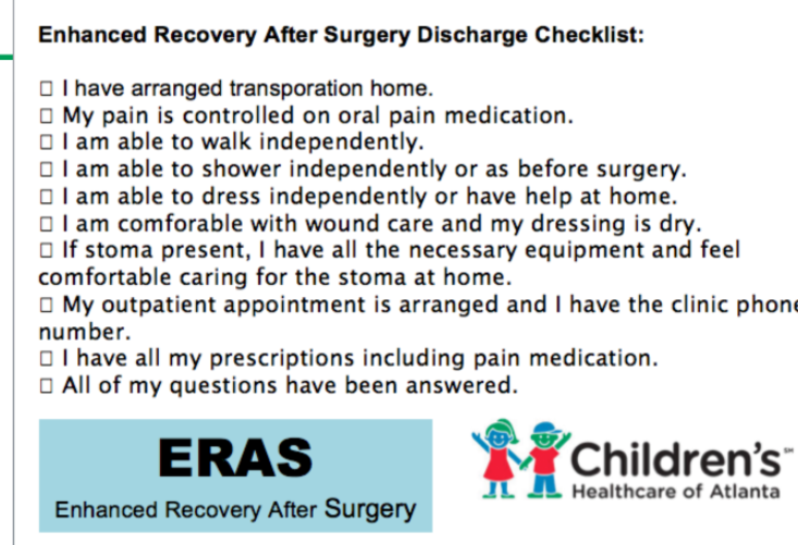
Fast Track Recovery/Enhanced Recovery After Surgery (ERAS is Awesome!)

At a recent ImproveCareNow population management meeting for our group, Dr. Kurt Heiss provided an update on the expanding use of ERAS.  In addition to colorectal surgery, uses at our hospital system have included bariatric surgery, craniofacial surgery, and umbilical hernia repairs. The results of this bundled care show fewer complications, less pain/less narcotics (more blocks), and shorter hospital stays (without increased readmission rates).

For those who are not as familiar as they would like (and for patients), I recommend a 7 minute Lego ERAS YouTube link: LEGO Surgery -Enhanced Recovery After Surgery

Related blog post: ERAS -Enhanced Recovery After Surgery (2016) With full slide set explaining ERAS further


Opiates, Inflammatory Bowel Disease and Mortality

A recent retrospective study (NE Burr et al. Clin Gastroenterol Hepatol 2018; 16: 534-41) with 3517 patient’s with Crohn’s disease (CD) and 5349 with ulcerative colitis (UC) examined the frequency of opioid prescriptions and the relationship to fatal outcomes.

Key findings:

  • Compared to 1990-93, the period of 2010-13 saw a sharp rise in the use of opiods in England: 10% compared to 30%.
  • Prescription of strong opioids (>3 prescriptions per calendar year) was associated with premature mortality: Hazard ratio 2.18 for CD and 3.3 for UC.

This study is in agreement with other data showing increasing use of opiate prescriptions worldwide for chronic noncancer pain (although there has been a drop in the past year).  As with other studies of patients with inflammatory bowel disease, this study shows an association between opioid use and mortality.

My take: Needing an opioid may be a marker for more severe disease. Whether the opioid use directly contributes to mortality remains unclear.


113 Recommendations for Crohn’s Disease Management from ACG

Full Text Link: ACG Clinical Guideline: Management of Crohn’s Disease. GR Lichtenstein et al. Am J Gastroenterol 2018; 113:481–517

A few of the recommendations from Table 1:

  • (Insurance companies –please read this one): #1 Fecal calprotectin is a helpful test that should be considered to help differentiate the presence of IBD from irritable bowel syndrome (IBS) (strong recommendation, moderate level of evidence).
  • #9 Perceived stress, depression, and anxiety, which are common in IBD, are factors that lead to decreased health-related quality of life in patients with
    Crohn’s disease, and lead to lower adherence to provider recommendations. Assessment and management of stress, depression, and anxiety should be
    included as part of the comprehensive care of the Crohn’s disease patient (strong recommendation, very low level of evidence)
  • #24, 25 Anti-TNF agents (infliximab, adalimumab, certolizumab pegol) should be used to treat Crohn’s disease that is resistant to treatment with corticosteroids (strong recommendation, moderate level of evidence). Anti-TNF agents should be given for Crohn’s disease refractory to thiopurines or methotrexate (strong recommendation, moderate level of evidence).
  • #26 Combination therapy of infliximab with immunomodulators (thiopurines) is more effective than treatment with either immunomodulators alone or
    inflximab alone in patients who are naive to those agents (strong recommendation, high level of evidence).
  • #27 For patients with moderately to severely active Crohn’s disease and objective evidence of active disease, anti-integrin therapy (with vedolizumab) with
    or without an immunomodulator is more effective than placebo and should be considered to be used for induction of symptomatic remission in patients with
    Crohn’s disease (strong recommendation, high level of evidence).
  • #30 Ustekinumab should be given for moderate-to-severe Crohn’s disease patients who failed previous treatment with corticosteroids, thiopurines, methotrexate, or anti-TNF inhibitors or who have had no prior exposure to anti-TNF inhibitors (strong recommendation, high level of evidence).
  • #46 Oral 5-aminosalicylic acid has not been demonstrated to be effective for maintenance of medically induced remission in patients with Crohn’s disease,
    and is not recommended for long-term treatment (strong recommendation, moderate level of evidence).
  • # 58 In high-risk patients, anti-TNF agents should be started within 4 weeks of surgery in order to prevent postoperative Crohn’s disease recurrence
    (conditional recommendation, low level of evidence).

From Table 2:

  • #9 Symptoms of Crohn’s disease do not correlate well with the presence of active inflammation, and therefore should not be the sole guide for therapy. Objective evaluation by endoscopic or cross-sectional imaging should be undertaken periodically to avoid errors of under– or over treatment.
  • #23 Routine use of serologic markers of IBD to establish the diagnosis of Crohn’s disease is not indicated.
  • #30 Small bowel imaging should be performed as part of the initial diagnostic workup for patients with suspected Crohn’s disease.
  • #44 Insufficient data exist to support the safety and efficacy of switching patients in stable disease maintenance from one biosimilar to another of the same biosimilar molecule.

Disclaimer: These blog posts are for educational purposes only. Specific dosing of medications/diets (along with potential adverse effects) should be confirmed by prescribing physician/nutritionist.  This content is not a substitute for medical advice, diagnosis or treatment provided by a qualified healthcare provider. Always seek the advice of your physician or other qualified health provider with any questions you may have regarding a condition.

Eliminating Gluten Challenge for the Diagnosis of Celiac Disease

Many patients receive a gluten-free diet (GFD) prior to a definitive diagnosis of celiac disease.  The diagnostic yield of serology can significantly decrease within a month after institution of a GFD.  A recent study (VK Sarna et al. Gastroenterol 2018; 154:886-96) has identified an HLA-DQ-Gluten Tetra

mer Blood test which can accurately identify celiac disease despite the implementation of a GFD.  This test quantifies HLA-DQ-gluten tetramer binding to T cells with flow cytometry. Key findings:

  • For patients receiving a GFD, the sensitivity was 97% and the specificity was 95% for the diagnosis of celiac disease
  • For patients not receiving a GFD, the sensitivity was 100% and the specificity was 90% for the diagnosis of celiac disease

My take: An accurate test to determine if celiac disease is present for those who have started a GFD would be quite helpful.  This HLA-DQ-Gluten Tetramer blood test still needs further validation in more patient populations. This test is NOT commerically-available at this time.

Morgan Falls

Related blog posts:

Disclaimer: These blog posts are for educational purposes only. Specific dosing of medications (along with potential adverse effects) should be confirmed by prescribing physician.  This content is not a substitute for medical advice, diagnosis or treatment provided by a qualified healthcare provider. Always seek the advice of your physician or other qualified health provider with any questions you may have regarding a condition.

Chattahoochee river -Morgan Falls

POEM for Achalasia in Children

Briefly noted: S Miao et al. JPGN 2018; 66: 257-62.  In this retrospective study, the authors examined the use of peroral endoscopic myotomy (POEM) in children.  They , describe a successful outcome in all 21 patients (range 11 months to 18 years).  Complications included subcutaneous emphysema (n=4), pneumoperitoneum (n=1), mediastinal emphysema ((n=4), pneumonia (n=1) and mucosal injury (n=1). The authors: “although Heller myotomy is still widely accepted as the standard treatment for achalasia in children, POEM …may provide a better treatment …due to less hospitalization, less trauma, …and long-term efficacy.”

Related blog posts:

Amber Cove, Dominican Republic

Capsule Endoscopy More Sensitive than MRE for Crohn’s Disease

Briefly noted: B Gonzalez-Suarez et al. IBD 24: 775-80.

In 47 patients with established (n=32) or suspected Crohn’s disease (n=15), MRE was first performed to exclude strictures and then subsequently capsule endoscopy (CE) (with patency capsule in 10 patients). Key finding: Small bowel lesions were found in 36 of 47 with CE compared with 21 of 47 with MRE (76.6% vs 44.7%, P=0.001)

Related blog post: Head-to-Head: Capsule endoscopy compared to colonoscopy

Time Will Tell: Granulomatous Upper GI Inflammation

A recent retrospective study (K Queliza et al. JPGN 2018; 66: 620-23) describes seven patients with granulomatous disease in the upper GI tract who were diagnosed with ulcerative colitis.

This study examined patients at a single center between 2007-2016 with ages ranging from 2 years to 17 years.  Median time of followup is not provided.  Two patients required colectomy.  All patients had non-casseating granulomas identified in either the stomach or duodenum (or both) along with moderate to severe pancolitis.  All of the patients had extensive investigations, generally cross-sectional imaging (MRE or CT) or capsule endoscopy

Key point::

  • “The final classification of IBD was based on expert opinion from gastroenterologists, radiologists, and pathologists upon thorough review of the medical records.”

My take: This study highlights the confusion of the essentially binary classification of IBD into either Crohn’s disease or ulcerative colitis, when in fact there are hundreds of genetic mutations which give rise to inflammatory bowel disease.  Given that granulomas are a hallmark of Crohn’s disease and there are no pathognomic features of ulcerative colitis, only time will tell if these patients have an ulcerative colitis phenotype.  I wonder how many centers would take exception to this classification and describe these patients as ‘indeterminate’ colitis/IBDU (IBD unclassified).

Related blog posts:

Lactobacillus rhamnosus GG Associated with Increased Necrotizing Enterocolitis in Observational Study

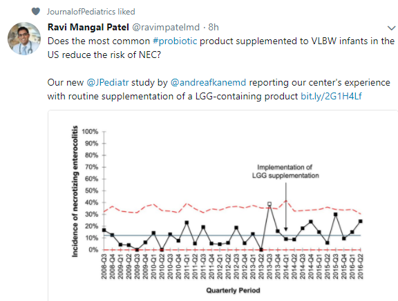
A recent retrospective study (AF Kane et al. J Pediatr 2018; 195: 73-9) with 640 VLBW infants found that the probiobiotic, Lactobacillus rhamnosus GG (LGG), was associated with an increased risk of necrotizing enterocolitis (NEC).

LGG supplementation was started at a median age of 6 days at a dose of 2.5 to 5 x 10 to the 9th CFU/day.

Key finding:

  • LGG group had an aOR of 2.10 for developing NEC.  LGG group NEC incidence was 16.8% whereas NEC incidence was 10.2% prior to institution of LGG.

The authors note their findings are in contrast to findings from 38 randomized trials (10,520) which have found that probiotics lowered the risk of NEC.

My take: This study reinforces the need for further studies to identify which factors and probiotic strains are likely to lead to reduced rates of NEC.

Related blog posts:

Joint Mobility –Not Associated with Increased Functional GI Disorders

According to a recent study (M Saps et al. JPGN 2018; 66: 387-90), joint hypermobility is not associated with an increased risk of functional gastrointestinal disorders (FGIDs).

From a school-based study of 654 children from a public school in Cali, Columbia, 148 (22.6%) were identified as having an FGID. Among this group, 136 children participated in the study along with 136 age/sex-matched healthy controls. Joint laxity was assessed to establish a Beighton score.

Key finding:

  • There was no significant difference in joint laxity between the FGID group and the control group, with OR of 1.03.

The implication of this study is that previous associations between joint hypermobility (JH) and FGIDs could be due to selection bias at tertiary care centers.  Alternatively, “it is possible that the association between FGIDs and JH exists, but it is only limited to a subset of patients that consult at specialized clinics.”

My take: This article challenges the idea that JH increases the risk of FGID.  Based on this study, if JH is a risk factor, it is hard to detect in a general population.

Related blog post:


Amber Cove, Dominican Republic