
Vitamin D and Ulcerative Colitis Remission

A recent study (J Gubatan et al. Clin Gastroenterol Hepatol 2017; 15: 240-6) examined a prospective study of 70 patients with ulcerative colitis (UC).  These patients (average age 48.6 yrs) were initially in clinical remission.  Key findings:

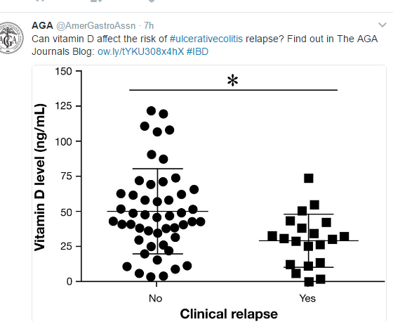
  • Mean baseline vitamin D (25-OH) level was lower among patients with subsequent relapse (29.5 ng/mL) than those without relapse (50.3 ng/mL)
  • Over 12 months, a 25-OH D value <35, was associated with a small increased risk of relapse (odds ratio1.25). 20% of patients with a value <35 had clinical relapse compared with 9% (P= .003) who had values >35.

Because vitamin D levels are inversely related to UC disease activity, this study is particularly intriguing.  By enrolling patients prospectively while in remission, this study suggests that good vitamin D levels may directly have immunoprotective and anti-inflammatory properties.

The AGA Journals blog provides an excellent summary of this study: Can Vitamin D Affect Risk of Ulcerative Colitis Relapse?

“In an editorial that accompanies the article, Stephen Hanauer reminds readers that the mean vitamin D level in the entire cohort was 44 ng/mL, and 60% of the subjects were taking vitamin D supplements. A normal vitamin D level is considered to be 20–40 ng/mL in healthy individuals, and the 35 ng/mL cut-off level used in the study was within this range.

Hanauer also mentions that in assessing the confidence intervals for risk of relapse at lower or higher vitamin D levels, there does not appear to be a dose–response effect in the odds ratios according to levels. Based on these findings, Hanauer says it would be premature to target a level of 35 ng/mL. He states that the best predictors of clinical relapse are still endoscopic and histologic markers of inflammation.”

My take: At this time, trying to maintain a normal vitamin D level is likely to be worthwhile; though, values obtained during acute flares remain unreliable.

Related blog posts:

Disclaimer: These blog posts are for educational purposes only. Specific dosing of medications/diets (along with potential adverse effects) should be confirmed by prescribing physician/nutritionist.  This content is not a substitute for medical advice, diagnosis or treatment provided by a qualified healthcare provider. Always seek the advice of your physician or other qualified health provider with any questions you may have regarding a condition.
screenshot-99

Complexity in Cystic Fibrosis Diagnosis

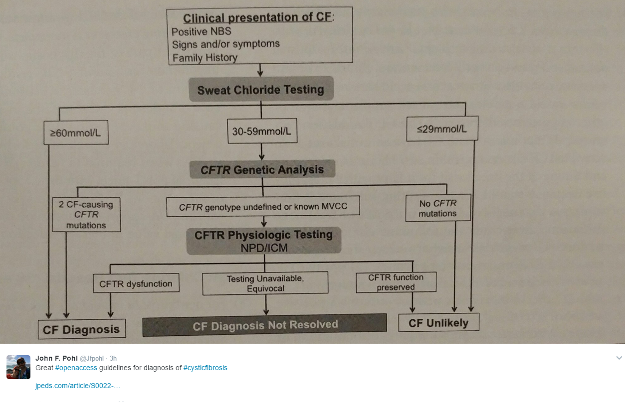
The availability of multiple diagnostic techniques for cystic fibrosis has increased the complexity and created areas of uncertainty.  A recent supplement (J Pediatr 2017; 181S: 1-55) delve into these issues.

“The diagnosis of CF has become increasingly complex, as CFTR mutations resulting in a wide spectrum of dysfunction have been increasingly identified.”

On page S6, 27 consensus recommendations are given.

The article S45-51, reviews cystic fibrosis transmembrane conductance regulator-related metabolic syndrome (CRMS) and cystic fibrosis screen positive, inconclusive diagnosis (CFSPID).  Key points:

  • CRMS and CFSPID are equivalent entities with CRMS being the preferred terminology in the US
  • CRMS/CFSPID are relatively frequent; for every 3 to 5 cases of CF, there is one case of CRMS/CFSPID.
  • The majority of CRMS/CFSPID do NOT develop CF. Approximately 10-20% develop clinical features concerning for CF.

screenshot-116

Related blog posts:


Triglyceride Levels and Pancreatitis

A recent study (JAMA Intern Med 2016; 176: 1834-42) suggests that even mild to moderate hypertriglyceridemia may increase risk of pancreatitis.

Among two large cohorts that were followed prospectively for a median of 6.7 years, 434 out of 116,550 patients developed acute pancreatitis.

Key finding (which persisted after adjustment of multiple potential confounding factors):

screenshot-140

More complete summary at GI & Hepatology News: “Mild, moderate hypertriglceridemia tied to pancreatitis”


Consensus Pancreatitis Recommendations

The INSPPIRE Group (CE Gariepy et al. JPGN 2017; 64: 95-103) has published consensus recommendations for acute recurrent pancreatitis (ARP) and chronic pancreatitis (CP).

While the authors acknowledge the need for high-level evidence/further research, they provide a large number of consensus recommendations.  These recommendations are succinctly summarized in Table 1 and Table 2.  From a reader’s perspective, my preference would have been to separate the recommendations for ARP and CP rather than to intermix them (though many of the recommendations are the same for both conditions).

ARP specific recommendations:

  • “Initial evaluation should include AST,ALT, GGT, Total bilirubin (fractionate if elevated), fasting lipids, and total serum calcium.”
  • Evaluate for fat-soluble vitamin deficiency, and pancreatic exocrine insufficiency at least annually

ARP and CP recommendations:

  • Consider ammonia and urine organic acids if there is a concern for undiagnosed metabolic disease.
  • Check for celiac disease.
  • Check for O&P if immunosuppressed, travel to endemic areas of Ascaris, or if peripheral eosinophilia.
  • Evaluation of genetic causes: should include sweat chloride test and PRSS1 gene testing. Consider SPINK1, CFTR, and CTRC evaluation.
  • Evaluate with MRCP (not ultrasound) acutely if GGT >2 x ULN or if direct bilirubin is elevated.
  • Non-acutely, MRCP recommended to evaluate pancreatic ductal abnormalities.  “When available, secretin-enhanced MRCP …should be obtained.” sMRCP can provide dynamic images of the pancreatic duct allowing differentiation of fixed from nonfixed lesions; this technique has not been widely adopted by pediatric radiologists compared with adult radiologists.

CP specific recommendations:

  • Evaluate for fat-soluble vitamin deficiency, pancreatic exocrine insufficiency, and pancreatic endocrine insufficiency at least annually

The authors did not recommend checking serum IgG4 in the absence of associated systemic disease or suggestive imaging for autoimmune pancreatitis.

Briefly noted: J-H Choi et al. Clin Gastroenterol Hepatol; 2017: 15: 86-92.  This study indicated that vigorous hydration with lactated ringer’s (LR) reduces risk of pancreatitis after ERCP.  A potential inference would be that LR would be an optimal fluid for pancreatitis more broadly. (Related: Why an ERCP Study Matters to Pediatric Care | gutsandgrowth)

Related blog posts:

Dragon Point, Labadee

Dragon Point, Labadee

Ileocecal Resection in Pediatric Crohn’s Disease

A recent retrospective study (K Diederen et al. Inflamm Bowel Dis 2017; 23: 272-82) provides data on the likelihood of complications and recurrence following ileocecal resection in pediatric Crohn’s disease (n=122).

Key findings:

  • Severe postoperative complications were noted in 9.8%.  Risk factors included colonic disease (Odds ratio 5.6), microscopically positive resection margins (OR 10.4), and emergency surgery (OR 6.8)
  • Overall complication rate was reported as 29.5% which is similar to rates reported in adults
  • Clinical recurrence rates after 1, 5, and 10 years: 19%, 49%, and 71%
  • Surgical recurrence rates after 1, 5, and 10 years: 2%, 12%, and 22%
  • Immediate postoperative therapy reduced the risk of clinical recurrence (HR 0.3) and surgical recurrence (HR 0.5)
  • “In this study, postoperative catch-up growth was found in patients younger than 16 years in the year after surgery.” Thus, surgery could be an important to reverse growth retardation.

Complications within 30 days of surgery were categorized with the Clavien-Dindo classification. Those with grade ≥III which required either surgical, endoscopic or radiologic intervention were considered severe.  In this population, the complications included intraabdominal septic complications and/or anastomotic leakage.

My take: In some patients, ileocecal resection should NOT be a last resort.  Waiting too late, increases the risk of complications.  The task at hand is prospectively identifying those who merit surgery sooner and then convincing the family to proceed.

screenshot-118

Related blog posts:

Clostridium difficile Infection in Inflammatory Bowel Disease: Expert Updates

A recent clinical practice update (S Khanna et al. Clin Gastroenterol Hepatol; 2017; 15: 166-74) provides some succinct recommendations regarding Clostridium difficile infection (CDI) in Inflammatory Bowel Disease (IBD).

Background: In 2011, the authors note that CDI was associated with 29,000 deaths and is now the most lethal enteric pathogen in the U.S.

Differences in pathogenesis of C diff in IBD compared to those without IBD:

  • Younger age
  • Less frequent antibiotic exposure
  • More often community onset (rather than hospital onset)
  • Higher recurrence (may be related to dysbiosis)

Key recommendations:

  • In patients with IBD flare, test for CDI
  • In patients with CDI and IBD, clinicians should consider “using vancomycin instead of metronidazole.”
  • In patients with recurrent CDI and IBD, consider fecal microbiota transplantation

Figure 4 proposes a management algorithm (for adults).  If uncomplicated CDI, recommended dose of vancomycin was 125 mg q6h. If no improvement in 3-4 days, then “consider escalation of immunosuppression.” For complicated CDI, consider oral vancomycin at 500 mg q6h and IV metronidazole 500 mg q8.  In addition, consider rectal vancomycin and surgery consult.

Complicated CDI includes ICU admission, hypotension, T >38.5, ileus/megacolon, mental status changes, leukocyte count >35,000  or < 2000, or lactate >2.2 mmol/L

Another review article (Y Chen et al. Inflamm Bowel Dis 2017; 23: 200-07) is a meta-analysis that identified six studies.  One of these studies was a case-control study with nearly 400,000 patients (and about 7000 cases of C diff). Key finding: CDI results in nearly a doubling of the risk of colectomy (OR 1.90), mainly in patients with ulcerative colitis.

Related blog posts

Disclaimer: These blog posts are for educational purposes only. Specific dosing of medications (along with potential adverse effects) should be confirmed by prescribing physician.  This content is not a substitute for medical advice, diagnosis or treatment provided by a qualified healthcare provider. Always seek the advice of your physician or other qualified health provider with any questions you may have regarding a condition.


Rare Tragic Reaction to Infliximab

A recent post on the pediatric GI Listserv pointed out a troubling case report: “Fatal Central Nervous System Disease Following First Infliximab Infusion in a Child With Inflammatory Bowel Disease,” FM. Baumer et al; Pediatric Neurology 2016; 57: 91-94.

“A seven-year-old boy diagnosed with ulcerative colitis and primary sclerosing cholangitis received infliximab. Six hours following his uneventful infusion, he awoke with headache and emesis and rapidly became obtunded…Cranial computed tomography revealed hypodense lesions in the cerebral hemispheres, cerebellum, and pons accompanied by hemorrhage…Within four days he met criteria for brain death.”

The authors note that “the close temporal association between our patient’s presentation and the infliximab infusion raises concern for a drug-related cause for his cerebral injury.” While this case report is terribly sad, severe and fatal reactions can unfortunately be encountered with a wide range of medications, including commonly used antibiotics.

My take: Thus, while the vast majority of pediatric patients with inflammatory bowel disease will benefit from infliximab therapy, there are rare tragic outcomes.  img_3954


Gastrojejunostomy Complications Frequent

Gastrojejnostomy (GJ) placement allows enteral feeds to bypass the stomach.  When a gastrostomy is already in place, GJ placement may allow patients to avoid surgery (eg. fundoplication).  Most practitioners would consider the risk of GJ placement to be low, but a recent report (J Moorse et al. JPS 2017; http://dx.doi.org/10.1016/j.jpedsurg.2017.01.026) suggests that it is higher than expected.  The abstract and link are below.

Link: Gastrojejunostomy tube complications — A single center experience and systematic review

Abstract

Purpose

Gastrojejunostomy tubes (GJTs) enable enteral nutrition in infants/children with feeding intolerance. However, complications may be increased in small infants. We evaluated our single-institution GJT complication rate and systematically reviewed existing literature.

Methods

With REB approval, a retrospective single-institution analysis of GJT placements between 2009 and 2015 was performed. For the systematic review, MOOSE guidelines were followed.

Results

At our institution, 48 children underwent 154/159 successful insertions primarily for gastroesophageal reflux (n = 27; 55%) and aspiration (n = 11; 23%). Median age at first GJT insertion was 2.2 years (0.2–18). Thirty-five (73%) had an index insertion when ≤10 kg. GJTs caused 2 perforations and 1 death. The systematic review assessed 48 articles representing 2726 procedures. Overall perforation rate was estimated as 2.1% (n = 36 studies, 23/1092, 95% CI: 1.0–3.2). Perforation rates in children <10 kg versus ≥10 kg were estimated as 3.1%/procedure (95% CI: 1.1%–5.0%) and 0.1%/procedure (95% CI: 0%–0.3%), respectively. The relative risk of perforation was 9.4 (95% CI: 2.8–31.3). Overall mortality was estimated as 0.9%/patient (n = 39 studies; 95% CI: 0.2–1.6%). Most perforations (19/23; 83%) occurred ≤30 days of attempted tube placement.

Conclusion

Gastrojejunostomy tubes are associated with significant complications and frequently require revision/replacement. Insertion in patients <10 kg is associated with increased perforation risk. Caution is warranted in this subgroup.

With regard to the methodology

  • ~90% of the procedures were performed by interventional radiology and the interventionist had a median of 6.6 years of experience
  • Most GJs were 16 French in width and most were either 15 cm or 22 cm in length

My take: This report highlights the significant risks associated with GJ placement, particularly in smaller patients (<10 kg).  Despite these risks, GJ placement is often the safest option.

Costa Maya, Mexico

Costa Maya, Mexico


Expert Advice on Clostridium difficile and Inflammatory Bowel Disease

Link: Management of Clostridium difficile Infection in Inflammatory Bowel Disease: Expert Review from the Clinical Practice Updates Committee of the AGA Institute

Abstract: The purpose of this expert review is to synthesize the existing evidence on the management of Clostridium difficile infection in patients with underlying inflammatory bowel disease. The evidence reviewed in this article is a summation of relevant scientific publications, expert opinion statements, and current practice guidelines. This review is a summary of expert opinion in the field without a formal systematic review of evidence.

Best Practice Advice 1: Clinicians should test patients who present with a flare of underlying inflammatory bowel disease for Clostridium difficile infection.

Best Practice Advice 2: Clinicians should screen for recurrent C difficile infection if diarrhea or other symptoms of colitis persist or return after antibiotic treatment for C difficile infection.

Best Practice Advice 3: Clinicians should consider treating C difficile infection in inflammatory bowel disease patients with vancomycin instead of metronidazole.

Best Practice Advice 4: Clinicians strongly should consider hospitalization for close monitoring and aggressive management for inflammatory bowel disease patients with C difficile infection who have profuse diarrhea, severe abdominal pain, a markedly increased peripheral blood leukocyte count, or other evidence of sepsis.

Best Practice Advice 5: Clinicians may postpone escalation of steroids and other immunosuppression agents during acute C difficile infection until therapy for C difficile infection has been initiated. However, the decision to withhold or continue immunosuppression in inflammatory bowel disease patients with C difficile infection should be individualized because there is insufficient existing robust literature on which to develop firm recommendations.

Best Practice Advice 6: Clinicians should offer a referral for fecal microbiota transplantation to inflammatory bowel disease patients with recurrent C difficile infection.


Related blog posts:

screenshot-94

Patterns and Puzzles with Very Early Onset Inflammatory Bowel Disease

A recent review (S Chandrakasan, S Venkateswaran, S Kugathasan [corrresponding author]. Pediatr Clin N Am 2017; 64: 139-160) provides a surprisingly easy read on very early onset (VEO) inflammatory bowel disease (IBD) (Thanks to Kathleen McNamara for sharing).  Because of the myriad of genetic defects (>50 monogenetic defects), the topic of VEO-IBD can be quite confusing.  The authors of this summary make a number of key points.

  • VEO-IBD is increasing in incidence
  • Many patients with VEO-IBD have an underlying primary immune defect.  Identification of these underlying disorders may allow targeted therapy.
  • In some patients, hematopoietic stem cell transplantation could result in definitive cure
  • VEO features: more often involves colon and often severe course with poor response to conventional immunosuppressives

Besides idiopathic IBD, the differential can be subdivided into subgroups:

  • T-cell defects (IPEX and IPEX-like) (gene defects: FOXP3, LRBA CTLA4, STAT1, STAT3, STAT5B, CD25, CTLA4, )
  • Defects in IL-10 signaling (IL10RA, IL10RB, IL-10)
  • Hyperinflammtory/autoinflammatory disorders (XIAP/SAP (BIRC4), Mevalonate kinase deficiency (MVK), PLCG2, Familial Mediterranean Fever, Familial HLH Type 5: STXBP2, Hermansky-Pudlak: HPS1, HPS4, HPS6)
  • Defects in neutrophil function/phagoycte function (chronic granulomatous disease (CGD) genes: CYBB, CYBA, NCF1, NCF2, NCF4,, Leukocyte Adhesion Defect (LAD) ITGB2, GSD type 1bSLC37A4, congenital neutropenia G6PC3)
  • Epithelial barrier defect (X-linked ectodermal dysplasia and immunodeficiency (NEMO), TTC7A, ADAM17, dystrophic epidermolysis bullosa (COL7a1), Kindler syndrome (FERMT1), mutations in guanylate cyclase c, telomere biology defects like  DKC and RTEL1 )
  • T/B cell defects (X-linked agammaglobulin (BTK), SCID/Omenn (RAG1, RAG2, IL-7Ra, IL-2RG), CVID, IL21, Wiskott-Aldrich (WAS) HIES, HIMS)

The authors provide some vignettes of a typical presentation of each subset along with some commentary.  For example, with T-cell defects: “mutations in FOXP3 result in either absent or decreased Treg cell numbers or a qualitative defect…results in broader immune dysregulation, resulting in multisystem autoimmunity with autoimmune endocrinopathy, autoimmune cytopenia, autoimmune hepatitis, and severe eczema.”  Other IPEX-like mutations include gain of function in STAT-1/STAT-3, LRBA deficiency, and CTLA-4 haploinsufficiency.

The authors recommend an initial immune evaluation in VEO-IBD:

  • CBC
  • peripheral smear evaluation
  • immunoglobulin levels, lymphocyte subsets with T-cell
  • B-cell, and NK-cell enumeration
  • CD45RA/RO enumeration and B-cell panel for class-switched memory B cells
  • neutrophil oxidative burst
  • T regulatory cell (CD4+CD25+FOXP3+) cell enumeration.

Due to the increasing complexity of the immune evaluation, the necessity of a pediatric immunologist is apparent.  In addition, the role of genetic panels that test for all of these disorders simultaneously is becoming routine.  Genetic testing can help improve diagnosis and allow for early targeted intervention.  With the emergence of new defects, selecting the right lab with an up-to-date panel is another caveat.

Examples of targeted therapies include the potential role of anakinra for CGD, abatacept for LRBA deficiency, toclizumab (IL-6 receptor blocking antibody) for STAT3 gain-of-function mutation, and sirolimus for Treg disorders.  Hematopoietic stem cell transplantation is an established therapy for IL-10 signaling defects.

My take: Collaboration with immunology is an important consideration in young children with IBD.

Related blog posts:

Disclaimer: These blog posts are for educational purposes only. Specific dosing of medications (along with potential adverse effects) should be confirmed by prescribing physician.  This content is not a substitute for medical advice, diagnosis or treatment provided by a qualified healthcare provider. Always seek the advice of your physician or other qualified health provider with any questions you may have regarding a condition.

I liked this t-shirt

I liked this t-shirt