Celiac Disease: Pro Tips (Part 4)

In June 2024 (special issue), Gastroenterology published an entire issue (193 pages) focused on celiac disease. There was a lot of useful information on almost every aspect of this disease. Below I have summarized some of the points –this is the last of 4 posts on this issue.

In addition to these articles, another good update on aspects of CeD was the Bowel Sounds podcast with Arun Singh. Link: Clinical Conundrums in Celiac Disease.

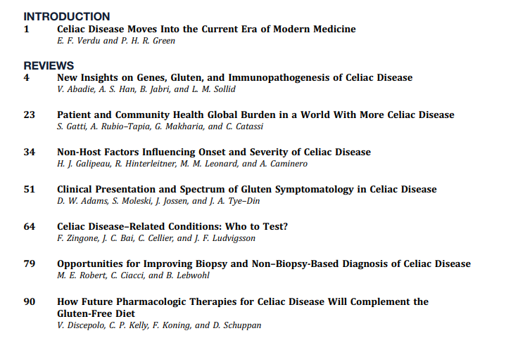
DW Adams et al. Gastroenterol 2024; 167: 51-63. Clinical Presentation and Spectrum of Gluten Symptomatology in Celiac Disease

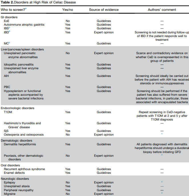
This article highlights the myriad presentations of CeD. Due to screening of at-risk groups. fewer patients are presenting with malabsorption and many patients at diagnosis are overweight or obese. In adults, 15-28% are overweight and 7-11% are obese.

  • Hematology: Among adults with iron-deficiency anemia, 1 in 31 have histologic evidence of CeD
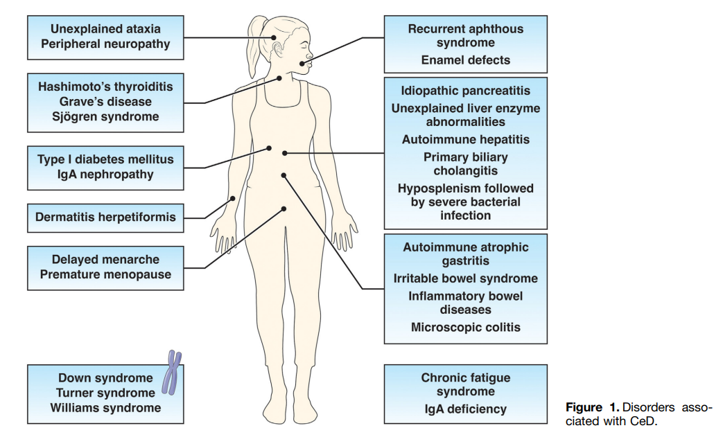
  • Skin: Dermatitis herpetiformis
  • Neurologic: Headache, brain fog, peripheral neuropathy, ataxia
  • Oral: dental enamel defects, aphthous stomatitis
  • Musculoskeletal/rheumatologic: osteopenia/osteoporosis/fractures, arthralgias/arthritis. Low bone mass with “z-scores less than -2 SD for age is present in 6-16% of children” with CeD
  • Growth in children: pubertal delay, short stature, growth failure
  • Fertility: female infertility, spontaneous abortions, premature births, hypogonadism (both sexes)
  • GI: stomach pain, diarrhea, bloating/distention, constipation, weight loss
  • Liver: elevated liver enzymes in up to 40% of patients with CeD at diagnosis. 1 in 20 patients with cryptogenic cirrhosis have CeD. In children, ~25% have “modest elevations in liver enzymes” at diagnosis. “Normalization …is generally seen within 6-12 months of a GFD.”
  • Endocrine: higher rates of type 1 diabetes and autoimmune thyroid disease
  • Asymptomatic: “10-27% of CeD patients are asymptomatic at diagnosis…Asymptomatic patients with CeD were taller, had lower TTG-IgA levels, and had less severe intestinal damage.” However, “patients reporting to be asymptomatic often record improvement” with GFD.

The authors detail how gluten exposure triggers symptoms. Gluten ingestion activates gluten-specific T cells and acute increases in interleukin-2 (IL-2). IL-2 is postulated to cause neuro-enteric symptoms and leads to release of serotonin. Serum IL-2 is a biomarker for acute gluten exposure.

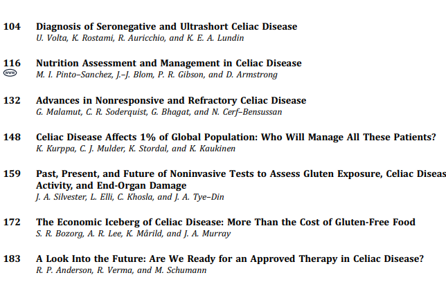
MI Pinto-Sanchez et al. Gastroenterol 2024; 167: 116-131. Nutrition Assessment and Management in Celiac Disease

  • This article provides specific nutritional recommendations for patients with CeD including involvement of a dietitian. The authors note that some studies in children have found low iron, vitamin D, calcium, vitamin B12, vitamin B6, folate, zinc and magnesium in their GFD. They recommend checking for iron, ferritin, folate, vitamin B12, vitamin D, vitamin A, zinc, selenium, copper, and chromium at diagnosis.
  • The adoption of a GFD “has been associated with an increased risk of MASLD likely related to the nutritional composition of packaged gluten-free foods.” (see: Gluten-Free Diet Can Be Unhealthy)
  • 30% of CeD adult patients experience persistent symptoms. The most common cause is continued gluten exposure (often inadvertent). Gluten contamination elimination diet can be implemented in this setting “though not feasible as a long-term solution.” (See: What To Do For Pediatric Patients with Non-Responsive Celiac Disease)

My take: More recent studies have indicated that more limited nutrient testing is appropriate in the pediatric population (see: How to Provide More Cost-Effective Celiac Care and Nutrient Deficiencies with Celiac Disease). Many nutrient deficiencies (in pediatrics) improve with institution of GFD and other nutrient deficiencies are similar in frequency as the general population.

U Volta et al. Gastroenterol 2024; 167: 104-115. Diagnosis of Seronegative and Ultrashort Celiac Disease

  • “Seronegative villous flattening is a very rare condition” in children…and is “often attributable to other diagnosis (eg. inflammatory bowel disease…). The authors provide a diagnostic algorithm. In this setting, first step is to check HLA-DQ2/DQ8. A negative test “virtually rules out CeD.” A positive test indicates CeD is possible if other conditions are excluded. This includes autoimmune enteropathy, immune deficiencies, medication effects, infections, and other conditions. (See:Seronegative Villous Atrophy and@AmyOxentenkoMD: Celiac Disease and Mimics and How Many Cases of Celiac Disease Are We Missing?)
  • CeD confined to 1st portion of duodenum is considered ultra-short CeD. Algorithm for ultra-short CeD (Figure 2). The disease is supported by positive serology and positive HLA DQ2/DQ8. If serology and/or HLA DQ2/DQ8 are negative, it is very unlikely to be CeD and other diagnosis need to be identified (eg. peptic duodenitis).
Flowers at Getty
Angeles Crest (near Los Angeles)

Celiac Disease: Pro Tips (Part 2)

In June 2024 (special issue), Gastroenterology published an entire issue (193 pages) focused on celiac disease. There was a lot of useful information on almost every aspect of this disease. Below I have summarized some of the points.

V Disepolo et al. Open Access! How Future Pharmacologic Therapies for Celiac Disease Will Complement the Gluten-Free Diet

  • This article focuses on the emerging pharmacologic treatments
One of the treatment strategies has been to try to sequester gluten. This has included using enzymes to degrade dietary gluten. Other approaches include genetically modifying diet, tight junction modulation, immune modulation and tolerance induction.
Some therapies being developed in adults may have unacceptable risk profiles for children. This could include blocking cytokine signaling (anti–IL-15, anti–IL-15/IL-21) and blocking intestinal T-cell recruitment

Related blog posts:

G Malamut et al. Advances in Nonresponsive and Refractory Celiac Disease

Celiac Disease: Pro Tips (Part 1)

In June 2024 (special issue), Gastroenterology published an entire issue (193 pages) focused on celiac disease. There was a lot of useful information on almost every aspect of this disease. Below I have summarized some of the points.

F Zingone et al. Open Access: Celiac Disease–Related Conditions: Who to Test?

  • The authors detail disorders with increased risk for CeD and which merit screening (see Table 2 below). Some disorders that merit screening that are more obscure include idiopathic pancreatitis, autoimmune hepatitis, delayed menarche, and chronic fatigue.
  • They note that type 1 diabetes mellitus could require serial screening. “Because CeD can manifest at any time and with greater frequency during the initial 5 years, conducting additional screenings in CeD-negative T1DM patients 2 and 5 years after T1DM diagnosis and those who later develop gastrointestinal (GI) symptoms may be advisable.” In addition, it is important to recognize that CeD serology testing is less reliable in patients with T1DM.

S Gatti et al. Patient and Community Health Global Burden in a World With More Celiac Disease, describes the worldwide burden of celiac disease and how to improve detection.

  • They note that the worldwide prevalence is between 0.7% and 2.9%. In this issue, most authors estimate the prevalence to be about 1% with more than 50% undetected.
  • There are many places with higher rates. In U.S. “children in Colorado had a 2.5-fold higher risk compared to Washington State…similar regional differences were seen …in Sweden, Finland, and Germany.”
  • They note the burden before and after diagnosis. Before diagnosis/undetected, there can be persistent symptoms, complications (eg. osteoporosis, decreased fertility) and impaired quality of life. Afterwards, there are increased costs of a GFD and psycho-social burden of GFD.
  • In terms of generalized screening compared to case-finding, the authors note that given the number of at-risk groups, the case-finding approach could entail screening >50% of the population.

V Abadie et al. New Insights on Genes, Gluten, and Immunopathogenesis of Celiac Disease, reviews the intricate details of genetic, biochemical, and immunologic studies, which together have revealed mechanisms of gluten peptide modification and HLA binding, thereby enabling a maladapted anti-gluten immune response.

  • What I was most interested in was the mechanisms behind ‘potential’ celiac disease (PCeD) in which patients have autoimmunity (+serology) but normal histology. In potential CeD, the anti-CD4+ T-cell response is present but decoupled from tissue cytotoxicity. However, notably, IL-21, a cytokine produced by gluten-specific CD4+ T cells in active CeD, is not up-regulated in potential CeD…In addition, patients with potential CeD lack the presence of an epithelial stress response associated with IL-15, HSP70, and HSP27 upregulation in epithelial cells.” “The presence of epithelial stress is a crucial prerequisite for the development of tissue damage.”

How Closely Related Are Eosinophilic Gastrointestinal Disorders To Isolated Eosinophilic Esophagitis

H Sato et al. Clin Gastroenterol Hepatol 2024; 22: 1531-1534. (Research Letter) Eosinophil Involvement Outside the Esophagus in Eosinophilic Esophagitis

The authors retrospectively studied a cohort of children (n=782) with eosinophilic gastrointestinal disorders (EGID) including 592 with isolated eosinophilic esophagitis (EoE), 190 with EGID which included esophageal involvement (“EI”) and 35 EGD without esophageal involvement (“Non-EI”).

Key findings:

  • Abdominal pain was more frequent in EI than isolated EoE: 61% vs 45%, p=.002)
  • EI patients had more dense esophageal eosinophil infiltrate (peak) than isolated EoE: 115 vs 92; P=.036) and higher peripheral blood eosinophil level (0.44 vs 0.38; p=0.027)
  • 94-Gene expression profiles from esophageal biopsies from a 168 subgroup showed that EoE and EI were not significantly different. The heat map for upregulated and downregulated gene expression was different based on disease activity but not significantly different between EoE or EI in either state.

My take (borrowed from authors): The similar molecular transcriptome between EI and EoE are indicative of a shared pathogenesis.

Related blog posts:

Colorado River near Moab

Tirzepatide for Metabolic Dysfunction–Associated Steatohepatitis (MASH) & Uptick in GLP1 Use

R Loomba et al. NEJM 2024; 391; 299-310. Tirzepatide for Metabolic Dysfunction–Associated Steatohepatitis with Liver Fibrosis

F Kanwal. N Engl J Med 2024;391:371-372. Dual Agonists for Management of Metabolic Dysfunction–Associated Steatohepatitis (Associated editorial)

Background: The efficacy and safety of tirzepatide, an agonist of the glucose-dependent insulinotropic polypeptide and glucagon-like peptide-1 receptors, in patients with MASH and moderate or severe fibrosis is unclear.

Methods: This was a phase 2, dose-finding, multicenter, double-blind, randomized, placebo-controlled trial involving participants with biopsy-confirmed MASH and stage F2 or F3 (moderate or severe) fibrosis. Participants were randomly assigned to receive once-weekly subcutaneous tirzepatide (5 mg, 10 mg, or 15 mg) or placebo for 52 weeks. 

My take: Until very recently, there were no effective pharmacologic agents for steatotic liver disease (SLD). Now it appears that there will be multiple effective agents. When newer agents for hepatitis C became available (Harvoni was FDA approved 10 years ago), there were concerns about the high cost. With SLD, this is an even bigger concern give the indefinite treatment period.

From editorial: “Overall, these data are encouraging. Clinicians providing care for patients with MASH will probably have an increasing number of options in their armamentarium. With a growing menu of effective treatments, harms and unacceptable side effects will be important considerations in making treatment decisions.”

Related blog posts:

ACG Review (Zobair Younassi, MD): NAFLD and NASH

Related article: YH Yeo et al. Annals of Intern Med 2024; https://doi.org/10.7326/M24-00. Shifting Trends in the Indication of Glucagon-like Peptide-1 Receptor Agonist Prescriptions: A Nationwide Analysis

Liver Briefs: Hereditary Angioedema/Liver Transplantation, Bulevirtide/PEG for HDV, and AASLD Cystic Fibrosis Guidance

NE Peters et al. NEJM 2024; 391; 56-59. Normalization of C1 Inhibitor in a Patient with Hereditary Angioedema

“An infant with genetically confirmed hereditary angioedema and low C1 inhibitor levels (but without previous episodes of angioedema) underwent liver transplantation for biliary atresia, an unrelated condition. Liver transplantation led to normalization of the C1 inhibitor level and function. To our knowledge, this represents the first patient to be potentially cured of hereditary angioedema.” This case report shows that liver-directed therapy can reverse hereditary angioedema.

Related blog posts:

T Asselah et al. NEJM 2024; 391:133-143. Bulevirtide Combined with Pegylated Interferon for Chronic Hepatitis D

Key finding: At 24 weeks after the end of treatment, HDV RNA was undetectable in 17% of the patients in the peginterferon alfa-2a group, in 32% of those in the 2-mg bulevirtide plus peginterferon alfa-2a group, in 46% of those in the 10-mg bulevirtide plus peginterferon alfa-2a group, and in 12% of those in the 10-mg bulevirtide group. 

My take: This is a long (48 weeks) and difficult treatment (2 injection meds and lots of peginterferon side effects). However, there is a fairly good response rate.

Related blog post: Image Only: World Hepatitis Day Infographic

ZM Sellers et al. Hepatology 2024; 79(5):p 1220-1238, May 2024. Open Access! Cystic fibrosis screening, evaluation, and management of hepatobiliary disease consensus recommendations

This link to the article also has links to related AASLD guidelines (eg. management of portal hypertension). Table 2 summarizes the ~34 recommendations which include yearly evaluation with labs, ultrasound at least every 2 years in pediatric patients (age 3 yrs and older) and against routine use of ursodeoxycholic acid.

Thanks: Updated Long-Term PPI Use Smartphrase

AK Kamboj et al. Clin Gastroenterol Hepatol 2024; 22: 1373-1376. Long-Term Proton Pump Inhibitor Use: Review of Indications and Special Considerations

This article provides a good review on long-term use of PPIs. Table 1 provides indications that may require long-term use of PPIs:

  • Erosive esophagitis: symptomatic Los Angeles Grade B or Maintenance for Grade C-D
  • Symptomatic Nonerosive GERD
  • Peptic stricture
  • Biopsy-proven Barrett’s esophagus
  • PPI-responsive esophageal mucosal diseases: eosinophilic esophagitis or lymphocytic esophagitis
  • Peptic ulcer disease without modifiable risk factor
  • High-risk patients receiving antiplatelet therapy
  • Zollinger-Ellison
  • Idiopathic pulmonary fibrosis (per pulmonologist)

Table 2 provides customary advice: ensure patient has a good indication for PPI otherwise consider deprescribing, and use lowest effective dose. It also summarizes potential adverse effects/management.

My take: The author’s “take-home message” is appropriate as a smartphrase for counseling patients (slightly modified below):

  • Although proton pump inhibitor (PPI) use is common, only a few conditions warrant its long-term use. These conditions include severe erosive esophagitis, PPI-responsive eosinophilic esophagitis, chronic esophageal mucosal diseases, and peptic ulcer disease with risk of recurrence (and others).
  • When PPIs are required long-term, efforts should be made to use the lowest possible dosing necessary to manage patient symptoms and underlying condition.
  • In patients that do not meet indications for long-term PPIs, efforts should be made to deprescribe these medications.
  • Although PPIs are often linked to various adverse conditions, these potential associations are largely based on low-quality studies and do not prove an increase risk for these conditions. Multiple larger-scale studies have also demonstrated results showing no such associations besides a marginal increase in enteric infections.
  • In general, routine testing should not be performed on long-term PPIs unless risk factors for specific conditions exist. In those with risk factors, monitoring could be needed for low magnesium, vitamin B12 deficiency, chronic kidney disease, and osteoporosis.

Related blog posts:

Bryce Canyon National Park

Proliferation of Formula Lawsuits In Necrotizing Enterocolitis

AAP Statement 7/27/24: AAP Statement In Response to NEC Lawsuit Verdict

Yesterday, the AAP put out a statement (see below) regarding the numerous lawsuits related to necrotizing enterocolitis. The lawsuits allege that formula companies did not provide physicians and patients warning that their products could increase the risk of necrotizing enterocolitis relative to breast milk or donated human milk.

This has led to huge verdicts. In March, a jury in Illinois awarded a mother $60 million (3/15/24 Reuters: Reckitt unit hit with $60 million verdict in Enfamil baby formula case in Illinois). This past week, a jury in Missouri awarded a women $95 million in compensatory damages and $400 million in punitive damages (CNBC 7/26/24: Abbott must pay $95 million in premature infant formula trial, jury finds).

Currently, when one searches for “NEC and lawsuits,” it is difficult to find these news reports due to dozens of law firm websites trying to attract clients. Per CNBC article, “close to 1,000 lawsuits have been filed against Abbott, Enfamil formula maker Reckitt Benckiser or both in federal or state courts.”

My take: These lawsuits are likely to exponentially increase the cost of formulas for all infants with little justification. It has been well-recognized for decades that there are many factors contributing to NEC; infants receiving human milk also develop NEC. For many infants in the NICU, sometimes there is no availability of human milk. What is going to happen to them if formula companies stop making premature formulas?

The most promising therapeutic to try to reduce the risk of NEC would be a safe probiotic. However, the FDA closed off this avenue (see: End of the Line for Probiotics for Preterm Infants). Even if the FDA reversed its position, no company would be willing to try to develop these products due to fear of litigation.

Related blog posts:

Impact of Gene Mutation on Juvenile Polyposis Syndrome

S Cohen et al. J Pediatr Gastroenterol Nutr.2024;79:161‐167. Open Access! Juvenile polyposis syndrome in children: The impact of SMAD4 and BMPR1A mutations on clinical phenotype and polyp burden

One of the authors on this paper is Dr. Erdman (see yesterday’s post: Dr. Steve Erdman: Perplexing Polyposis Patients: a Case-Based Discussion).

Background/Methods: A constitutional disease‐causing variant (DCV) in the SMAD4 or BMPR1A genes is present in 40%–60% of patients with juvenile polyposis syndrome (JPS). A total of 124 children with JPS were included: 69 (56%) DCV‐negative and 55 (44%) DCV‐positive (53% SMAD4 and 47% BMPR1A) with a median (interquartile range) follow‐up of 4 (2.8–6.4) years

Key findings:

  • DCV‐positive children were diagnosed at an older age compared to DCV‐negative children [median 12 years vs. 5 years, respectively, p < 0.001], had a higher frequency of family history of polyposis syndromes (50.9% vs. 1.4%, p < 0.001), experienced a greater frequency of extraintestinal manifestations (27.3% vs. 5.8%, p < 0.001), and underwent more gastrointestinal surgeries (16.4% vs. 1.4%, p = 0.002). All operations in the DCV‐positive group were performed in patients with SMAD4 mutations
  • DCV-positive children had more frequent new polyps: average of 12.2 versus 2 new polyps for every year of follow‐up
  • DCV-positive children had a lower frequency of rectal bleeding (56% compared to 93%) which could be a factor in later presentation
  • There was no difference in the burden of polyps between patients with SMAD4 and BMPR1A mutations. A higher frequency of gastric polyps was observed in the SMAD4 group (55.3%vs. 9.1% for the patients in the BMPR1A group ,p = 0.004). . HHT was observed only in the SMAD4 mutations group (20.7% vs. 0 in the BMPR1Agroup. p = 0.024)

My take: Children with DCV-positive JPS likely require more frequent surveillance than DCV-negative JPS.

Related blog posts:

Arches National Park -Fiery Furnace Hike
Just outside Arches -Moab bridge that crosses Colorado River

Dr. Steve Erdman: Perplexing Polyposis Patients: a Case-Based Discussion

Recently, Dr. Steve Erdman gave our group a great update on polyposis disorders.  My notes below may contain errors in transcription and in omission. Along with my notes, I have included many of his slides.

Key points:

  • There has been breath-taking progress in understanding of polyposis disorders. It is important to have genetic counselors participate to optimize testing and evaluation
  • In patients with suspected polyposis syndromes, a genetic diagnosis is very important and can help guide management
  • Family history is very important. If several family members have had GI or other cancers at a young age, more aggressive interventions are usually indicated. However, individual family members can have a wide variation in presentation
  • In patients with many polyps, it is worthwhile to alert family to the fact that some polyps can be missed on colonoscopy and to contact medical team if there are recurrent symptoms like rectal bleeding
  • Some disorders, like juvenile polyposis syndrome (JPS), the connection between polyp presence and cancer risk is not clear. The increased risk for colon cancer my remain after polypectomy.
  • For most individuals with adenomatous polyps, removal of the polyps prevents cancer development (in the GI tract) as there is a well-described adenoma-to-cancer sequence that typically takes 7-10 years  to progress from adenoma to colon cancer
  • With FAP, the severity is related in part to the specific mutation. Mutations in the mutation cluster region are associated with an aggressive phenotype and mutations causing attenuated FAP are less aggressive
  • For FAP, timing of potential colectomy involves factors including severity as well as social factors. In teenagers in which there is a concern about being lost to follow-up, this is a factor that could influence earlier intervention
  • Many times a 2nd opinion in pathology can be helpful, especially if colon cancer is reported. However, histologic dysplasia can be tricky as well
  • Isolated CHRPE usually does not require evaluation. Dr. Erdman noted that sometimes genetic testing is offered to a family for reassurance. He discouraged colonoscopy in this setting unless a genetic diagnosis has been established or symptoms like rectal bleeding are present.  The penetrance of APC mutations (development of polyps) can be  quite variable (especially with attenuated form)
  • For JPS, after all polyps have been removed, consider surveillance every 3-5 years or for active symptoms (related post: ESPGHAN Juvenile Polyposis Syndrome in Children –Position Paper)

Case #1 presented a 14 yo with 50+ multilobulated pedunculated polyps which histologically were tubulovillus adenomas. Initial diagnosis was elusive despite extensive testing

Case#2 presented 8 yo twins. Aggressive management was indicated as 5 family members developed colorectal cancer prior to age 20 years.

Case#2 Improvements in testing allowed identification of a point mutation in the 1B promoter region of the APC gene

Case#3 presented a 16 yo with anemia and pain who was found to have a colonic mass related to mismatch repair mutation. Dr. Erdman indicated that obtaining adequate tissue for a diagnosis (“dig a hole”) is important. (As an aside, other colleagues have had the experience of tumors which were highly vascular and it is important to keep this possibility in mind)

Amsterdam II Criteria for Lynch Syndrome

Case#4 presented a 12 yo with neurofibromatosis (NF-1) who developed CRC and ultimately diagnosed with CMMR-D. This is a highly aggressive cancer susceptibility disorder with a very poor prognosis (see post: Are you familiar with CMMR-D?)

  • Case#5 presented two siblings (13 yo, 17 yo) who had half-sibling who died from CRC at age 25 yrs. This case illustrated “genetic anticipation” as each generation in this family with Lynch syndrome tended to develop CRC earlier in life. Amsterdam criteria can be helpful in identifying Lynch syndrome

Related blog posts:

Disclaimer: This blog, gutsandgrowth, assumes no responsibility for any use or operation of any method, product, instruction, concept or idea contained in the material herein or for any injury or damage to persons or property (whether products liability, negligence or otherwise) resulting from such use or operation. These blog posts are for educational purposes only. Specific dosing of medications (along with potential adverse effects) should be confirmed by prescribing physician.  Because of rapid advances in the medical sciences, the gutsandgrowth blog cautions that independent verification should be made of diagnosis and drug dosages. The reader is solely responsible for the conduct of any suggested test or procedure.  This content is not a substitute for medical advice, diagnosis or treatment provided by a qualified healthcare provider. Always seek the advice of your physician or other qualified health provider with any questions you may have regarding a condition.