A Bajer, E Levine. NEJM 2025;393: e23. Stercoral Colitis
This young adult presented to the ED with left-sided abdominal pain and chronic constipation. A CT scan was consistent with a diagnosis of stercoral colitis. “In stercoral colitis, chronically impacted feces distend the colon, resulting in inflammation. In some cases, the fecaloma may lead to focal-pressure necrosis or perforation.”
My take: Most often a CT is not needed in this setting. However, it is important to recognize that a severe impaction can lead to complications.
S Zeneddin et al. J Pediatr Gastroenterol Nutr. 2025;81:960–966. Acid suppression after esophageal atresia repair: Some infants do benefit
Methods: The authors performed a retrospective study using the Pediatric Health Information System for infants undergoing EA/TEF repair between 2010 and 2022 (n=1445 infants). Acid suppression was defined as receipt of an H2 blocker or proton pump inhibitor on the day of discharge or longer than 30 inpatient days. Complex EA/TEF repair was defined as delayed repair (>7 days), G-tube placement before repair (likely a sign of a long gap or type A anomaly), prolonged hospitalization (>60 days), or multiple inpatient fluoroscopies. The authors defined stricture solely if it required intervention.
Key findings:
257 (17.8%) required dilation by 1 year. Of the 688 (47.6%) infants who met criteria for complex EA/TEF, 126 (18.6%) required a dilation.
At 1 year, stricture rate was similar in infants with simple EA/TEF, with or without acid suppression (17.5% vs. 17.0%, p = 0.90)
In infants with complex EA/TEF, stricture rates were lower among those who received acid suppression compared to those who did not (15.3% vs. 26.0%, p = 0.001).
The associated editorial (D George, DK Robie. J Pediatr Gastroenterol Nutr. 2025;81:911–912) reviews some of the limitations of the study but does not provide clear recommendations on utilization of acid suppression therapy: the decision should be “should be individualized, weighing the potential benefits against the risks.”
My take: It is not surprising that more complex EA would have higher stricture rates. In my training (in the 1990s!), it was routine practice to use indefinite acid suppression. This article indicates that patients with low risk EA likely do not need acid suppression. In high risk patients, the algorithm by Yasuda et al (see post below J Am Coll Surg 2024; 238: 831-843) provides their approach to weaning acid suppression.
Related blog posts:
Evidence-Based Algorithm for Surveillance in Esophageal Atresia PatientsJL Yasuda et al. J Am Coll Surg 2024; 238: 831-843. The algorithms above suggest that at minimum, EA patients should have endoscopy every 5 years (likely starting between 12-18 months). More frequent endoscopy (every 2-3 years) may be worthwhile in those with risk factors (e.g. long gap EA, hiatal hernia, and prior esophagitis) and follow-up endoscopy is needed sooner if change in therapy (stricture dilation, esophagitis treatment or treatment de-escalation).
Disclaimer: This blog, gutsandgrowth, assumes no responsibility for any use or operation of any method, product, instruction, concept or idea contained in the material herein or for any injury or damage to persons or property (whether products liability, negligence or otherwise) resulting from such use or operation. These blog posts are for educational purposes only. Specific dosing of medications (along with potential adverse effects) should be confirmed by prescribing physician. Because of rapid advances in the medical sciences, the gutsandgrowth blog cautions that independent verification should be made of diagnosis and drug dosages. The reader is solely responsible for the conduct of any suggested test or procedure. This content is not a substitute for medical advice, diagnosis or treatment provided by a qualified healthcare provider. Always seek the advice of your physician or other qualified health provider with any questions you may have regarding a condition.
Methods: Meta-analysis of 17 randomized controlled trials in 8871 adults with moderate-severe UC. The authors calculatedthe ratio of odds ratio of achieving remission with active drug vs placebo, in TNF antagonist–naïve vs TNF antagonist–exposed patients.
Key findings:
JAK inhibitors: Less efficacious in TNF antagonist–naïve vs exposed patients (6 trials; ratio of OR, 0.47)
IL-23 antagonists: No significant difference was observed in efficacy of selective interleukin-23 antagonists vs placebo in TNF antagonist–naïve vs exposed patients (6 trials; ratio of OR, 1.07)
Lymphocyte trafficking inhibitors: More efficacious in TNF antagonist–naïve vs exposed patients (5 trials; odds ratio [OR], 1.88)
Discussion:
This study “confirmed prior observations that exposure to TNF antagonists significantly reduces the efficacy of lymphocyte trafficking inhibitors in inducing remission, including both vedolizumab and S1P receptor modulators, by approximately 50%.In contrast, prior exposure to TNF antagonists was associated with a significant increase in the efficacy of JAK inhibitors in inducing remission, with 2-fold higher efficacy in TNF antagonist–exposed vs TNF antagonist–naïve patients”
In the SELECT-COMPARE trial in patients with rheumatoid arthritis, there was also an improved response to upadacitinib in patients with prior adalimumab.
“The current findings raise the intriguing possibility that exposure to TNF antagonists could result in lasting effects on the immune system that differentially alter responsiveness to therapies with distinct mechanisms of action”
My take: This study suggests that JAK inhibitors are a good choice for secondary therapy after anti-TNF agents. Other factors, besides efficacy, including safety, extraintestinal manifestations, and cost, have to be considered as well.
Disclaimer: This blog, gutsandgrowth, assumes no responsibility for any use or operation of any method, product, instruction, concept or idea contained in the material herein or for any injury or damage to persons or property (whether products liability, negligence or otherwise) resulting from such use or operation. These blog posts are for educational purposes only. Specific dosing of medications (along with potential adverse effects) should be confirmed by prescribing physician. Because of rapid advances in the medical sciences, the gutsandgrowth blog cautions that independent verification should be made of diagnosis and drug dosages. The reader is solely responsible for the conduct of any suggested test or procedure. This content is not a substitute for medical advice, diagnosis or treatment provided by a qualified healthcare provider. Always seek the advice of your physician or other qualified health provider with any questions you may have regarding a condition.
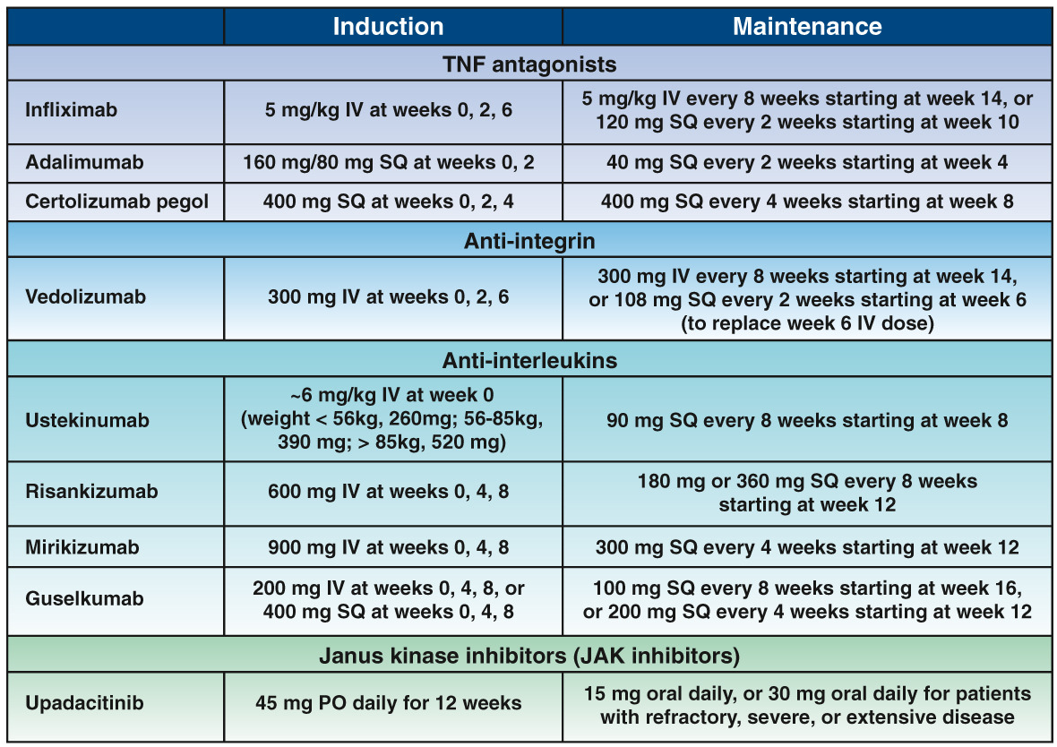
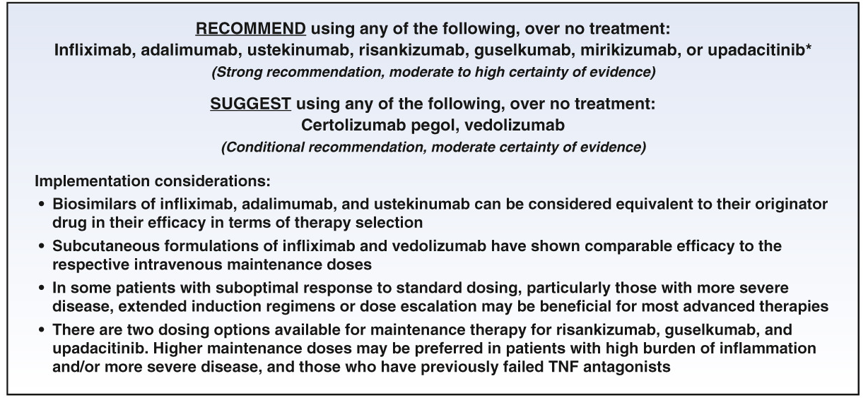
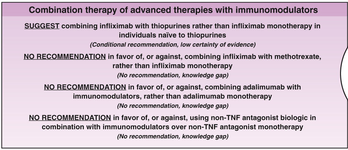
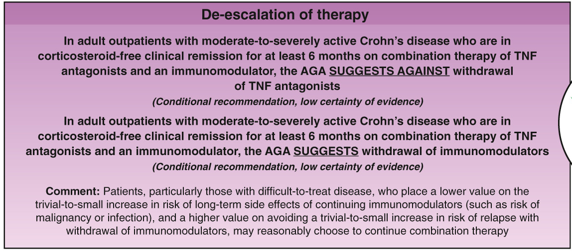
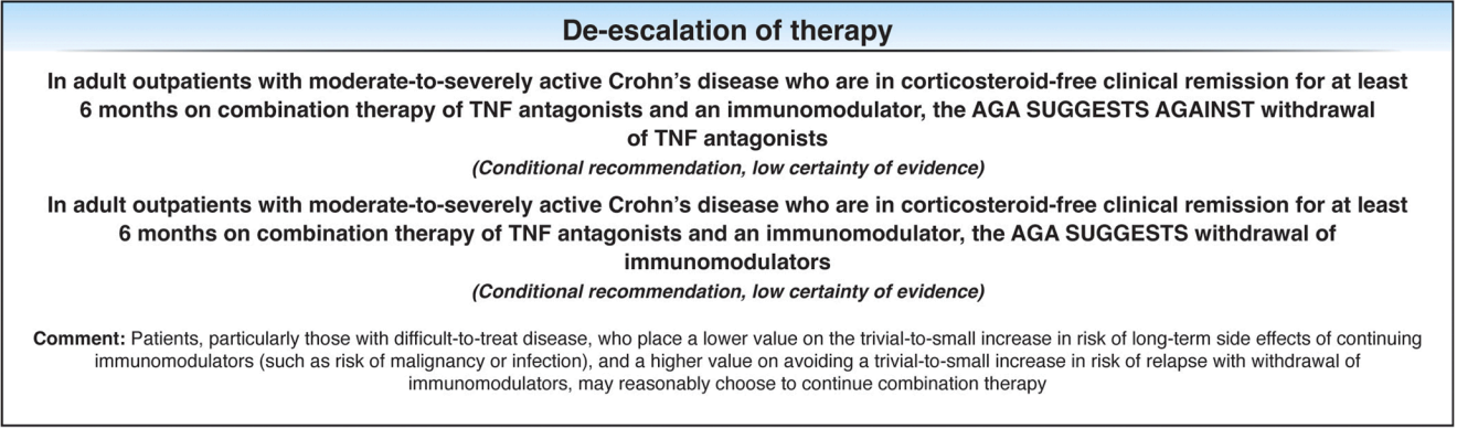
Yesterday and Today I am highlighting two adult clinical guidelines both of which are equivalent to up-to-date textbook chapters with specific recommendations; both are open access. In addition, the articles have accompanying author podcasts. Thanks to Ben Gold for these references.
Selected Management Recommendations:
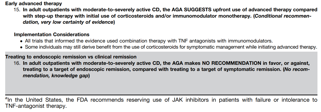
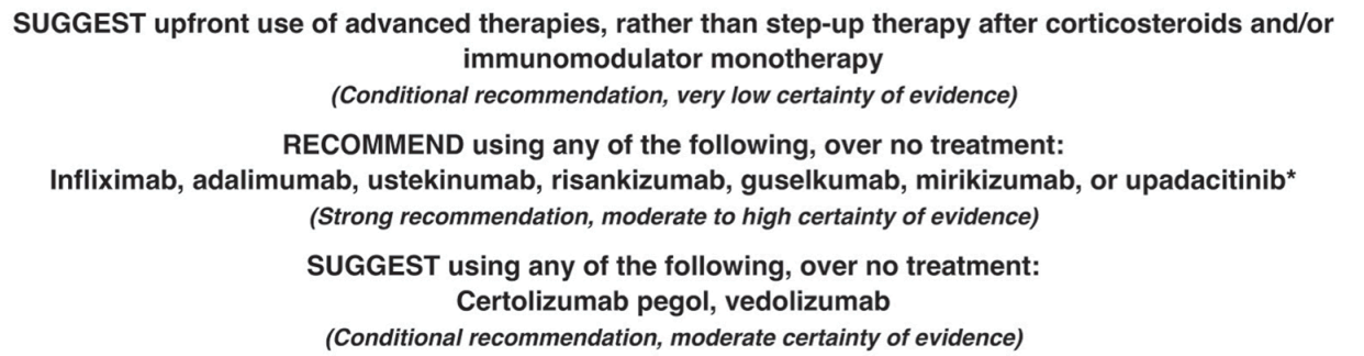
Table 1, #3: We suggest against requiring failure of conventional therapy before initiation of advanced therapy for the management of CD
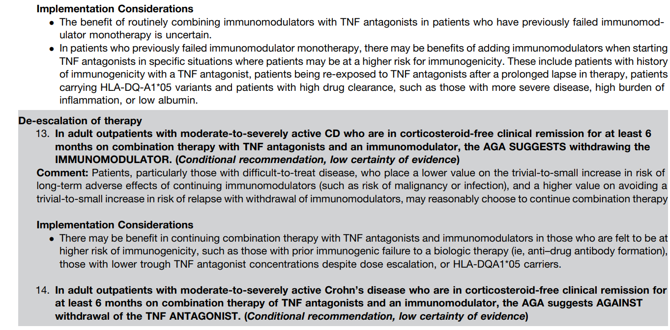
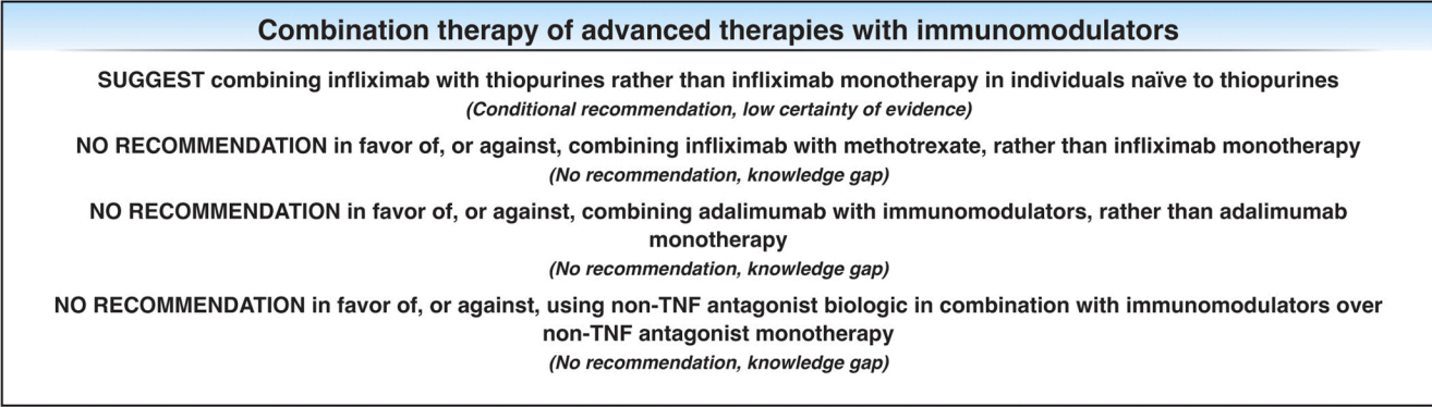
Table 1, #13: We recommend combination therapy of intravenous infliximab with immunomodulators (thiopurines) as compared with treatment with either immunomodulators alone or intravenous infliximab alone in patients with CD who are naive to those agents
Table 1, #33: In patients with high-risk CD, we recommend anti-TNF therapy to prevent postoperative endoscopic recurrence
Key Concepts:
Table 2, #9: Symptoms of CD do not correlate well with the presence of active inflammation and therefore should not be the sole guide for therapy. Objective evaluation by endoscopic or cross-sectional imaging should be undertaken periodically to avoid errors of under- or over-treatment.
Table 2, #14: The 10-year cumulative risk of major abdominal surgery in CD is 40%–55%, although recent studies performed in the biologic era suggest that the 10-year risk may have decreased to 30%. The 10-year risk of a second resection after the first is 35%, although again more recent studies suggest that this may have dropped to closer to 30%.
Table 2, #15: In CD, the 5-year rate of symptomatic postoperative recurrence is ∼50%.
Table 2, #29: Small bowel imaging should be performed as part of the initial diagnostic workup for patients with suspected CD.
Table 2, #31: Because of the absence of radiation exposure, magnetic resonance enterography should be used preferentially in young patients (younger than 35 years) and in patients in whom it is likely that serial exams will need to be performed.
Table 2, #38: Mucosal healing as determined by endoscopy is a goal of therapy. Scoring systems are available to measure the endoscopic disease activity and may be used to monitor response to therapy.
Table 2, #41: Antibiotics are not an effective treatment for luminal inflammatory CD and should not be used as a primary therapy.
My take: Given the rapid changes in available therapies, it would be optimal to make these collaborative guidelines (AGA, ACG, NASPGHAN) available online with frequent updates (similar to HCVguidelines.org).
Today and tomorrow I am highlighting two adult clinical guidelines both of which are equivalent to up-to-date textbook chapters with specific recommendations; both are open access. In addition, the articles have accompanying author podcasts. Thanks to Ben Gold for these references.
Table 2 in the UC guideline makes 54 recommendations and Table 3 provides 57 key concepts.
Selected Management Recommendations:
Table 2, #4: We recommend treating patients with UC to achieve endoscopic improvement (Mayo score 0 or 1) to increase the likelihood of sustained steroid-free remission and to prevent hospitalization and surgery
Table 2, #5: We recommend the use of FC (fecal calprotectin) in UC to assess response to therapy, to evaluate suspected relapse, and during maintenance
Table 2, #33: When infliximab is used as induction therapy for patients with moderately to severely active UC, we recommend combination therapy with a thiopurine
Table 2, #43: Recommend continuing tofacitinib or upadacitinib as compared with no treatment for maintenance of remission in patients with prior moderately to severely active UC now in remission after induction with tofacitinib or upadacitinib. **The authors recommend continuing each biologic that achieved remission with induction therapy (#38-#43)
Table 2, #51: In patients with ASUC failing to adequately respond to intravenous corticosteroids (IVCS) by 3 days, we recommend medical rescue therapy with infliximab or cyclosporine (Strong recommendation, moderate quality of evidence).
Key concepts:
Table 3, #29: Patients who are primary nonresponders to an anti-TNF (defined as lack of therapeutic benefit after induction and despite sufficient serum drug concentrations) should be evaluated and considered for alternative mechanisms of disease control (e.g., in a different class of therapy) rather than cycling to another drug within the anti-TNF class.
Table 3, #31: Subcutaneous infliximab and vedolizumab are considered equivalent to the standard intravenous maintenance dosing of these agents. The equivalence of the subcutaneous formulations for induction or as substitution for escalated doses of these therapies has not been robustly established.
Table 3, #41: Patients with UC should have available all medical options as recommended by their doctor and healthcare team. Third-party payers and requirements for step therapy should not come between the patient and their healthcare team in making decisions about treatment for UC.
Table 3, #48: All patients with ASUC should undergo a flexible sigmoidoscopy within 72 hours and preferably within 24 hours of admission. This should be used to assess endoscopic severity of inflammation and to obtain biopsies to evaluate for cytomegalovirus (CMV) colitis.
Table 3, #51: Nonsteroidal anti-inflammatory drugs (NSAIDs), narcotics, and medications with anticholinergic side effects should be avoided in ASUC.
Table 3, #57: In patients with ASUC initiating infliximab, dose intensification should be considered for those patients with low serum albumin (<2.5 g/dL).
My take: This article does an excellent job of summarizing current available evidence and good practice. Many of the recommendations may be helpful in garnering approval from third party payers.
R Smith-Bindman et al. NEJM 2025; 393: 1269-1278. Medical Imaging and Pediatric and Adolescent Hematologic Cancer Risk
Methods: This was a retrospective cohort of 3,724,623 children born between 1996 and 2016 in six U.S. health care systems and Ontario, Canada, until the earliest of cancer or benign-tumor diagnosis, death, end of health care coverage, an age of 21 years, or December 31, 2017.
Key findings:
During 35,715,325 person-years of follow-up (mean, 10.1 years per person), 2961 hematologic cancers were diagnosed, primarily lymphoid cancers (2349 [79.3%]), myeloid cancers or acute leukemia (460 [15.5%]), and histiocytic- or dendritic-cell cancers (129 [4.4%]).
The excess cumulative incidence of hematologic cancers by 21 years of age among children exposed to at least 30 mGy (mean, 57 mGy) was 25.6 per 10,000
The authors estimated that 10.1% of hematologic cancers may have been attributable to radiation exposure from medical imaging, with higher risks from the higher-dose medical-imaging tests such as CT
Cumulative Incidence of Hematologic Cancer According to Attained Age and Radiation Dose to Bone Marrow among Children without Down’s Syndrome
Discussion Points:
“A 15-to-30-mGy exposure equivalent to one to two CT scans of the head was associated with an increased risk by a factor of 1.8”
“Although CT and other radiation-based imaging techniques may be lifesaving, our findings underscore the importance of carefully considering and minimizing radiation exposure during pediatric imaging to protect children’s long-term health”
“Research on Japanese atomic-bombing survivors showed that leukemia rates peaked 6 to 8 years after exposure, with excess risk lasting for more than five decades, particularly for acute myeloid leukemia”
This study tried to avoid concerns about reverse causation — in which imaging is performed because of existing cancer symptoms –by lagged exposures by 6 and 24 months
“The increasing use of low-value imaging in children and excessive radiation doses in CT are well documented…In many cases, reducing the imaging dose or substituting magnetic resonance imaging or ultrasonography may be more feasible than avoiding imaging altogether”
While the risks in aggregate appear quite substantial, the editorial (L Morton. NEJM 2025; 393; 1337-1339.Studying Cancer Risks Associated with Diagnostic Procedures –Interpret Wisely) makes the point that the risks for the individual are very small. “Fewer than 1% of youths in this study accumulated doses of 30 mGy or more from medical imaging and even at this exposure level, the excess cumulative incidence of hematologic cancers was low (25.6 per 10,000)…we need to ensure that all involved in medical imaging…wisely interpret the results…to understand the balance of the very small risks and the notable benefits of necessary imaging examinations to provide optimal patient care.”
My take: This study is a reminder to carefully evaluate the benefits, risks and alternatives when using ionizing radiation studies.
This study explored a treatment with botulinum toxin (aka. Botox) for people who can’t belch due to a condition called retrograde cricopharyngeus dysfunction (R-CPD), which causes gas-related discomfort. Researchers used high-resolution manometry (HRM) with carbonated drink provocation to diagnose R-CPD. Then, they tested a treatment involving botulinum toxin injections into the cricopharyngeal muscle.
Out of 65 participants, 52 received the treatment, and 92% of those who received the treatment were able to belch after three months, significantly improving their symptoms and quality of life. In contrast, the control group, which included participants who deferred or declined treatment, saw no improvement. After 3 months, 43/51 (84%) of the treatment group reported being satisfied or very satisfied with therapeutic outcome.
10/20/25 NY Times: Peanut Allergies Have Plummeted in Children, Study Shows “The new study, published Monday in the journal Pediatrics, found that food allergy rates in children under 3 fell after those guidelines were put into place — dropping to 0.93 percent between 2017 and 2020, from 1.46 percent between 2012 and 2015. That’s a 36 percent reduction in all food allergies, driven largely by a 43 percent drop in peanut allergies.”
10/16/25 ABC News: Obesity remains high in the US., but more states showing progress, a new report finds “For the first time in more than a decade, the number of states with rates of obesity of 35% or more dropped, an encouraging sign that America’s epidemic of excess weight might be improving. But cuts to federal staff and programs that address chronic disease could endanger that progress, according to a new report released Thursday. Nineteen states had obesity rates of 35% or higher in 2024, down from 23 states the year before, according to an analysis of the latest data collected by the U.S. Centers for Disease Control and Prevention”
In this single center retrospective study from Ireland, the authors examined 122 patients (93 with Crohn’s disease [CD], 18 with ulcerative colitis [UC], 1 with IBDU) who received infliximab and had prospectively-collected data. The earlier cohort 2018-2019 received 5 mg/kg/dose and the later group 10 mg/kg/dose. Both groups had proactive therapeutic drug monitoring (pTDM).
Key findings:
The 5 mg/kg group, compared to the 10 mg/kg group, was less likely to have target pre-third TLs (6% vs 80%, P < .001) with the stated goal of >/= 15 microgm/mL
Fewer patients in the 5 mg/kg than 10 mg/kg group had pre-fourth TLs ≥5 µg/mL (6/48 [12.5%] vs 28/50 [56%], P < .001; mean [SD] TL 3.5 [6.3] vs 10.0 [9.9], P < .001)
Concurrent immunomodulator therapy was more common in the 5 mg/kg group (43% compared to 24%)
80% of patients were still receiving infliximab at 1 year including 87% of patients with CD and 54% with UC
The higher dose group had a lower CRP at 1 year followup. 26% of patients receiving the lower dose had a CRP > 5 mg/L compared with 9% in the higher dose group.
Some other measures of long term outcome (eg. IFX durability, clinical remission) were slightly better but did not reach statistical significance (see below)
Discussion Points:
“Our data show higher rates of below-target infliximab levels during and after induction in the 5 mg/kg group. Higher rates of dose escalation in this group during the first year resulted in similar dosing regimens…Thus, the similar infliximab durability and clinical outcomes at 1-year follow-up reflect early-dose optimization leading to dose equalization between the 2 groups, rather than a lack of benefit to higher dosing regimens”
“Our data affirm that proactive TDM with pre-emptive dose escalation restores below-target infliximab TLs and sustains clinical response…Indeed, in our cohort, some patients with low IFX levels pre-third dose were given their fourth dose 6 weeks later, rather than the standard 8 weeks. Without proactive TDM results, our rate of suboptimal TLs pre-fourth and during maintenance therapy would have been higher in both groups”
“Rates of immunomodulator use in the 10 mg/kg group were lower than in the earlier cohort of 5 mg/kg, reflecting changes in clinical practice over time”
My take:
This study shows that 94% of pediatric patients did NOT achieve adequate levels of infliximab at the pre-third dose with “standard” therapy. This was true even with 43% of the lower dose cohort receiving combination therapy (which often helps improve pharmacokinetics)
Proactive therapeutic drug monitoring helped mitigate the clinical outcomes, especially in the lower dosed cohort
“Children with IBD treated with the historic standard dose of 5 mg/kg induction are at increased risk of pharmacokinetic treatment failure related to high rates of suboptimal TLs”
Disclaimer: This blog, gutsandgrowth, assumes no responsibility for any use or operation of any method, product, instruction, concept or idea contained in the material herein or for any injury or damage to persons or property (whether products liability, negligence or otherwise) resulting from such use or operation. These blog posts are for educational purposes only. Specific dosing of medications (along with potential adverse effects) should be confirmed by prescribing physician. Because of rapid advances in the medical sciences, the gutsandgrowth blog cautions that independent verification should be made of diagnosis and drug dosages. The reader is solely responsible for the conduct of any suggested test or procedure. This content is not a substitute for medical advice, diagnosis or treatment provided by a qualified healthcare provider. Always seek the advice of your physician or other qualified health provider with any questions you may have regarding a condition.
A total of 21 experts (authors of article), recommended by ESNM, The European Association for Gastroenterology, Endoscopy and Nutrition (EAGEN) and The European Society for Primary Care Gastroenterology (ESPCG), from different countries agreed to participate as the International Working Group for the European Consensus on Bloating to vote on the Delphi statements.
This article regarding bloating/distension in adults is summarized in two tables. Table 1 has 75 statements. Table 2 is a summary –here are many of its recommendations:
Patients with functional bloating and abdominal distention should receive a lactose‐limiting diet trial based on their self‐reported symptoms or the presence of intolerance during a breath test after ingestion of a defined lactose load
A low FODMAP diet is effective in reducing functional bloating and abdominal distention
Rifaximin may be useful for the treatment of functional bloating and abdominal distention with efficacy
Among antispasmodic agents, pinaverium and otilonium bromide have been shown to be the most effective drugs for the treatment of functional bloating and abdominal distension
Lubiprostone, plecanatide and linaclotide are effective in improving constipation associated with functional bloating and abdominal distension
Linaclotide is the most effective secretagogue for functional bloating, although limited data is available for lubiprostone and plecanatide as well
Selective serotonin reuptake inhibitors (SSRI’s) are effective in reducing symptoms of functional bloating
Tricyclic antidepressants (TCA) such as amitriptyline are effective in reducing symptoms of functional bloating
In patients with discrete episodes of visible abdominal distension, biofeedback‐guided techniques to re‐educate abdominothoracic muscular activity are safe and effective for correction of abdominal distention and are associated with improvement in the subjective sensation of abdominal bloating
“Hypnotherapy improves symptoms of bloating in patients with IBS. However, its effect on functional bloating and abdominal distension was not explored and cannot be recommended”
Figure 1 provides an algorithm. For workup, it suggests checking the following in all patients: TSH, HgbA1c, CBC, CRP, TTG IgA, IgA, Glucose
In those with alarm features (eg. anemia, wt loss, suspicion of organic disease), more extensive evaluation is recommended
My take: One of my colleagues would often say that if there are a lot of treatments for a disease it usually indicates that none of them are very good.
Disclaimer: This blog, gutsandgrowth, assumes no responsibility for any use or operation of any method, product, instruction, concept or idea contained in the material herein or for any injury or damage to persons or property (whether products liability, negligence or otherwise) resulting from such use or operation. These blog posts are for educational purposes only. Specific dosing of medications (along with potential adverse effects) should be confirmed by prescribing physician. Because of rapid advances in the medical sciences, the gutsandgrowth blog cautions that independent verification should be made of diagnosis and drug dosages. The reader is solely responsible for the conduct of any suggested test or procedure. This content is not a substitute for medical advice, diagnosis or treatment provided by a qualified healthcare provider. Always seek the advice of your physician or other qualified health provider with any questions you may have regarding a condition.