Will Bariatric Surgery Become an Endoscopic Procedure?

A recent study (BK A Dayyeh et al. Clin Gastroenterol Hepatol 2017; 15: 37-43) provides evidence that endoscopic sleeve gastoplasty can be an effective treatment for obesity.

AGA Website Summary Endoscopic Sleeve Gastroplasty: A Promising New Weight Loss Procedure

An excerpt:

In the fight against obesity, bariatric surgery is currently the most effective treatment; however, only 1 to 2 percent of qualified patients receive this surgery due to limited access, patient choice, associated risks and the high costs. A novel treatment method — endoscopic sleeve gastroplasty — might offer a new solution for obese patients. Endoscopic sleeve gastroplasty is a minimally invasive, safe and cost-effective weight loss intervention, according to a study1 published online in Clinical Gastroenterology and Hepatology, the official clinical practice journal of the American Gastroenterological Association…

In this study of 25 patients with obesity who underwent the procedure at the Mayo Clinic in Rochester, MN, endoscopic sleeve gastroplasty reduced excess body weight by 54 percent at one year. Further, the procedure delayed solid food emptying from the stomach and created an earlier feeling of fullness during a meal, which resulted in a more significant and long-lasting weight loss.

Endoscopic sleeve gastroplasty was well tolerated as an outpatient treatment, requiring less than two hours of procedure time. Patients resumed their normal lifestyle within one to three days. The treatment was performed using standard “off-the-shelf” endoscopic tools as opposed to specific weight loss devices or platforms. The cost of endoscopic sleeve gastroplasty is roughly one-third that of bariatric surgery.

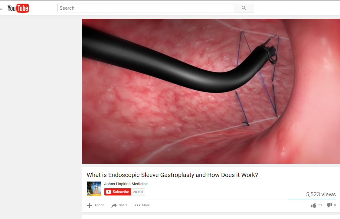
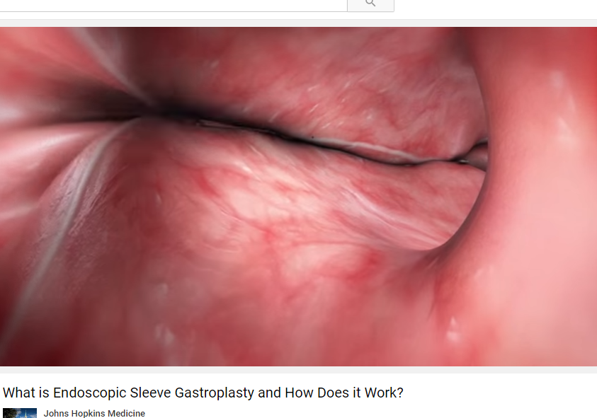
4 minute YouTube description from Johns Hopkins: What is Endoscopic Sleeve Gastroplasty and How Does it Work?

screenshot-73

screenshot-74 screenshot-75

Consensus Pancreatitis Recommendations

The INSPPIRE Group (CE Gariepy et al. JPGN 2017; 64: 95-103) has published consensus recommendations for acute recurrent pancreatitis (ARP) and chronic pancreatitis (CP).

While the authors acknowledge the need for high-level evidence/further research, they provide a large number of consensus recommendations.  These recommendations are succinctly summarized in Table 1 and Table 2.  From a reader’s perspective, my preference would have been to separate the recommendations for ARP and CP rather than to intermix them (though many of the recommendations are the same for both conditions).

ARP specific recommendations:

  • “Initial evaluation should include AST,ALT, GGT, Total bilirubin (fractionate if elevated), fasting lipids, and total serum calcium.”
  • Evaluate for fat-soluble vitamin deficiency, and pancreatic exocrine insufficiency at least annually

ARP and CP recommendations:

  • Consider ammonia and urine organic acids if there is a concern for undiagnosed metabolic disease.
  • Check for celiac disease.
  • Check for O&P if immunosuppressed, travel to endemic areas of Ascaris, or if peripheral eosinophilia.
  • Evaluation of genetic causes: should include sweat chloride test and PRSS1 gene testing. Consider SPINK1, CFTR, and CTRC evaluation.
  • Evaluate with MRCP (not ultrasound) acutely if GGT >2 x ULN or if direct bilirubin is elevated.
  • Non-acutely, MRCP recommended to evaluate pancreatic ductal abnormalities.  “When available, secretin-enhanced MRCP …should be obtained.” sMRCP can provide dynamic images of the pancreatic duct allowing differentiation of fixed from nonfixed lesions; this technique has not been widely adopted by pediatric radiologists compared with adult radiologists.

CP specific recommendations:

  • Evaluate for fat-soluble vitamin deficiency, pancreatic exocrine insufficiency, and pancreatic endocrine insufficiency at least annually

The authors did not recommend checking serum IgG4 in the absence of associated systemic disease or suggestive imaging for autoimmune pancreatitis.

Briefly noted: J-H Choi et al. Clin Gastroenterol Hepatol; 2017: 15: 86-92.  This study indicated that vigorous hydration with lactated ringer’s (LR) reduces risk of pancreatitis after ERCP.  A potential inference would be that LR would be an optimal fluid for pancreatitis more broadly. (Related: Why an ERCP Study Matters to Pediatric Care | gutsandgrowth)

Related blog posts:

Dragon Point, Labadee

Dragon Point, Labadee

“Addressing Physician Burnout”

In the last few years, there have been increasing reports of physician burnout.  A recent report (JAMA. Published online February 9, 2017. doi:10.1001/jama.2017.0076) (thanks to Ben Gold for this reference) provides a concise review of the reasons and potential mitigating strategies.

Full Text Link: “Addressing Physician Burnout”

An excerpt:

Physicians also have to navigate a rapidly expanding medical knowledge base, more onerous maintenance of certification requirements, increased clerical burden associated with the introduction of electronic health records (EHRs) and patient portals, new regulatory requirements (meaningful use, e-prescribing, medication reconciliation), and an unprecedented level of scrutiny (quality metrics, patient satisfaction scores, measures of cost).

These challenges have taken a toll on US physicians. Burnout is a syndrome of exhaustion, cynicism, and decreased effectiveness at work…The first large, national study of burnout among US physicians across all specialties did not occur until 2011. That study of 7288 participating physicians documented that approximately 45% reported at least 1 symptom of burnout and that burnout was more common among physicians than US workers in other fields…

The first large, national study of burnout among US physicians across all specialties did not occur until 2011. That study of 7288 participating physicians documented that approximately 45% reported at least 1 symptom of burnout and that burnout was more common among physicians than US workers in other fields…

Physician burnout has been linked to self-reported errors, turnover, and higher mortality ratios in hospitalized patients…

The current burden of documentation related to the clinical encounter required to meet billing requirements, quality reporting, and separate justification for each test ordered individually is unsustainable…

Individual physicians must also do their part…Individual physicians have a professional responsibility to take care of themselves. Adequate sleep, exercise, and attending to personal medical needs should be considered a minimal standard for self-care. Physicians must also proactively identify personal and professional priorities and take deliberate steps to integrate their personal and professional lives.

Related blog posts:

birthoutcomesaftertnt

Ileocecal Resection in Pediatric Crohn’s Disease

A recent retrospective study (K Diederen et al. Inflamm Bowel Dis 2017; 23: 272-82) provides data on the likelihood of complications and recurrence following ileocecal resection in pediatric Crohn’s disease (n=122).

Key findings:

  • Severe postoperative complications were noted in 9.8%.  Risk factors included colonic disease (Odds ratio 5.6), microscopically positive resection margins (OR 10.4), and emergency surgery (OR 6.8)
  • Overall complication rate was reported as 29.5% which is similar to rates reported in adults
  • Clinical recurrence rates after 1, 5, and 10 years: 19%, 49%, and 71%
  • Surgical recurrence rates after 1, 5, and 10 years: 2%, 12%, and 22%
  • Immediate postoperative therapy reduced the risk of clinical recurrence (HR 0.3) and surgical recurrence (HR 0.5)
  • “In this study, postoperative catch-up growth was found in patients younger than 16 years in the year after surgery.” Thus, surgery could be an important to reverse growth retardation.

Complications within 30 days of surgery were categorized with the Clavien-Dindo classification. Those with grade ≥III which required either surgical, endoscopic or radiologic intervention were considered severe.  In this population, the complications included intraabdominal septic complications and/or anastomotic leakage.

My take: In some patients, ileocecal resection should NOT be a last resort.  Waiting too late, increases the risk of complications.  The task at hand is prospectively identifying those who merit surgery sooner and then convincing the family to proceed.

screenshot-118

Related blog posts:

Clostridium difficile Infection in Inflammatory Bowel Disease: Expert Updates

A recent clinical practice update (S Khanna et al. Clin Gastroenterol Hepatol; 2017; 15: 166-74) provides some succinct recommendations regarding Clostridium difficile infection (CDI) in Inflammatory Bowel Disease (IBD).

Background: In 2011, the authors note that CDI was associated with 29,000 deaths and is now the most lethal enteric pathogen in the U.S.

Differences in pathogenesis of C diff in IBD compared to those without IBD:

  • Younger age
  • Less frequent antibiotic exposure
  • More often community onset (rather than hospital onset)
  • Higher recurrence (may be related to dysbiosis)

Key recommendations:

  • In patients with IBD flare, test for CDI
  • In patients with CDI and IBD, clinicians should consider “using vancomycin instead of metronidazole.”
  • In patients with recurrent CDI and IBD, consider fecal microbiota transplantation

Figure 4 proposes a management algorithm (for adults).  If uncomplicated CDI, recommended dose of vancomycin was 125 mg q6h. If no improvement in 3-4 days, then “consider escalation of immunosuppression.” For complicated CDI, consider oral vancomycin at 500 mg q6h and IV metronidazole 500 mg q8.  In addition, consider rectal vancomycin and surgery consult.

Complicated CDI includes ICU admission, hypotension, T >38.5, ileus/megacolon, mental status changes, leukocyte count >35,000  or < 2000, or lactate >2.2 mmol/L

Another review article (Y Chen et al. Inflamm Bowel Dis 2017; 23: 200-07) is a meta-analysis that identified six studies.  One of these studies was a case-control study with nearly 400,000 patients (and about 7000 cases of C diff). Key finding: CDI results in nearly a doubling of the risk of colectomy (OR 1.90), mainly in patients with ulcerative colitis.

Related blog posts

Disclaimer: These blog posts are for educational purposes only. Specific dosing of medications (along with potential adverse effects) should be confirmed by prescribing physician.  This content is not a substitute for medical advice, diagnosis or treatment provided by a qualified healthcare provider. Always seek the advice of your physician or other qualified health provider with any questions you may have regarding a condition.


Rare Tragic Reaction to Infliximab

A recent post on the pediatric GI Listserv pointed out a troubling case report: “Fatal Central Nervous System Disease Following First Infliximab Infusion in a Child With Inflammatory Bowel Disease,” FM. Baumer et al; Pediatric Neurology 2016; 57: 91-94.

“A seven-year-old boy diagnosed with ulcerative colitis and primary sclerosing cholangitis received infliximab. Six hours following his uneventful infusion, he awoke with headache and emesis and rapidly became obtunded…Cranial computed tomography revealed hypodense lesions in the cerebral hemispheres, cerebellum, and pons accompanied by hemorrhage…Within four days he met criteria for brain death.”

The authors note that “the close temporal association between our patient’s presentation and the infliximab infusion raises concern for a drug-related cause for his cerebral injury.” While this case report is terribly sad, severe and fatal reactions can unfortunately be encountered with a wide range of medications, including commonly used antibiotics.

My take: Thus, while the vast majority of pediatric patients with inflammatory bowel disease will benefit from infliximab therapy, there are rare tragic outcomes.  img_3954


Gastrojejunostomy Complications Frequent

Gastrojejnostomy (GJ) placement allows enteral feeds to bypass the stomach.  When a gastrostomy is already in place, GJ placement may allow patients to avoid surgery (eg. fundoplication).  Most practitioners would consider the risk of GJ placement to be low, but a recent report (J Moorse et al. JPS 2017; http://dx.doi.org/10.1016/j.jpedsurg.2017.01.026) suggests that it is higher than expected.  The abstract and link are below.

Link: Gastrojejunostomy tube complications — A single center experience and systematic review

Abstract

Purpose

Gastrojejunostomy tubes (GJTs) enable enteral nutrition in infants/children with feeding intolerance. However, complications may be increased in small infants. We evaluated our single-institution GJT complication rate and systematically reviewed existing literature.

Methods

With REB approval, a retrospective single-institution analysis of GJT placements between 2009 and 2015 was performed. For the systematic review, MOOSE guidelines were followed.

Results

At our institution, 48 children underwent 154/159 successful insertions primarily for gastroesophageal reflux (n = 27; 55%) and aspiration (n = 11; 23%). Median age at first GJT insertion was 2.2 years (0.2–18). Thirty-five (73%) had an index insertion when ≤10 kg. GJTs caused 2 perforations and 1 death. The systematic review assessed 48 articles representing 2726 procedures. Overall perforation rate was estimated as 2.1% (n = 36 studies, 23/1092, 95% CI: 1.0–3.2). Perforation rates in children <10 kg versus ≥10 kg were estimated as 3.1%/procedure (95% CI: 1.1%–5.0%) and 0.1%/procedure (95% CI: 0%–0.3%), respectively. The relative risk of perforation was 9.4 (95% CI: 2.8–31.3). Overall mortality was estimated as 0.9%/patient (n = 39 studies; 95% CI: 0.2–1.6%). Most perforations (19/23; 83%) occurred ≤30 days of attempted tube placement.

Conclusion

Gastrojejunostomy tubes are associated with significant complications and frequently require revision/replacement. Insertion in patients <10 kg is associated with increased perforation risk. Caution is warranted in this subgroup.

With regard to the methodology

  • ~90% of the procedures were performed by interventional radiology and the interventionist had a median of 6.6 years of experience
  • Most GJs were 16 French in width and most were either 15 cm or 22 cm in length

My take: This report highlights the significant risks associated with GJ placement, particularly in smaller patients (<10 kg).  Despite these risks, GJ placement is often the safest option.

Costa Maya, Mexico

Costa Maya, Mexico


PICC versus Broviac for TPN in Intestinal Failure

LinkA comparison of Broviac® and peripherally inserted central catheters in children with intestinal failure

Abstract:

Central venous catheters (CVCs) are a source of morbidity for children with intestinal failure (IF). Complications include infection, breakage, occlusion, and venous thrombosis. Broviacs® have traditionally been preferred, but peripherally inserted central catheters (PICCs) are gaining popularity. This study compares complications between Broviacs® and PICCs in children with IF.

Methods

After IRB approval, children with IF receiving parenteral nutrition (2012–2016) were reviewed. Complications were compared between Broviacs® and PICCs using the generalized estimation equation population-averaged Poisson regression model. P values <0.05 were considered significant.

Results

36 children (0.1–16 years) with IF were reviewed, accounting for 27,331 catheter days, 108 Broviacs® (3F–9F), and 54 PICCs (2-11F). Broviacs® had a significantly higher infection rate (4.2 vs. 2.6/1000 catheter days, p = 0.011), but PICCs were more likely to break (1.56 vs. 0.26/1000 catheter days, p = 0.002). When comparing same size catheters (3F), there were no significant differences in infection, breakage, or occlusion. Twelve children (33%) had central venous thrombosis, all after Broviac® placement. Three children (8%) had basilic vein thrombosis after PICC placement.

Conclusion

Although Broviacs® and PICCs had similar complication rates, there were fewer central venous thromboses associated with PICCs. This should be considered when choosing catheters for children with IF.

screenshot-113

My take: Despite some possible advantages of PICC in this study, a prospective randomized study is needed if one is to make a compelling argument regarding better outcomes.  Given the retrospective nature of this study, it could be that the sicker patients may have been more likely to receive a Broviac® and this could have skewed the results.

Chronic Diarrhea Recommendations

Recent guidelines for adults with chronic diarrhea (>4 weeks):

Full text link: LR Schiller, DS Pardi, JH Sellin. Clin Gastroenterol Hepatol 2017; 15: 182-193.

A few key points:

  • The authors advocate treatment, not testing, for adults who meet Rome criteria for irritable bowel syndrome (IBS) without alarm symptoms.
  • Dietary history is essential.
  • “True food allergies are rare causes of chronic diarrhea in adults”
One of my colleagues questioned whether this product could be part of an effective cleanout

One of my colleagues questioned whether this product could be part of an effective cleanout


Expert Advice on Clostridium difficile and Inflammatory Bowel Disease

Link: Management of Clostridium difficile Infection in Inflammatory Bowel Disease: Expert Review from the Clinical Practice Updates Committee of the AGA Institute

Abstract: The purpose of this expert review is to synthesize the existing evidence on the management of Clostridium difficile infection in patients with underlying inflammatory bowel disease. The evidence reviewed in this article is a summation of relevant scientific publications, expert opinion statements, and current practice guidelines. This review is a summary of expert opinion in the field without a formal systematic review of evidence.

Best Practice Advice 1: Clinicians should test patients who present with a flare of underlying inflammatory bowel disease for Clostridium difficile infection.

Best Practice Advice 2: Clinicians should screen for recurrent C difficile infection if diarrhea or other symptoms of colitis persist or return after antibiotic treatment for C difficile infection.

Best Practice Advice 3: Clinicians should consider treating C difficile infection in inflammatory bowel disease patients with vancomycin instead of metronidazole.

Best Practice Advice 4: Clinicians strongly should consider hospitalization for close monitoring and aggressive management for inflammatory bowel disease patients with C difficile infection who have profuse diarrhea, severe abdominal pain, a markedly increased peripheral blood leukocyte count, or other evidence of sepsis.

Best Practice Advice 5: Clinicians may postpone escalation of steroids and other immunosuppression agents during acute C difficile infection until therapy for C difficile infection has been initiated. However, the decision to withhold or continue immunosuppression in inflammatory bowel disease patients with C difficile infection should be individualized because there is insufficient existing robust literature on which to develop firm recommendations.

Best Practice Advice 6: Clinicians should offer a referral for fecal microbiota transplantation to inflammatory bowel disease patients with recurrent C difficile infection.


Related blog posts:

screenshot-94