Comparative Safety of Janus Kinase Inhibitors vs Tumor Necrosis Factor Antagonists For Inflammatory Bowel Disease Patients

D Ahuja et al. Clin Gastroenterol Hepatol 2026. 24: 794-804. Comparative Safety of Janus Kinase Inhibitors vs Tumor Necrosis Factor Antagonists in Patients With Inflammatory Bowel Diseases

Methods: This retrospective cohort study used an administrative claims database and identified patients with IBD who were new users of either JAK inhibitors (n=856) or TNF antagonists (n=9422) between 2016 and 2023. Mean age was 45 years.

Key findings:

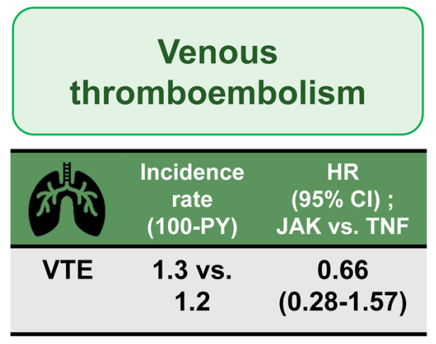
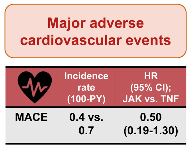
  • There was no difference in the risk of VTE (1.3 vs 1.2; HR, 0.66; 95% CI, 0.28–1.57) and MACE (0.4 vs 0.7; HR, 0.50; 95% CI, 0.19–1.30)
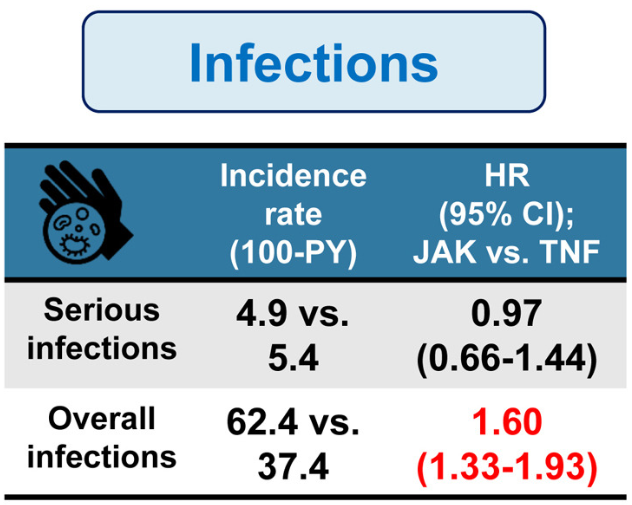
  • There was no difference in the risk of serious infections (4.9 vs 5.4; HR, 0.97); however, JAK inhibitors were associated with an increase risk of overall infections (incidence rate, 62.4 per 100 person-years [PY] vs 37.4 per 100 PY; hazard ratio [HR], 1.60)
The authors note that their findings differ from the ORAL study (Ytterberg et al. NEJM 2022; 386: 316-326.) which showed higher risk of MACE in patients receiving tofacitinib. In the current study, even in patients deemed to be at higher risk for MACE (age >50 years with at least 1 cardiovascular risk factor), JAK inhibitors were associated with lower incidence and risk of MACE compared with TNF antagonists (IR per 1000 PY, 0.4 vs 2.1, HR 0.10).

My take (borrowed from the authors) “It is unlikely that JAK inhibitors are associated with higher risk of VTE and MACE compared with TNF antagonists in most patients with IBD.”

Related blog posts:

“Real-World” Experience: High Dose Upadacitinib Recaptures IBD Response

AHY Ho et al. Clin Gastroenterol Hepatol 2026; 24: 763-771. Real-world Experience of Upadacitinib Reinduction and High-dose Maintenance Therapy in Inflammatory Bowel Disease

Methods: This was a prospective cohort study of patients (n=181 — 79 CD, 83 UC, 6 -IBD-U, 13 with IPAA) treated with UPA between April 2022 and November 2023. Included patients responded to UPA induction, had loss of response (LOR) after dose reduction, and subsequently received reinduction therapy with 45 mg QD. They were followed for a median duration 93 weeks.

Key findings:

  • Dose escalation to 45 mg QD for a median of 13 weeks (IQR, 8–36 weeks) recaptured clinical response in 80.4%
  • Among patients who recaptured response, 19 again reduced dose
  • 93.8% of patients on 45 mg QD maintained remission vs 21.1% who again dropped to 30 mg QD (P < .001)
  • Acne/rosacea was the most common adverse event (39%); there were no serious adverse events

In their discussion, the authors note that dose escalation with another JAK inhibitor, tofacitinib, also has been shown to reverse LOR (in about 50%). In addition, they note that “in our experience, prolonged exposure to 45 mg QDD UPA is safe.” Though, “a longer follow-up period…is required to address long-term safety of UPA in IBD, especially at a higher dose.”

My take: Many patients taking UPA have not responded to multiple other advanced therapy. As such, the potential to recapture response with a higher dose of UPA is an important finding. Dose intensification is an effective strategy for most of the advanced therapies.

Briefly noted: S Honap et al. Clin Gastroenterol Hepatol 2026; 24: 784-793. Open Access! Comparative Effectiveness of Tofacitinib vs Upadacitinib for the Treatment of Acute Severe Ulcerative Colitis In this retrospective study of 111 adults with ASUC, Between days 3 and 7 after treatment initiation, upadacitinib was associated with greater response rates (84% vs 54%), but response/remission was comparable at day 98 (45%/36% vs 55%/48%) and day
182 (29/29% vs 39/34%).

Related blog posts:

ADMIRE CD II: Stem Cell Therapy NOT More Effective Than Placebo for Complex Perianal Fistulas in Crohn’s Disease

J Colombel J et al. Gastroenterology, 2026. Open Access! Darvadstrocel in Patients With Crohn’s Disease With Complex Perianal Fistulas: The ADMIRE CD II Phase 3 Randomized Trial

Background: The ADMIRE CD II, a phase 3 trial of the efficacy and safety of darvadstrocel in patients with complex perianal fistulas (CPF) at weeks 24 and 52, conducted in more than twice as many sites and patients (n=568) as ADMIRE CD, from North America as well as Europe and Israel. This trial was conducted after the approval of darvadstrocel in Europe and Japan and therefore aimed to provide further confirmation of efficacy in patients with CPF.

Key findings:

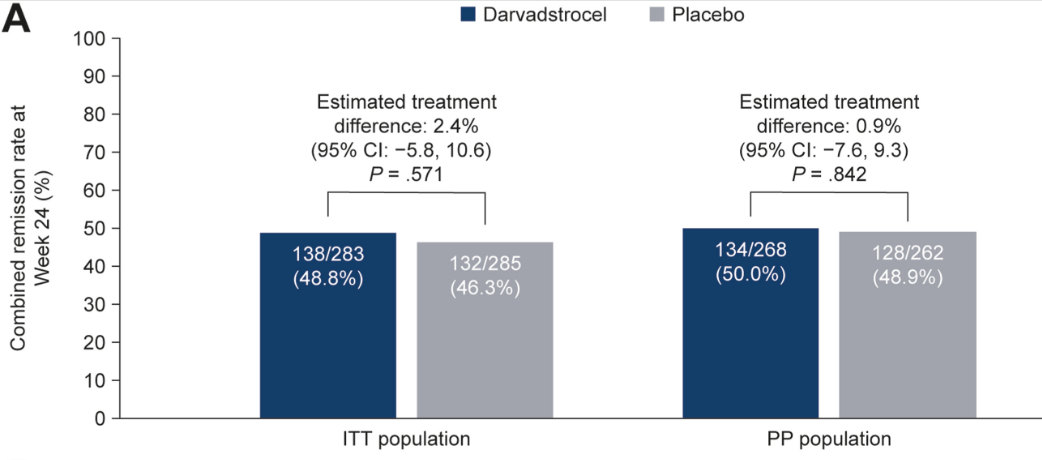
  • At week 24, combined remission was achieved in 138 of 283 (48.8%) patients in the darvadstrocel group and 132 of 285 (46.3%) in the placebo group 
  • There were no significant differences in key secondary endpoints for darvadstrocel vs placebo (clinical remission at week 24 [P = .515] and time to clinical remission [P = .374])
  • Treatment-emergent adverse events were infrequent and experienced by similar proportions of patients receiving darvadstrocel (203/278 [73.0%]) and placebo (201/274 [73.4%])
ITT (intention-to-treat), PP (per protocol) Results

In the discussion, the authors speculate on why ADMIRE CD found a significant response to darvadstrocel whereas the current larger ADMIRE CD II did not.

  1. ” In ADMIRE CD II, all patients had seton placement (mandatory at least 2–3 weeks before treatment administration), whereas in ADMIRE CD, seton placement was as clinically indicated, and 10 patients (2 darvadstrocel, 8 placebo) did not have a seton. The fact that all patients, including those in the control group, underwent fistula preparation before treatment administration may have enhanced response rates in the placebo group in ADMIRE CD II”
  2. “In patients with CPF, it is well established that higher trough levels of concomitant systemic therapies are associated with improved fistula healing outcomes compared with lower trough levels. These data were not collected during ADMIRE CD II or ADMIRE CD, but it is acknowledged that the possibility of higher trough levels in ADMIRE CD II compared with ADMIRE CD could have contributed to the difference in placebo response rates between trials… also relevant to consider the higher proportion of patients using background immunosuppressants or a combination of immunosuppressants and monoclonal antibodies in ADMIRE CD II compared with ADMIRE CD.”

My take (borrowed in part from the authors): These results “challenge the relevance of stem cell therapy for perianal fistula healing.” It appears that good surgical management along with optimized medical therapy achieve the same results.

Related blog posts:

Comparing Vedolizumab in “Early” and “Late” Crohn’s Disease

Lancet Gastroenterol Hepatol 2026; 11: 12-21. Vedolizumab in early and late Crohn’s disease (LOVE-CD): a phase 4 open-label cohort study

Methods: Eligible patients were adults aged 18–80 years with moderate to severe Crohn’s disease (Crohn’s Disease Activity Index [CDAI] 220–450, with ulcers at endoscopy). Patients were divided into two groups: those with early Crohn’s disease, n=86 (defined as a diagnosis less than 2 years ago and naive to advanced treatment [naive or only treated with corticosteroids or immunomodulators, or both]); and those with late Crohn’s disease, n=174 (defined as a diagnosis more than 2 years ago and previously treated with corticosteroids, immunomodulators, and anti-TNF agents). The primary endpoint was the proportion of patients with clinical and endoscopic remission (defined as CDAI ≤150 and SES-CD <4) at both week 26 and 52.

Key findings:

  • Clinical and endoscopic remission at both week 26 and 52 was achieved in 27 (31·4%) of 86 patients with early Crohn’s disease versus 15 (8·6%) of 174 patients with late Crohn’s disease (difference 22·8%, 95% CI 12·6–33·7)
  • Serious adverse events occurred in three (3·5%) of 86 patients with early Crohn’s disease versus 46 (26·4%) of 174 patients with late Crohn’s disease and included infections (one [1·2%] vs 13 [7·5%]), surgery (none vs eight [4·6%]), intestinal obstruction (none vs four [2·3%]), exacerbation of Crohn’s disease (one [1·2%] vs six [3·4%]), and malignancy (none vs three [1·7%])
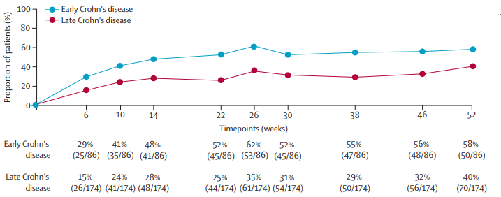
Corticosteroidfree clinical remission at all timepoints

Discussion:

  • “After 52 weeks of open-label treatment, almost 60% of patients with early disease achieved clinical remission and more than 50% were in endoscopic remission. By contrast, deep remission rates at both weeks 26 and 52 were observed in less than 10% of patients with late Crohn’s disease (ie, those with longstanding disease and previous exposure to anti-TNF
    agents).”
  • “Despite the earlier stage at which vedolizumab was initiated, disease severity was comparable betweenthe early and late Crohn’s disease groups, from a clinical (median CDAI 255 vs 259), endoscopic (median SES-CD 9 vs 12), and biochemical perspective (median serum C-reactive protein 9 mg/L vs 8 mg/L).”
  • “A pivotal observation in LOVE-CD was that dose intensification after week 26 (ie, doubling the dose) in patients without an endoscopic response did not lead to higher endoscopic remission rates, despite significantly higher serum vedolizumab concentrations. This
    finding suggests that the dosing schedule that was originally designed and approved is optimal for most patients and saturates the target. Patients who do not respond most likely have other dominant immune pathways that are activated and remain unaffected by
    vedolizumab.”
  • “All three classes of biologics approved for the treatment of Crohn’s disease perform better when initiated early in the disease course.”

My take: For Crohn’s disease (CD), vedolizumab should be mainly used in those without prior biologic therapy. In addition, changes in vedolizumab dosing based on concentrations is much less helpful than it is with anti-TNF agents.

Related blog posts:

Disclaimer: This blog, gutsandgrowth, assumes no responsibility for any use or operation of any method, product, instruction, concept or idea contained in the material herein or for any injury or damage to persons or property (whether products liability, negligence or otherwise) resulting from such use or operation. These blog posts are for educational purposes only. Specific dosing of medications (along with potential adverse effects) should be confirmed by prescribing physician. Because of rapid advances in the medical sciences, the gutsandgrowth blog cautions that independent verification should be made of diagnosis and drug dosages. The reader is solely responsible for the conduct of any suggested test or procedure. This content is not a substitute for medical advice, diagnosis or treatment provided by a qualified healthcare provider. Always seek the advice of your physician or other qualified health provider with any questions you may have regarding a condition.

Chronic Nonbacterial Osteomyelitis Associated with Pediatric Inflammatory Bowel Disease

M Matar et al. J Pediatr Gastroenterol Nutr. 2026;82:487–494. Chronic nonbacterial osteomyelitis associated with pediatric inflammatory bowel disease: : A multicenter retrospective study from the Paediatric inflammatory bowel disease Porto Group of ESPGHAN

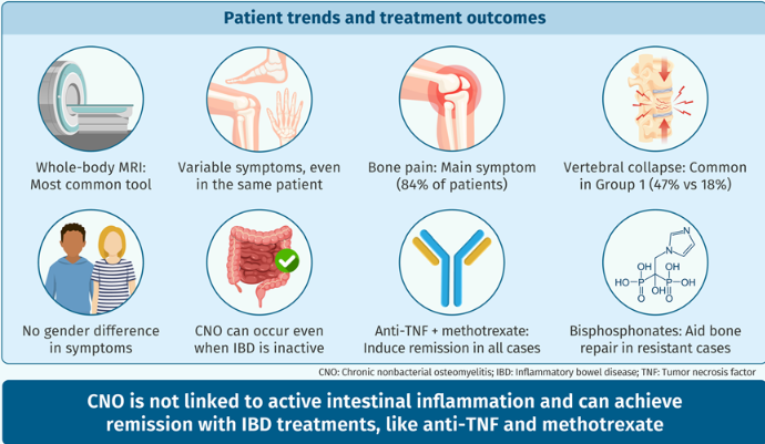
Methods: Retrospective study with 45 pediatric patients with inflammatory bowel disease (n=32 with Crohn’s disease, n=8 with ulcerative colitis, n=5 with IBD-U)

Key findings:

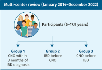
  • CNO presented in 15 patients (33%) within 3 months of IBD diagnosis, and in additional 20 (44%) patients after IBD diagnosis; in 10 (22%) patients CNO preceded the diagnosis of IBD with a median time 46 (25–248) weeks
  • 11 (24%) subjects displayed at least one additional extra-intestinal manifestations, including arthritis (6, 13%), erythema nodosum (4, 9%), sacro-ileitis (2, 4%), psoriasis (1, 2%), and pyoderma gangrenosum (1, 2%).
  • Complications occurred in six patients and included vertebral collapse, bone fracture, and bone deformity. In eight (18%) subjects vertebral collapse was present already at the time of diagnosis.
  • “While in most patients, diagnosis of CNO was associated with either clinical or laboratory indices of active IBD, especially CD, in some cases, the intestinal disease was quiescent.”
  • “All patients achieved remission of CNO at some point during follow-up, which may support the hypothesis that anti-TNF treatment is unlikely to promote CNO development and does not reinforce the theory of a paradoxical effect that has been suggested by Cordesse et al.22“

My take: This is a useful review highlighting the association of CNO with both active disease and quiescent IBD. The authors argue that their data does not support the development of CNO as a paradoxical effect. I disagree with this premise. Many of the extraintestinal manifestations, that anti-TNF agents can treat, rarely can be caused by them. Besides CNO, paradoxical reactions to anti-TNF agents have included the development of rheumatoid arthritis, psoriasis, hidradenitis suppurativa and autoimmune liver disease.

Related blog posts:

How to Improve the Value of Biologic Infusions: Reduce Lab Testing and Frequency

T Shah et al. JPGN Reports 2026;1–5. Responsible laboratory surveillance of pediatric patients with inflammatory bowel disease on biologic infusion therapy

This retrospective single-center study with 34 pediatric patients with inflammatory bowel disease examined the laboratory costs (2020-2021) associated with monitoring biologic therapy.

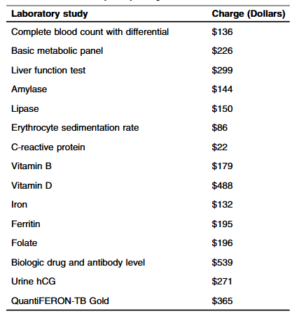
Methods: “Routine laboratory studies were defined as those part of the standardized infusion protocol at SBCH and were obtained with each scheduled infusion. The following laboratory studies were considered routine/standard: complete blood count with differential, basic metabolic panel, liver function tests, amylase, lipase, erythrocyte sedimentation rate, C-reactive protein, vitamin D, iron, ferritin, vitamin B12, folate, urine hCG (if a subject was female). Other laboratory studies that were collected, but not considered routine studies included QuantiFERON-TB, and biologic drug and antibody level.”

Key findings:

  • The average hospital charge for studies obtained per infusion was $1308.36 with an average annual cost of $9543.44 per patient
  • Fifteen (6%) instances of change in clinical management were found. “Only a limited subset of the 15 laboratory studies included were utilized in making changes: biologic drug, Vitamin D, and iron level”
  • During the study, 248 infusions were administered with a “total annual charge amongst all patients in the study was $324,447”

Discussion:

  • “Our study population had well controlled disease as evident by low PCDAI and PUCAI scores…Our observations suggest the utility of routine laboratory surveillance at each biologic infusion is minimal, favoring decreased testing for IBD patients, especially those in clinical remission.”
  • “We propose obtaining laboratory tests twice a year, or with every third infusion, for patients with mild disease or in remission based on their disease activity index scores. In our small cohort of patients, this change in practice would reduce the total annual costs by 66% ($214,154.82)”

My take: It has been my practice, for most patients with IBD, to obtain labs with every other infusion (~3 times per year). Typically, I will obtain a CBC/d, CMP and CRP and obtain other labs like Vit D, GGT, Quantiferon Gold and drug level monitoring less frequently. I rarely check Vit B12, ESR, Folate, Amylase, and Lipase.

Related blog posts:

Disclaimer: This blog, gutsandgrowth, assumes no responsibility for any use or operation of any method, product, instruction, concept or idea contained in the material herein or for any injury or damage to persons or property (whether products liability, negligence or otherwise) resulting from such use or operation. These blog posts are for educational purposes only. Specific dosing of medications (along with potential adverse effects) should be confirmed by prescribing physician. Because of rapid advances in the medical sciences, the gutsandgrowth blog cautions that independent verification should be made of diagnosis and drug dosages. The reader is solely responsible for the conduct of any suggested test or procedure. This content is not a substitute for medical advice, diagnosis or treatment provided by a qualified healthcare provider. Always seek the advice of your physician or other qualified health provider with any questions you may have regarding a condition.

VedoKids Study: Vedolizumab for Extraintestinal Manifestations of Inflammatory Bowel Disease

G D’Arcangelo et al. J Pediatr Gastroenterol Nutr. 2026;82:495–502. Open Access! Vedolizumab for extraintestinal manifestations in pediatric inflammatory bowel disease: Results from the VedoKids study

Background: “Since vedolizumab is a gut-selective anti-α4β7 integrin, its effect on EIMs has been a matter of debate, with relevant data lacking in pediatric IBD. A systematic review, which included three interventional studies, five non-interventional studies, and three case series, concluded that there is insufficient evidence supporting the efficacy of vedolizumab for treating pre-existing EIMs in adults.3“

Methods: This was a subgroup analysis of the pediatric VedoKids cohort, a multicenter, prospective “real-life” study of children (aged 0–18 years) with IBD treated with vedolizumab and followed through 54 weeks.

Key findings:

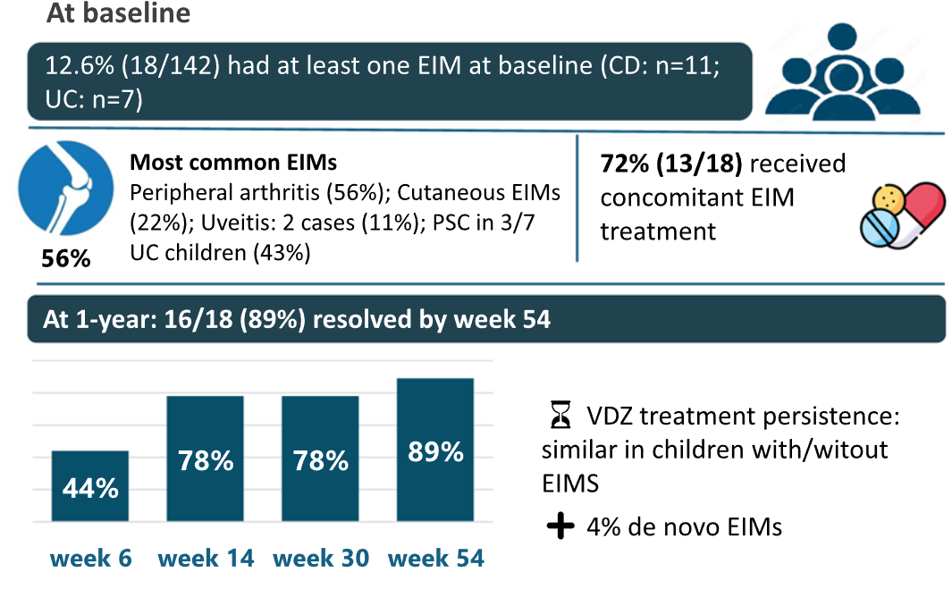
  • EIMs were identified in 18/142 (12.6%) children at baseline
  • Children with EIMs had an average age of diagnosis of 9 yrs compared to 12 yrs in those without EIMs
  • Children with EIMs had higher rate of pancolitis in UC and ileocolonic distribution in CD
  • Prior anti-TNF medication was noted in 16 (89%) of EIM cohort compared to 74 (60%) of non-EIM cohort
  • Concomitant medications were administered in 72% of EIM cases and to a similar number of non-EIM patients. For EIM patients, ASA were given in 7, steroids in 10, thiopurines in 4 and methotrexate in 2
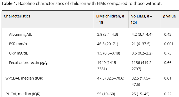
  • Children with EIMs had more active disease (see below)
  • EIM resolution rate of 89%, mainly occurring within the early weeks of vedolizumab treatment

My take: While this study has several limitations, including the high rate of concomitant medications, it shows that most patients receiving vedolizumab had resolution of their EIMs. In addition, it shows that patients with EIMs had a more severe IBD phenotype.

Related blog posts:

Methotrexate Tolerance and Toxicity in Pediatric Inflammatory Bowel Disease

E Vermeer et al. J Pediatr Gastroenterol Nutr. 2026;82:477–486. Open Access! Methotrexate toxicity and intolerance in paediatric inflammatory bowel disease: A retrospective cohort study

This was a  a retrospective single-center cohort study, including pediatric IBD patients (n=207) initiating MTX between 2010 and 2023. The median follow-up time was 303 days.

Key findings:

  • Methotrexate was used in combination with a biologic medication in 114 patients (55%)
  • 157 patients (75.8%) experienced at least one MTX-induced AE, with hepatotoxicity occurring in 84 patients (53.5%), myelotoxicity in 43 patients (27.4%), and nausea in 95 patients (60.5%). Most hepato- and myelotoxicity cases were categorized as grade 1 or mild (60.7% and 81.4%, respectively). 10 patients had grade 3 hepatotoxicity (ALT 195-780 U/L)
  • Nausea was reported in 46%. Fatigue was identified in 13, Headache in 6, and Alopecia in 6
  • MTX was discontinued in 60 out of 157 cases with an AE (38%), including 27 following nausea, 27 and 4 following hepatotoxicity
  • Sixty-five (43.0%) of all biochemical toxicities occurred within the first 3 months of MTX initiation
  • Strategies to manage AEs included reduced dosage, use of antiemetics or PPIs, and change in route of administration

Discussion:

  • The authors recommend biochemical testing after initiation “at 2, 4, 8, and 12 weeks, as most actionable toxicities occurred during this period. After 3 months, laboratory assessments could potentially be spaced out to every 4–6 months instead of every 3 months for stable patients, aligning with the new Dutch guideline for monitoring MTX toxicity in rheumatology.41 More frequent testing should be reserved for patients with risk factors such as renal dysfunction, hepatotoxic co-medications, or prior toxicity.42“
  • A study limitation was “the frequent use of combination therapy, leading to a heterogeneous study population and possible overestimation of AE rates”

My take: Methotrexate remains an important part of treatment, especially combination treatment to prevent or overcome immunogenicity. Careful monitoring and prophylactic treatments of nausea may improve durability.

Related blog posts:

Japanese Garden, Buenos Aires

Longer Adalimumab Dosing Intervals Associated with Worse Outcomes for Crohn’s Patients in Remission (LADI Trial)

LMA Van Lierop et al. Gastroenterol 2026; 170: 404-407. Open Access! Long-Term Outcomes of Increased Versus Conventional Adalimumab Dose Interval for Patients With Crohn’s Disease in Stable Remission: 3-Year Follow-Up of the Randomized Controlled LADI Trial

Methods: “The LADI trial enrolled adults with luminal CD in corticosteroid-free clinical (CFCR) and biochemical remission, on adalimumab, 40 mg every 2 weeks. After randomization in a 2:1 ratio, the intervention group started on a 3-week interval and increased to 4 weeks, if in clinical and biochemical remission at week 24. The control group remained on adalimumab biweekly…The primary end point in this long-term follow-up (LTFU) study was the proportion of patients in CFCR (Harvey Bradshaw Index [HBI] <5 or remission per Physician Global Assessment [PGA] without systemic corticosteroids) without complications at year 3, on the assigned adalimumab interval.”

Key findings:

  • The proportion of patients achieving the primary end point was 34 of 95 (35.8%, intervention) vs 41 of 48 (85.4%, control; P < 0.001).
  • At year 3, 39 of 95 (41.1%) in the intervention group remained on the randomized or further de-escalated adalimumab regimen
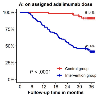
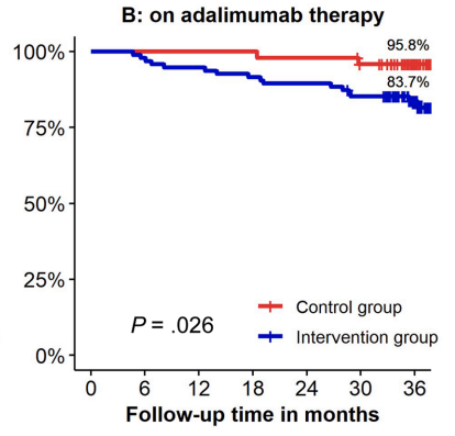
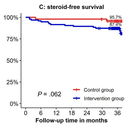
  • Kaplan-Meier analyses of secondary end points showed the following probabilities at year 3 (intervention vs control) (Figure 1): remaining on the assigned adalimumab dose, 41.4% vs 91.4% (P < .0001); remaining on adalimumab, 83.7% vs 95.8% (P = .026); corticosteroid-free survival, 87.4% vs 95.7% (P = .062); and complication-free survival, 83.2% vs 97.9% (P = .015)
Kaplan-Meier curves visualizing maintenance of assigned dosing at baseline, continued adalimumab therapy, and corticosteroid-free survival in both groups. (A) Probability of maintaining assigned adalimumab dosing interval of 3–4 weeks (intervention) vs 2 weeks (control).
(B) Probability of remaining on adalimumab therapy over time.
(C) Probability of remaining in corticosteroid-free remission.

My take: About 60% of patients were unable to de-escalate their adalimumab dosing interval. Suboptimal dosing increased the risk of complications and having adalimumab therapy become ineffective.

Related blog posts:

PREdiCCt Trial: Lower Calprotectin Targets in Crohn’s Disease and Ulcerative Colitis

Constantine-Cooke N, Gros B, Plevris N, et al Gut 2026. doi: 10.1136/gutjnl-2025-337846. (Open Access!) Associations between demographic, clinical and dietary factors and flares in inflammatory bowel disease: the PRognostic effect of Environmental factors in Crohn’s and Colitis (PREdiCCt) prospective cohort study

Methods: Multicentre, prospective cohort study conducted across 47 UK centres. Patients with Crohn’s disease (CD), ulcerative colitis (UC) or IBD unclassified (IBDU) in self-reported remission were prospectively followed up. 2629 participants (1370 CD; 1259 UC/IBDU) – followed up for a median of 4.1 years.

Key findings:

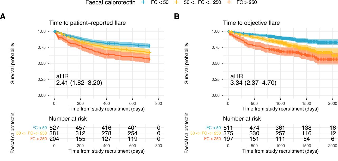
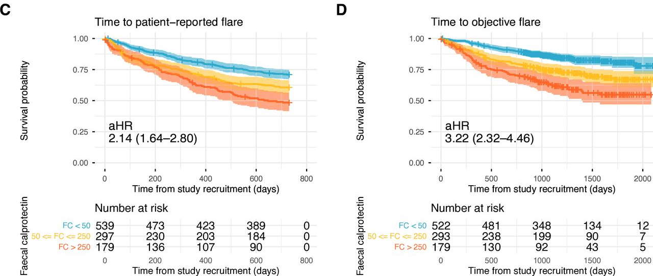
  • Baseline FC was strongly associated with patient-reported flares (FC ≥250 µg/g: adjusted HR (aHR) 2.22; FC 50–250 µg/g: aHR 1.52 (reference <50 µg/g)).
  • Baseline FC was also strongly associated with objective flares (FC ≥250 µg/g: aHR 3.25; FC 50–250 µg/g: aHR 1.98). Objective flares were “clinical flare plus C-reactive protein >5 mg/L and/or faecal calprotectin (FC) >250 µg/g with treatment escalation.” In ulcerative colitis, the probability of an objective flare within two years rose from 11% in those with baseline calprotectin below 50 µg/g to 34% in those above 250.
  • At 24 months, cumulative patient-reported and objective flare rates were 28% and 12% in CD, and 33% and 15% in UC/IBDU, respectively. Overall, patient-reported flares were more common (31%), while objective flares were less frequent (14%).
  • In UC, higher total meat intake was associated with increased risk of objective flares (highest versus lowest quartile: aHR 1.95, 95% CI 1.07 to 3.56). The absolute two-year risk rose from 12% in the lowest quartile of meat intake to 26% in the highest.
  • No consistent associations were observed for ultraprocessed foods, fiber or polyunsaturated fatty acids and flare.
Flares by faecal calprotectin (FC) stratified into FC < 50, 50 ≤ FC ≤ 250, and FC >250 μg/g. (A) Patient-reported flare in Crohn’s disease; (B) objective flare in Crohn’s disease; (C) patient-reported flare in ulcerative colitis/inflammatory bowel disease unclassified; (D) objective flare in ulcerative colitis/inflammatory bowel disease unclassified. aHR, adjusted hazard ratio.

My take: Lower calprotectin values, even in remission, are associated with better outcomes. Risk was meaningfully increased even in the 50–250 µg/g range, compared with levels below 50. Higher meat intake may increase the risk of flares for UC.

Summary of study information from Charlie Lees: The PREdiCCt Study: Can We Predict IBD Flares?

Related blog posts: