Gastroenterol 2024; 166: 87. Open Access! Spotlight (1 page summary)
Key recommendations
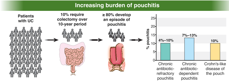
AGA recommends metronidazole and/or ciprofloxacin as preferred treatment of pouchitis with duration of treatment 2-4 weeks.
For Crohn’s-like disease of the pouch, AGA guideline recommends using either ileal-release budesonide or advanced immunosuppressive agents (eg. Biological therapies and small molecule therapies)
“In patients with cuffitis, topical therapies should be the first-line therapy, such as mesalamine suppositories, corticosteroid suppositories, or corticosteroid ointment applied directly to the cuff. Biological therapies and small molecule therapies are recommended in refractory cases
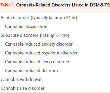
Two recent articles review cannabis-related disorders:
DA Gorelik. NEJM 2023; 389: 2267-2275. Cannabis-Related Disorders and Toxic Effects
Background: “In the United States, an estimated 52.4 million persons 12 years of age or older used cannabis in 2021, representing 18.7% of the community-dwelling population in that age group,5 and 16.2 million persons met the diagnostic criteria for cannabis use disorder, which has as its core feature the use of cannabis despite adverse consequences…Cannabis use poses a global disease burden, albeit substantially less than that posed by other psychoactive substances such as alcohol, tobacco (nicotine), opioids, and stimulants.10 The Global Burden of Disease project calculated that cannabis use in 2016 was responsible for an estimated 646,000 years of healthy life lost to disability.”
Key points:
“Cross-sectional surveys suggest that recent cannabis use increases the risk of motor vehicle crashes by 30 to 40%.26 By comparison, a blood alcohol concentration of 0.08% increases the risk of crashes by 250 to 300%.26“
“Cannabis use disorder, like other substance use disorders, is a chronic, relapsing condition.”
“A substantial reduction or a cessation of cannabis use after heavy or long-term use results in a withdrawal syndrome that is usually mild and self-limiting.39…Common psychological symptoms of cannabis withdrawal include depressed mood, anxiety, restlessness, irritability, decreased appetite, and sleep disturbance. Physical signs and symptoms are less common and include abdominal cramps, muscle aches, tremor, headache, sweating, chills, and weight loss. These signs and symptoms typically begin within 1 to 2 days, peak within 2 to 6 days, and last for several weeks…The prevalence of any withdrawal symptoms is almost 50% in persons who were using cannabis daily.”
Neonatal cannabis exposure: “Pregnant persons who use cannabis expose their neonates to cannabis. Such in utero exposure is associated with increased risk among newborns of having low birth weight, being small for gestational age, and being admitted to the neonatal intensive care unit.”
“Cannabinoid hyperemesis syndrome, a form of cyclic vomiting syndrome that is often accompanied by abdominal pain, occurs during or within 48 hours after frequent and heavy cannabis use.75 Cannabinoid hyperemesis syndrome is a major reason for cannabis-related visits to emergency departments, and it accounts for about 10% of patients with cyclic vomiting syndrome.76 Cannabinoid hyperemesis syndrome is distinguished from cyclic vomiting syndrome by its temporal association with cannabis use, relief with hot baths or showers, and resolution with extended abstinence from cannabis…The symptoms of cannabinoid hyperemesis syndrome are treated with benzodiazepines, haloperidol, and topical capsaicin. “
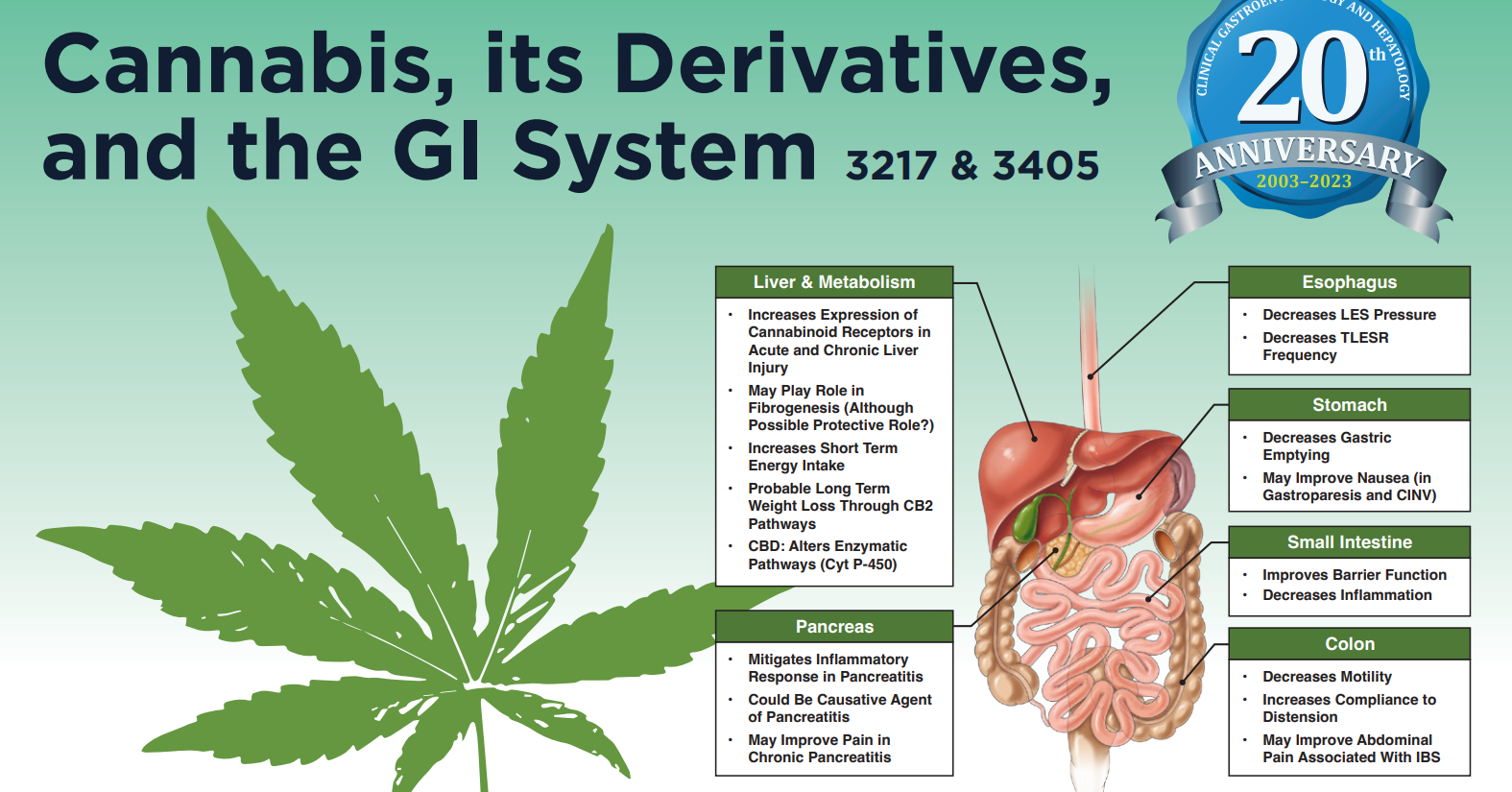
M Camilleri, T Zheng. Clin Gastroenterol Hepatol 2023; 21: 3217-3229. Cannabinoids and the Gastrointestinal Tract This article focuses more on the GI tract effects of cannabinoids.
At a lot of IBD conferences, there is often a lot of focus on health maintenance including discussions on optimizing immunization levels and nutrition. It is often striking how, in comparison, so little attention is focused on emotional health which seems to cause a much greater health burden.
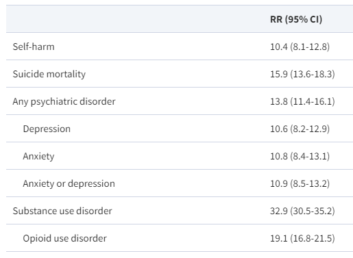
CS Tse et al. Inflamm BowelDis 2024; 30: 150-153. Increased Risks for Suicide, Self-Harm, Substance Use, and Psychiatric Disorders in AdultsWith Inflammatory Bowel Disease: A Nationwide Study in the United States From 2007 to2017
Background: Patients with IBD are also at an increased risk for chronic opioid use, depression, anxiety, sleep disturbance, and disease-related disability (eg, unemployment), all known risk factors for suicide
Methods: This cross-sectional study uses the Nationwide Emergency Department Sample of the Healthcare Cost and Utilization Project (HCUP) as a public domain representing 80% of the U.S. population. This analysis included more than 260 million emergency department visits across the United States from 2007 to 2017.
Key findings:
Inflammatory bowel disease conferred >10-fold risk for suicide deaths, self-harm, substance use, and psychiatric disorders.
The absolute numbers of self-harm rates were low (<1% of all-cause inflammatory bowel disease emergency department visits; total 56 suicide deaths). This amounts to about 5 suicide deaths per year (compared to 0.5 per year for patients with celiac disease.
The risk of self-harm was higher in patients with Crohn’s disease than ulcerative colitis (RR, 3.3; 95% CI, 1.2-5.4), though the suicide risk was not statistically different (RR, 2.3; 95% CI, 0.8-4.5).
From Table 2: RR of self-harm, suicide, psychiatric disorders, and substance use of adults with inflammatory bowel disease compared with celiac disease in the United States from 2007 to 2017. The authors found that rates of self-harm and suicide were the same for patients with celiac disease as the general population (RR 1.0).
My take: Attention to mental health is important component of good care for patients with inflammatory bowel disease.
There is little evidence that we are reducing diagnostic errors despite more lab testing and more imaging. “One of the important reasons for these errors is failure to consider the diagnosis when evaluating the patient.” This, in turn, may be related to brief office visits.
AI support to radiologists for a large mammography study “showed improvement in accuracy with a considerable 44% reduction of screen-reading workload.” The cancer detection rate was 6.1 per 1000 compared to 5.1 per 1000 in the control group.
In difficult NEJM CPC cases, large language AI model (LLM) outperformed clinicians (see slide below).” The LLM was nearly twice as accurate as physicians for accuracy of diagnosis, 59.1 versus 33.6%, respectively.”
“Likewise, the cofounder of OpenAI, Ilya Sutskever, was emphatic about AI’s future medical superintelligence: ‘If you have an intelligent computer, an AGI [artificial general intelligence], that is built to be a doctor, it will have complete and exhaustive knowledge of all medical literature, it will have billions of hours of clinical experience.’ “
My take (borrowed from Dr. Topol): “We are certainly not there yet. But in the years ahead, …it will become increasingly likely that AI will play an invaluable role in providing second opinions with automated, System 2 machine-thinking, to help us move toward the unattainable but worthy goal of eradicating diagnostic errors.”
Seven pediatric patients with perianal Crohn’s disease were treated with mesenchymal stem cells. Key finding: At 6 months, 83% had complete clinical and radiographic healing. This healing rate is higher than “the 50% efficacy reported by the only completed randomized control phase III clinical trial.[ADMIRE study].”
MA Baarslag et al. NEJM 2023; 389: 1790-1796. Severe Immune-Related Enteritis after In Utero Exposure to Pembrolizumab
This case report details severe immune-related gastroenterocolitis after in utero exposure to pembrolizumab, an anti–PD-1 agent; the infant presented at 4 months of life. Extensive testing did not identify any underlying causes of VEO-IBD. This infant required TPN for a short period, but subsequently responded to treatment with glucocorticosteroids and infliximab (with plans to continue until at least 3 years of age). Both programmed death 1 (PD-1) and cytotoxic T-lymphocyte-associated antigen 4 (CTLA-4) immune checkpoint inhibitors are negative regulators of T-cell immune function. Inhibition of these targets, resulting in increased activation of the immune system and can result in medication-induced colitis in the patients who take them and potentially in infants exposed to these agents in utero.
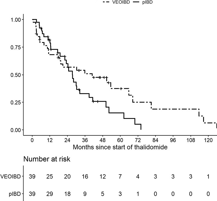
M Bramuzzo et al. Inflamm Bowel Dis 2024; 30: 20-28. https://doi.org/10.1093/ibd/izad018. Efficacy and Tolerance of Thalidomide in Patients With Very Early Onset Inflammatory Bowel Disease
This retrospective study with 39 patients with VEO and 39 patients with pediatric IBD.
Key findings:
The treatment persistence at 1, 2, and 3 years was 68.2%, 57.0%, and 50.9% for VEOIBD patients and 81.7%, 60.0% and 33.0% for pIBD patients, respectively
A significantly higher proportion of VEOIBD patients discontinued therapy due to lack of efficacy (48.2% vs 17.2%; P = .03), while AEs were the main reason for discontinuation in pIBD patients
A significatively lower number of VEOIBD patients experienced AEs compared with pIBD patients (14 [35.9%] vs 30 [76.9%]; P = .0005).
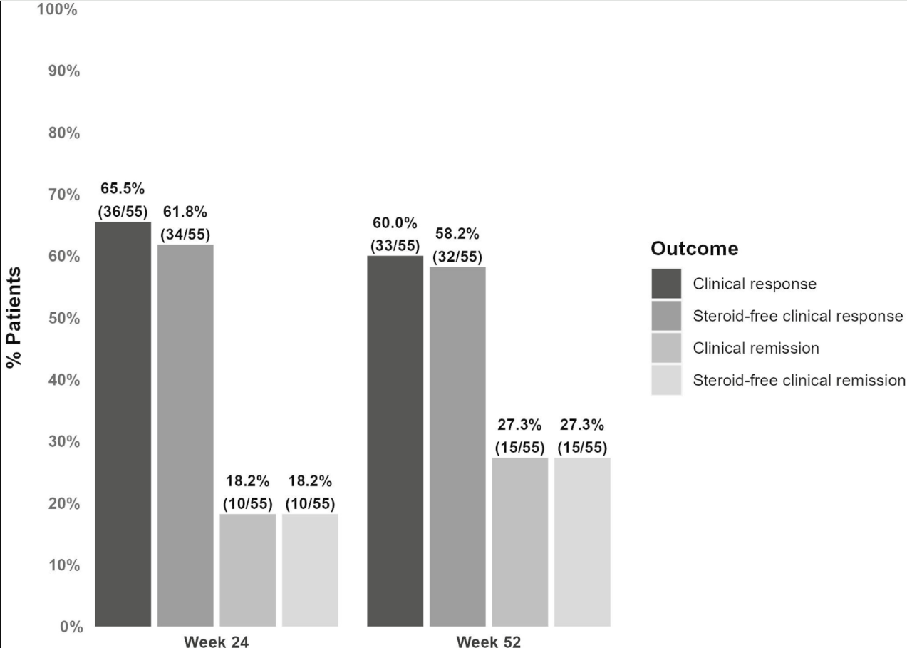
D Alsoud et al Inflamm Bowel Dis 2024; izad315, https://doi.org/10.1093/ibd/izad315. Real-world Effectiveness and Safety of Risankizumab in Patients with Moderate to Severe Multirefractory Crohn’s Disease: A Belgian Multicentric Cohort Study
Methods: Data from consecutive adult CD patients who started risankizumab before April 2023 were retrospectively collected at 6 Belgian centers. A total of 69 patients (56.5% female, median age 37.2 years, 85.5% exposed to ≥4 different advanced therapies and 98.6% to ustekinumab, 14 with an ostomy) were included.
Key findings:
At week 24, 61.8% (34 of 55) and 18.2% (10 of 55) of patients without an ostomy achieved steroid-free clinical response and remission, respectively.
At week 52, these numbers were 58.2% (32 of 55) and 27.3% (15 of 55), respectively. Endoscopic data were available in 32 patients, of whom 50.0% (16 of 32) reached endoscopic response within the first 52 weeks.
Results in patients with an ostomy were similar (steroid-free clinical response and remission, 42.9% and 14.3%, respectively).
20.3% (14 of 69) of patients underwent CD-related intestinal resectionsand 18.8% (13 of 69) of patients discontinued risankizumab during followup (median 68 weeks).
Risankizumab was well tolerated with no safety issues.
Discussion points: “98.6% of patients in the current study were exposed to ustekinumab compared with less than 20% in the registration trials. This indicates that a previous lack or loss of response to the inhibition of the p40 subunit common to IL-12 and IL-23 does not preclude a potential response from subsequent selective inhibition of IL-23. “
My take: This study shows that risankizumab can be effective in refractory patients, even in those who have received similar type medications (eg. ustekinumab).
Disclaimer: This blog, gutsandgrowth, assumes no responsibility for any use or operation of any method, product, instruction, concept or idea contained in the material herein or for any injury or damage to persons or property (whether products liability, negligence or otherwise) resulting from such use or operation. These blog posts are for educational purposes only. Specific dosing of medications (along with potential adverse effects) should be confirmed by prescribing physician. Because of rapid advances in the medical sciences, the gutsandgrowth blog cautions that independent verification should be made of diagnosis and drug dosages. The reader is solely responsible for the conduct of any suggested test or procedure. This content is not a substitute for medical advice, diagnosis or treatment provided by a qualified healthcare provider. Always seek the advice of your physician or other qualified health provider with any questions you may have regarding a condition.
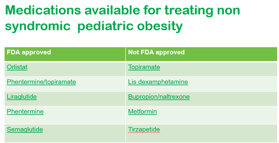
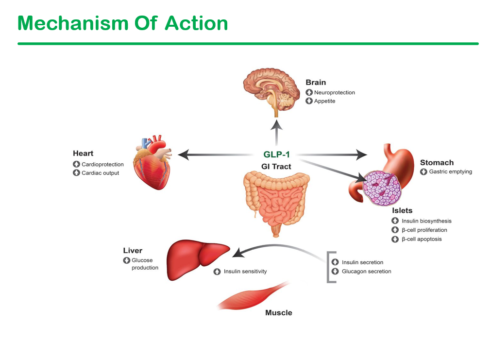
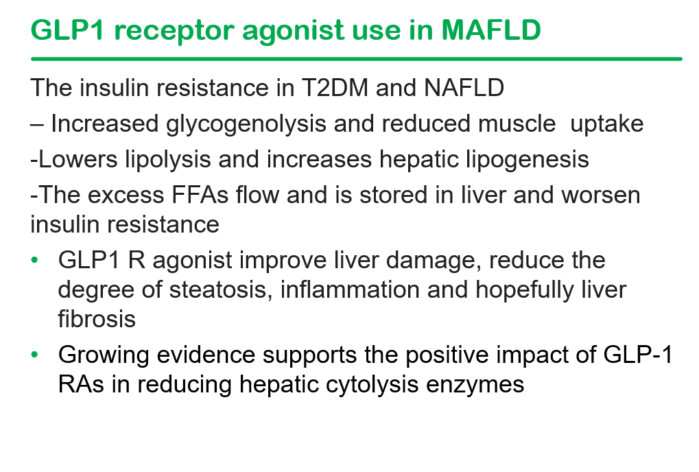
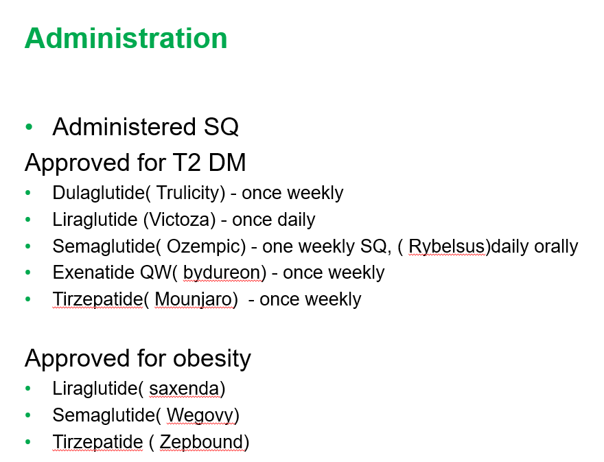
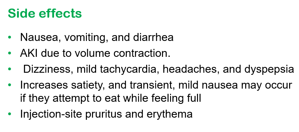
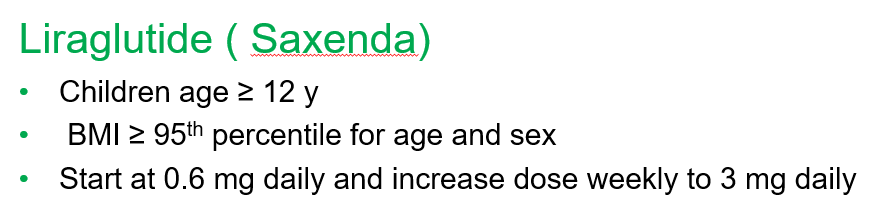
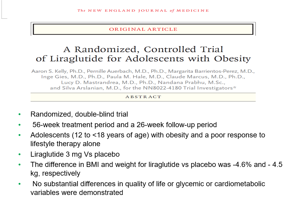
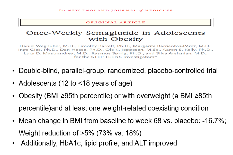
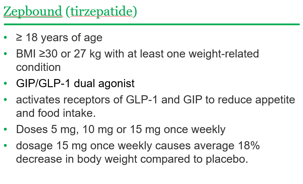
Recently, Dr. Shruthi Arora, an Emory Pediatric Endocrinologist and part of CHOA’s Strong4Life team, provided a terrific review of pediatric obesity pharmacology for our group.
Here are a few slides from Dr. Arora’s lecture:
General points from this lecture:
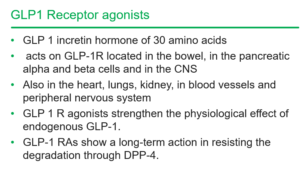
GLP-1 agents are a huge advance but currently limited by affordability (frequently there is a lack of insurance coverage if there is not T2DM) and availability. In addition, most individuals will regain weight loss when these agents are stopped.
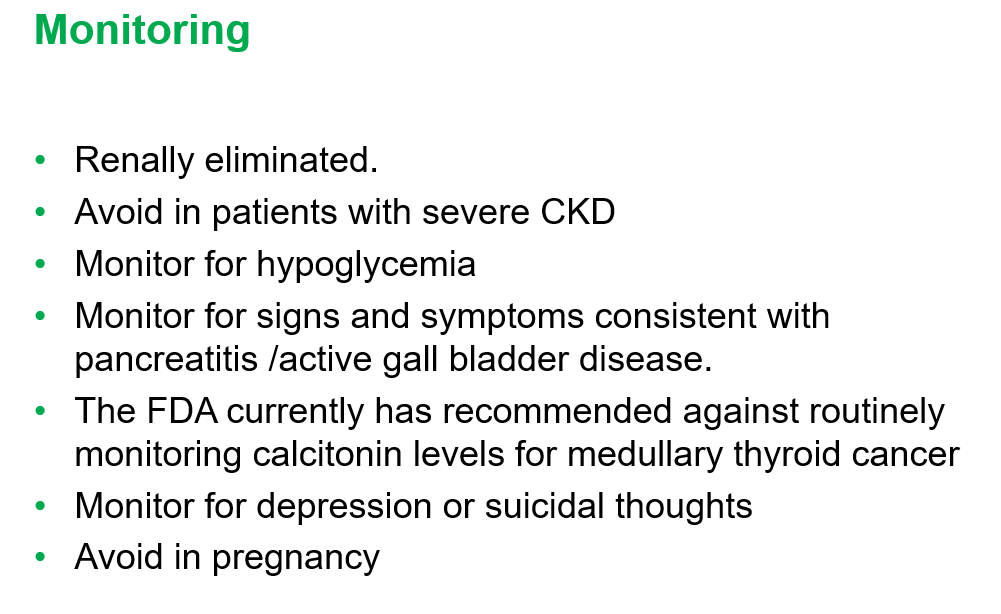
GLP-1 agents are not recommended in the following: patients with gastroparesis, and patients with a personal or family history significant for MEN 2 A /MEN 2 B/ Medullary thyroid cancer
Long-term data is still needed. These agents have been associated with muscle and bone loss; thus, working to assure a good diet is still very important
——————————————————————————
NASPGHAN has a good review/webinar on this topic as well: Pediatric MASLD in the Current Era of Pharmacological and Surgical Obesity Treatment Options. For members, after sign in, you can register and login to this webinar (look under clinical practice tab). This webinar made a lot of useful points (many covered by Dr. Arora too).
For GLP-1 agents, due to effects on gastric emptying, they are generally held prior to anesthesia. If they are given weekly, then hold 1 week prior to anesthesia. If it is a daily medication, hold for 1 day prior to anesthesia.
Surgery definitely helps improve MASH -though variable responses in patients. SLEEVE gastrectomy is currently the most frequent bariatric surgery
There is trouble getting GLP-1 medications.
Limited knowledge regarding long-term effects of cycling of GLP-1 agents.
Obesity is a long-term disease –>anticipate long-term treatment
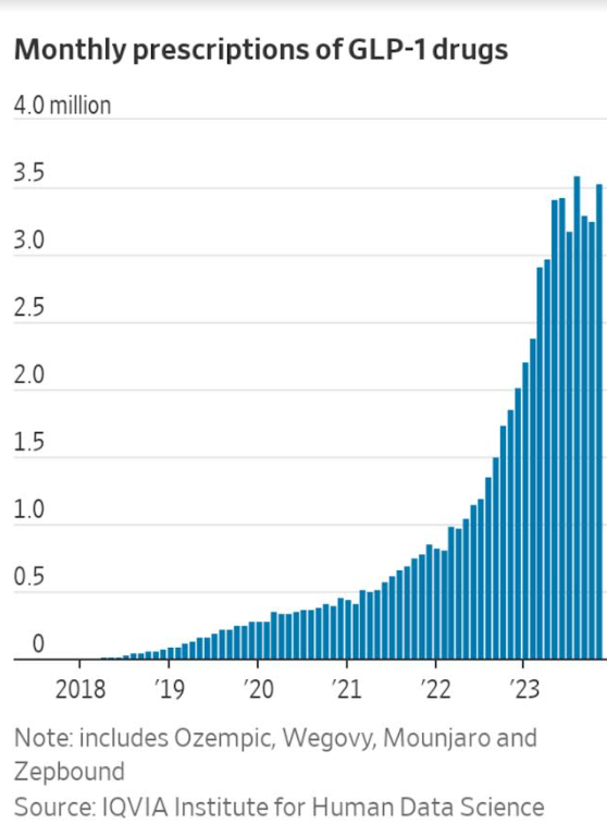
The Wall Street Journal recently published a personal account of using the newer obesity medications. Bradley Olson, 1/12/24: A Weight-Loss Drug Changed My Life. Will It Solve My Problem? (behind a paywall). This article discusses the dramatic improvement experienced by the writer along with his concerns about the cost of the medication and potential for rebound when he can no longer afford it. Two of the figures:
Disclaimer: This blog, gutsandgrowth, assumes no responsibility for any use or operation of any method, product, instruction, concept or idea contained in the material herein or for any injury or damage to persons or property (whether products liability, negligence or otherwise) resulting from such use or operation. These blog posts are for educational purposes only. Specific dosing of medications (along with potential adverse effects) should be confirmed by prescribing physician. Because of rapid advances in the medical sciences, the gutsandgrowth blog cautions that independent verification should be made of diagnosis and drug dosages. The reader is solely responsible for the conduct of any suggested test or procedure. This content is not a substitute for medical advice, diagnosis or treatment provided by a qualified healthcare provider. Always seek the advice of your physician or other qualified health provider with any questions you may have regarding a condition.
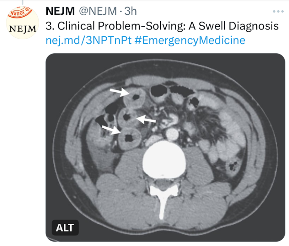
J Allam et al. NEJM 2024; 390-71-76.A Swell Diagnosis
This clinical problem-solving case report describes a previously healthy 19 yo male with sudden-onset severe diffuse abdominal pain. His ED evaluation was unremarkable (including labs [CBC/d, CMP, Lipase, CMP, thryotropin, ESR, CRP and UA], and EKG). He was discharged after 3 hours. Over the next 10 years, he presented to the ED on numerous occasions with the same symptom complex and normal labs/mostly CT scans. One CT scan showed small-bowel thickening thought to be due to AGE. Ultimately, his symptoms increased to every 2 weeks. Extensive evaluations (multiple panendoscopies, MRCP, MRE, U/S, and infectious workup) were undertaken and numerous treatments were given without benefit. Ultimately, a CT scan showed remarkable circumferential wall thickening in the jejunum (see below). This led to evaluation for hereditary angioedema.
The article serves as a good review of this disorder and of the differential diagnosis.
Key points:
Background: Hereditary angioedema, which is due to a deficiency of functional C1 inhibitor protein, is a rare autosomal dominant genetic disorder that affects approximately 1 in 50,000 persons worldwide.1
Mean age of onset : 8 to 12 years, and symptoms often worsen during puberty.1
Presentation: The disease is characterized by recurrent episodes of swelling in various parts of the body and can be severely debilitating. The hallmark symptom of hereditary angioedema is localized swelling of the skin and submucosal tissues (in the face, lips, throat, hands, feet, or genitalia) that is nonpitting, nonpruritic, and not accompanied by urticaria. Triggers include emotional stress, physical trauma, infections, physical exertion, and surgery and other medical procedures.
Abdominal pain: In a series of 149 patients with hereditary angioedema who had 521 attacks, 49% of the episodes were characterized by isolated abdominal pain.5 Abdominal attacks are generally not associated with fever, peritoneal signs, or leukocytosis.
Laryngeal edema occurs in approximately 0.9% of all attacks7 and may be life-threatening, leading to asphyxiation and death.
Differential Diagnosis: Some of the rare diagnosis that were discussed: acute intermittent porphyria, familial Mediterranean fever (FMF), mastocytosis, and eosinophilic gastroenteritis.
Pathophysiology: Deficiency of C1 inhibitor protein: The majority of cases of hereditary angioedema are caused by either decreased levels (type I) or reduced functionality (type II) of C1 inhibitor. A third subtype (type III) that is associated with different mutations but the same clinical features is characterized by normal quantitative and functional C1 inhibitor levels.2
The best screening test: measurement of the level of C4 (which is exhausted as a result of uncontrolled activation of the complement pathway when C1 inhibitor is deficient or dysfunctional); results may be normal in 10% of patients between attacks. Quantification of C1 inhibitor levels can then be performed to differentiate between the low levels in hereditary angioedema type I and the normal levels in hereditary angioedema type II.
Treatment: Food and Drug Administration–approved agents include human plasma–derived C1 inhibitor concentrate, recombinant human C1 inhibitor, icatibant (bradykinin B2 receptor antagonist), and ecallantide (kallikrein inhibitor).9 Each has been shown in randomized, controlled trials to decrease the median time to symptom relief.10-14 The first three therapies may be administered by the patient, whereas ecallantide requires support from a health care professional for management of possible anaphylaxis, which is reported in up to 4% of patients.9 Most treatments are used on-demand, though some patients benefit from long-term prophylaxis.
Preprocedure prophylaxis: Preprocedural prophylaxis is recommended for patients undergoing procedures that may trigger an attack (e.g., dental surgery, endotracheal intubation, or an endoscopic procedure). C1 inhibitor concentrate can be administered intravenously before the procedure, or treatment with an anabolic androgen (e.g., danazol or stanozolol) can be started 5 days before and continued for 2 to 5 days after the procedure9; …Fresh frozen plasma may be used as a second-line therapy when these therapies are not available
My take: This case “highlights the importance of maintaining a high clinical suspicion for hereditary angioedema in patients with episodic severe abdominal pain and negative workup for other illnesses” even in patients without other manifestations.
Methods: “Åkerström et al7 conducted a multi-national population-based cohort study of all patients with a diagnosis of Barrett’s esophagus in the national patient registries of Denmark (2012–2020), Finland (1987–1996 and 2010–2020), Norway (2008–2020), and Sweden (2006–2020). Patients with Barrett’s who underwent anti-reflux surgery (ARS) were compared with non-operated patients, most of whom were presumed to be using anti-reflux medication. The cohort consisted of 33,939 patients with Barrett’s esophagus, 542 (1.6%) of whom had undergone anti-reflux surgery.” Followup was up to 32 years. Mean age in the cohort was 64.3 years.
Key finding: “The main findings of the study were: 1) that the HR of developing EAC [esophageal adenocarcinoma] was actually greater in the ARS [anti-reflux surgery] cohort than in the non-operated cohort HR (adjusted HR 1.9, 95% CI 1.1–3.5); and 2) that the HR did not stabilize with a longer period of follow-up, but instead continued to increase, going from 1.8 (95% CI 0.6–5.0) within 1–4 years of follow-up to 4.4 (95% CI 1.4–13.5) after 10–32 years of follow-up.”
Discussion points:
The editorial suggests that ARS may not actually increase the risk of EAC, but instead the difference may be related to a selection of bias of choosing patients with more severe Barrett’s to have surgery.
For a patient at age 25 years with Barrett’s the cumulative risk of 50 years would translate to a 12% lifetime risk of EAC
The editorial reviews the use of PPIs from the AspECT trial and noted that “high-dose PPI plus low-dose aspirin was more effective than low-dose PPI alone in preventing that composite end point [all-cause mortality, EAC, or high-grade dysplasia]…a large part of the treatment effect was attributable to reduction in all-cause mortality (including cardiovascular) rather than EAC or high-grade dysplasia”
From editorial: Patient’s main reason for undergoing laparoscopic anti-reflux surgery in a community practice: 83 patients in a managed care environment. Data from Vakil et al.6
My take: This article provides good evidence that reflux surgery does not reduce the risk of esophageal adenocarcinoma in those at highest risk. For pediatric patients with severe reflux, this information is helpful for accurate counseling.
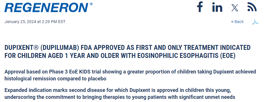
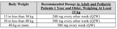
“Regeneron Pharmaceuticals, Inc. (NASDAQ: REGN) and Sanofi today announced that the U.S. Food and Drug Administration (FDA) has approved Dupixent® (dupilumab) for the treatment of pediatric patients aged 1 to 11 years, weighing at least 15 kg, with eosinophilic esophagitis (EoE).”
Disclaimer: This blog, gutsandgrowth, assumes no responsibility for any use or operation of any method, product, instruction, concept or idea contained in the material herein or for any injury or damage to persons or property (whether products liability, negligence or otherwise) resulting from such use or operation. These blog posts are for educational purposes only. Specific dosing of medications (along with potential adverse effects) should be confirmed by prescribing physician. Because of rapid advances in the medical sciences, the gutsandgrowth blog cautions that independent verification should be made of diagnosis and drug dosages. The reader is solely responsible for the conduct of any suggested test or procedure. This content is not a substitute for medical advice, diagnosis or treatment provided by a qualified healthcare provider. Always seek the advice of your physician or other qualified health provider with any questions you may have regarding a condition.