Methods: Using National Swedish registries, the authors evaluated a matched cohort study, 1378 individuals with PSC and 13,549 general population comparators and their first-degree relatives.
Key findings:
After excluding inflammatory bowel disease and autoimmune hepatitis, the prevalence of autoimmune disease was 18% in PSC and 11% in comparators, OR: 1.77
Highest odds were seen for celiac disease [OR: 4.3], sarcoidosis [OR: 2.74], diabetes type 1 [OR: 2.91], and autoimmune skin disease [OR: 2.15]
First-degree relatives of individuals with PSC had higher odds of developing IBD [OR: 3.25], autoimmune hepatitis [OR: 5.94], and any autoimmune disease than relatives of the comparators [OR: 1.34]
My take: Keep an eye out for other autoimmune diseases in patients (& their 1st-degree relatives) with PSC.
H Szajewska et al. JPGN 2024; https://doi.org/10.1002/jpn3.12280. Open Access! Early diet and the risk of coeliac disease. An update 2024 position paper by the ESPGHAN special interest group on coeliac disease
Key points:
Breastfeeding, whether any amount, exclusive, or of any duration, does not reduce the risk of developing CD
Introducing gluten into an infant’s diet at any time between completed 4 months (≥17 weeks) and 12 months of age does not affect the cumulative incidence of CD
In medical school, I distinctly remember my anatomy professor discussing how a test’s accuracy was affected by the underlying population tested. He said, ‘Well if you do an RPR test for syphilis among prostitutes and it is abnormal, then it is likely accurate as the test performs better in a population with a higher risk of the condition. Whereas if you run the same test on the faculty, the test would perform with much less accuracy. Well, actually, may be the faculty here are not the best example…’
This discussion comes to mind with the recent publication regarding the No-Biopsy Approach for Celiac disease in adults:
In this systematic review and meta-analysis with 12,103 adult patients from 18 studies, the key findings:
The pooled prevalence of biopsy-proven celiac disease in the included studies was 62%.The proportion of patients with IgA-tTG ≥10×ULN was 32%.
The summary sensitivity of IgA-tTG ≥10×ULN was 51%, and the summary specificity was 100%
However, the positive predictive value of the no-biopsy approach to identify patients with celiac disease was 65%, 88%, 95%, and 99% if celiac disease prevalence was 1%, 4%, 10%, and 40%, respectively in hypothetical cohorts.
The duodenal biopsy, long the cornerstone of a celiac disease diagnosis, has lost some of its luster in recent years. As data emerged that the specificity of tissue transglutaminase (TTG) IgA for duodenal villus atrophy increases in accordance with the degree of antibody elevation in children, in 2012 the European Society for Pediatric Gastroenterology and Nutrition adopted a biopsy-free pathway for the diagnosis of celiac disease in symptomatic children who met stringent criteria, including a ≥10-fold TTG IgA elevation and an elevated endomysial antibody on a separate blood draw.1 With further data supporting the positive predictive value of a highly elevated TTG IgA2 these guidelines were modified in 2020 to now include asymptomatic children.3
..the specificity of an elevated TTG IgA seems to be lower in people with type 1 diabetes.8
Studies employing a serial serology strategy, such as a ≥10-fold TTG IgA elevation that persists over a number of months, might yield specificities that are so high that that they are impervious to decreases in positive predictive values wrought by low underlying prevalences…
Most patients with celiac disease do not mount a sufficiently high TTG IgA to rely exclusively on a serological diagnosis. And the minority who do meet the stringent criteria of a biopsy-free diagnosis might wisely choose to confirm their life-long diagnosis with a biopsy.
My take: Ironically, this study, which showed that a TTG IgA ≥10 x ULN was highly indicative of celiac disease in adult patients with a 100% specificity and a positive predictive value of 98%, makes an argument for ongoing duodenal biopsies in those at lower risk for celiac disease. However, the lower positive predictive values (in some groups) are based on hypothetical cohorts. Shared decision-making is important and relying on a single elevated lab test is particularly problematic.
The summary with nine “best practice advice” statements is not very helpful. However, Figure 2 and Table 1 are very useful.
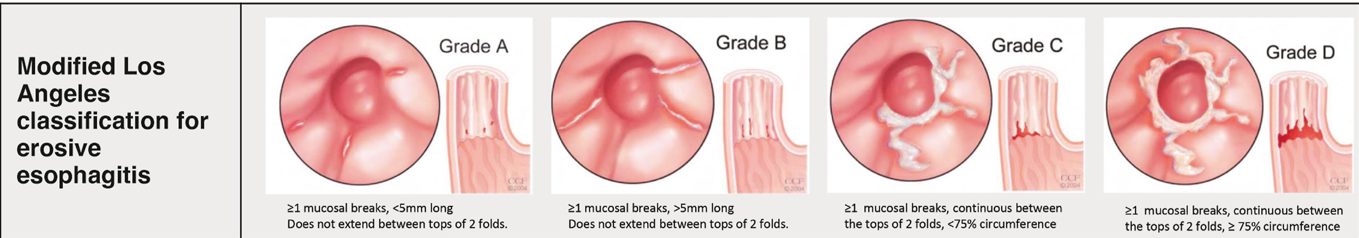
From Figure 2 -not shown below (but in article) are Prague classification for Barrett’s and EREFS for eosinophilic esophagitis. The remaining parts of this figure include the Los Angeles classification for erosive esophagitis, the Hill classification of the gastroesophageal flap, and the Forrest classification of peptic ulcers:
From Table 1:
Table 1 also gives guidance for biopsies with peptic ulcer disease, Barrett’s esophagus, gastric preneoplasia, and for gastric polyps.
My take: When suspicious of underlying disease, this article recommends taking more biopsies and in more jars.
Best Approach for Identifying Eosinophilic Esophagitis Prior studies have shown higher yield when taking 5 or 6 biopsies rather than fewer biopsies; thus, the location of biopsies may not be as important as the number of specimens. Also, prior studies have shown that having another pathologist review the slides can increase the yield by ~20%; this indicates that careful review of specimens by itself is helpful. Perhaps, more specimen containers will increase the time that a pathologist reviews the biopsies.
A recent study (AR Lee et al. Nutrients; 2019, 11, 399). Open access: Persistent Economic Burden of the Gluten Free Diet) quantifies the additional costs of a gluten free diet (GFD) in the U.S. Thanks to Kipp Ellsworth for this reference.
The authors conducted a “market basket” study to establish the cost of a GFD. “A market basket is a group of products that are purchased by consumers …for this study, the market basket was food that would necessitate a GF substitute, including staple foods, snack foods, and commonly used ready-made or convenience meals.”
Key findings:
GF products were more expensive, overall the increase was 183%. This is an improvement from a 2006 study which found the increase overall at 240% (adjusted for inflation).
Mass-market products were 139% more expensive than wheat-based versions
Discussion:
Cost is identified as a frequent reason for nonadherence with diet, cited by 33% in one study
Overall, the burden of GFD is more frequently related to the restrictive nature of the diet which leads to a negative impact on quality of life. According to the authors, in one study (Am J Gastroenterol 2014; 109: 1304-11), treatment burden for celiac was ranked higher than for diabetes hypertension, and congestive heart failure
My take: This study shows the significant economic burden of a GFD. In Italy, the “government offers celiac patients vouchers to buy gluten-free food — up to 140 euros per month.” (NPR: Italy, Land of Pizza and Pasta)
Related blog posts:
Lost Boys (& Girls) of Celiac This blog documents frequent occurrence of patients being lost to followup. It is likely that cost is a factor in this situation as well.
A recent cross-sectional study (K Gerasimidis et al. JPGN 2018; 67: 356-60) examined the use of fecal gluten immunogenic peptide (GIP) to assess for adherence with gluten free diet (GFD) in biopsy-proven celiac disease (CD).
GIP reflects recent gluten consumption. There is a commercially-available kit available (Ivydal GIP Testing) –though I am uncertain about how its reliability compares to the GIP measured in this study.
In the study, the authors note that GIP positivity can occur with as little as 100 mg of gluten/day ingestion. GIP is a 33-mer peptide from α2-gliadin that is stable against breakdown by gastric, pancreatic, and intestinal brush border enzymes.
Key findings of this study:
GIP was detectable in 16% of patients with previous CD diagnosis (N=67)
GIP was detectable in 95% of newly-diagnosed CD patients (n=19) and was detectable in 27% at 1 year afterwards.
When compared with traditional indicators of GFD adherence (eg. TTG levels, Biagi score, clinical assessment), 4 out of 5 children with detectable GIP were missed
My take: Fecal GIP for celiac disease adherence has similar potential as a biomarker as calprotectin has for IBD. A normal GIP appears to be much more sensitive at detecting gluten ingestion.
A recent study (L Norsa et al. JPGN 2018; 67: 361-6) examines data from 197 patients with celiac disease (CD) (out of a cohort of 337) who had a diagnosis established before 1985. The authors examined three groups: lifelong strict GFD (n=133), discontinued GFD (n=29), and no GFD (22). A total of 63 had follow-up endoscopy data available, with 29 in lifelong GFD, 20 in discontinued GFD, and 14 in no GFD.
Key findings:
In those with followup endoscopy, in those with lifelong GFD 27 of 29 (93%) had no atrophy (Marsh 0-1-2) on histology, in those with discontinued GFD 12 of 20 (60%) had no atrophy on histology, and in those with no GFD 8 of 14 (57%) had no atrophy on histology.
Thus, among the group with long-term poor adherence to gluten-free diet, almost two-thirds showed no recurrence of villous atrophy on duodenal biopsies.
In the entire cohort of 197, there were no apparent differences in autoimmune diseases between those receiving lifelong GFD (26%) compared to the other two groups, 17% and 23% respectively.
Limitations:
retrospective design.
initial diagnosis was more than 30 years ago & there are significant differences in the diagnostic approach currently
sample size
My take: This study indicates that some individuals who have been diagnosed with celiac disease may be OK with ongoing gluten consumption. Those who maintained a GFD were much more likely to have no villous atrophy on duodenal biopsies.
Another study (SP Paul et al. JPGN 2018; 66: 641-44) has shown that high anti-TTG IgA levels are reliable in establishing the diagnosis of celiac disease in asymptomatic children from high-risk groups. In this study with prospectively-collected data from 2007-2017, 84 of 157 children had anti-TTG titers >10x ULN. 75 of these 84 were from high-risk groups, mainly type 1 diabetes (36), and first degree relatives (24)
Key finding:
All 75 with high titers from high-risk groups had histologic evidence of celiac disease.
Related study: R Mandile et al. JPGN 2018; 66: 654-56. This prospective study showed that 19 of 35 (54%) patients with potential celiac disease had a complete clinical response on a gluten-free diet to symptoms like abdominal pain and diarrhea. Thus, in many patients with potential celiac disease, a gluten-free diet will not be effective.
This retrospective study of 487 pediatric patients shows that it takes a long time to normalize celiac serology/anti-tissue transglutaminase antibody (TTG). The median time was 407 days for the 80.5% of patients that normalized their serology in the study time frame. The time was 364 days for those who were considered adherent to a gluten-free diet. Patients with type 1 diabetes were less likely to normalize their TTG levels. Faster normalization occurred in those with lower titers at baseline.
In this chart review, among 135 children, normal ESR and CRP were observed in 28% of children with Crohn disease and 42% of children with ulcerative colitis.
This guideline paper details 31 recommendations (some with multiple parts) for the evaluation and management of children with neurologic impairment. The recommendations include detailed evaluations including knee heights, skinfold thickness measures, DXA scan, routine micronutrient bloodwork, along with a low threshold for oropharyngeal dysphagia assessment. The paper has recommendations for evaluations of reflux, constipation, and dental problems. The authors suggest “considering use of enteral feeding if total oral feeding time exceeds 3 hours per day.”
A recent study (GJ Lee et al. JPGN 2016; 63: 340-3) adds a little bit more information regarding hypertransaminasemia in newly diagnosed celiac disease. Some previous information was summarized in a previous blog: Celiac Hepatopathies (2013)
In this retrospective, single center study, 185 children had transaminases obtained at the time of celiac diagnosis (185/388 = 47.7%).
Key findings:
Among this group, 28 (15.1%) had elevated transaminases, with an average of ALT 2.52 x ULN and AST 1.87 x ULN.
Patients with elevated liver transaminases tended to be younger (mean 6.3 yrs compared with 11.0 without elevation). Among those who had followup blood testing available, 15/21 (71.4%) normalized their values over an average of 210 days.
For the 6 who had persistent elevation of transaminases, 3 were suspected to have poor adherence, 1 was thought to have a fatty liver, 1 had only mild elevation, and 1 remained unexplained.
My take: This study indicates that elevated transaminases are common in children with celiac, particularly younger children. As with other studies, the majority resolve on a gluten-free diet. As there is a recognized association with autoimmune hepatitis, in those with elevated ALT, followup after institution of a gluten free diet seems prudent.