Long-term Use of Proton Pump Inhibitors for Eosinophilic Esophagitis

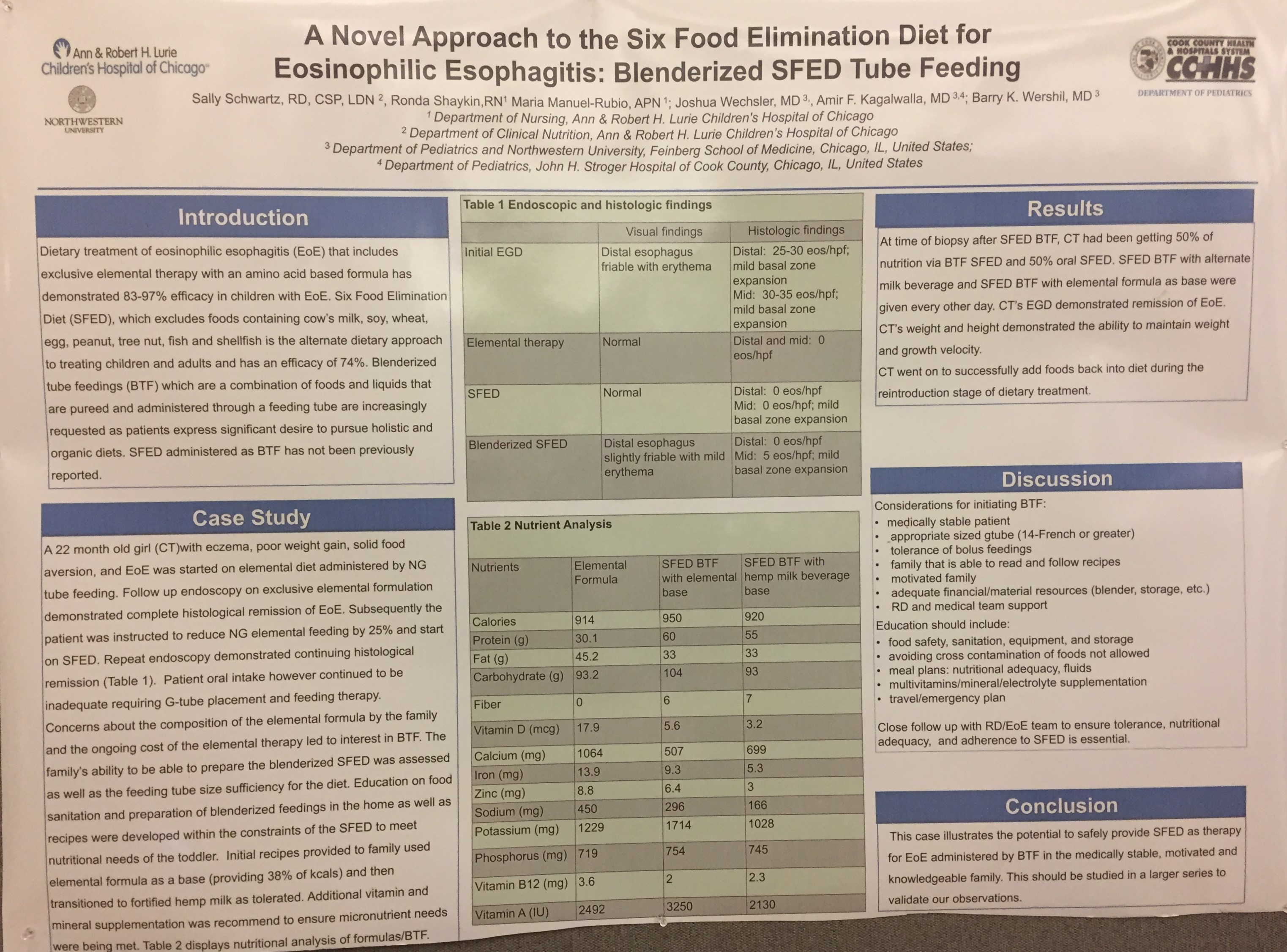
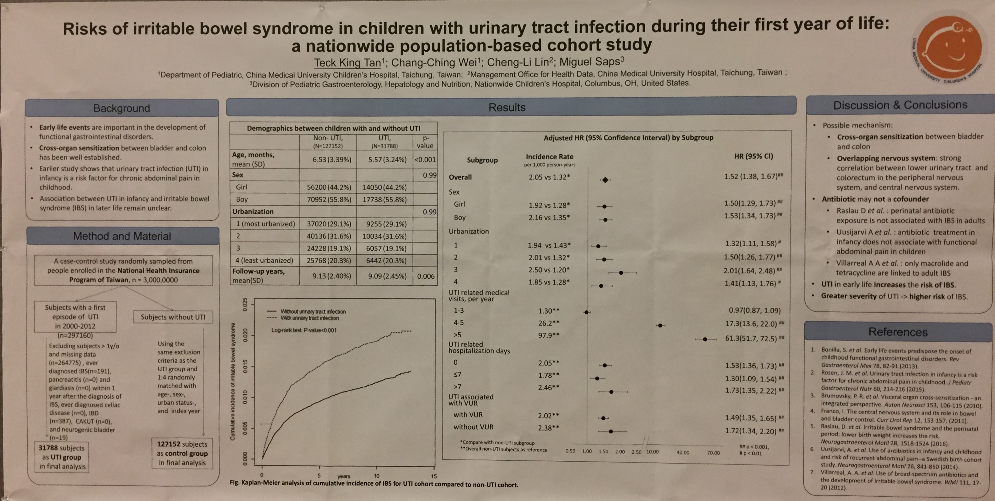
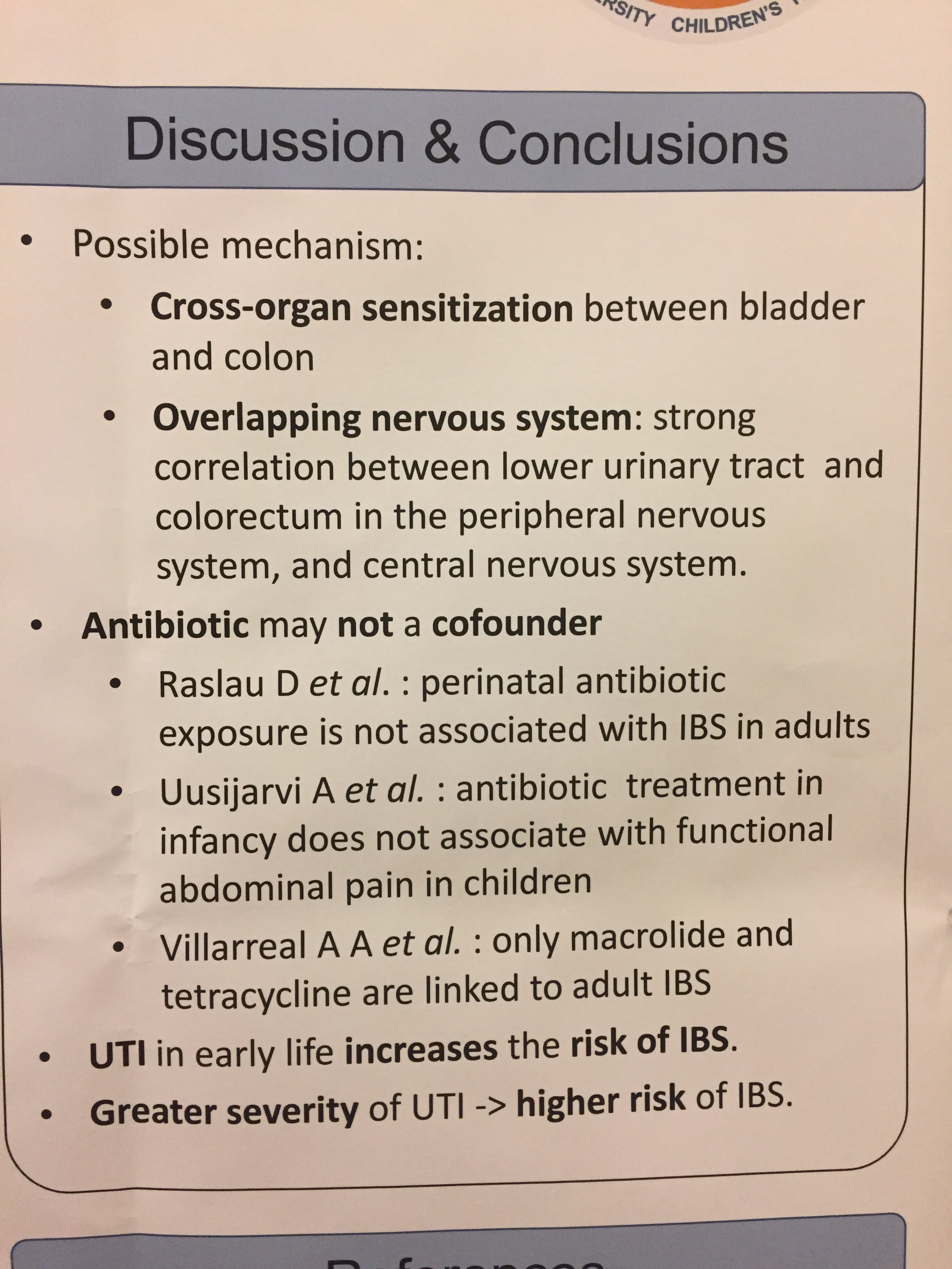
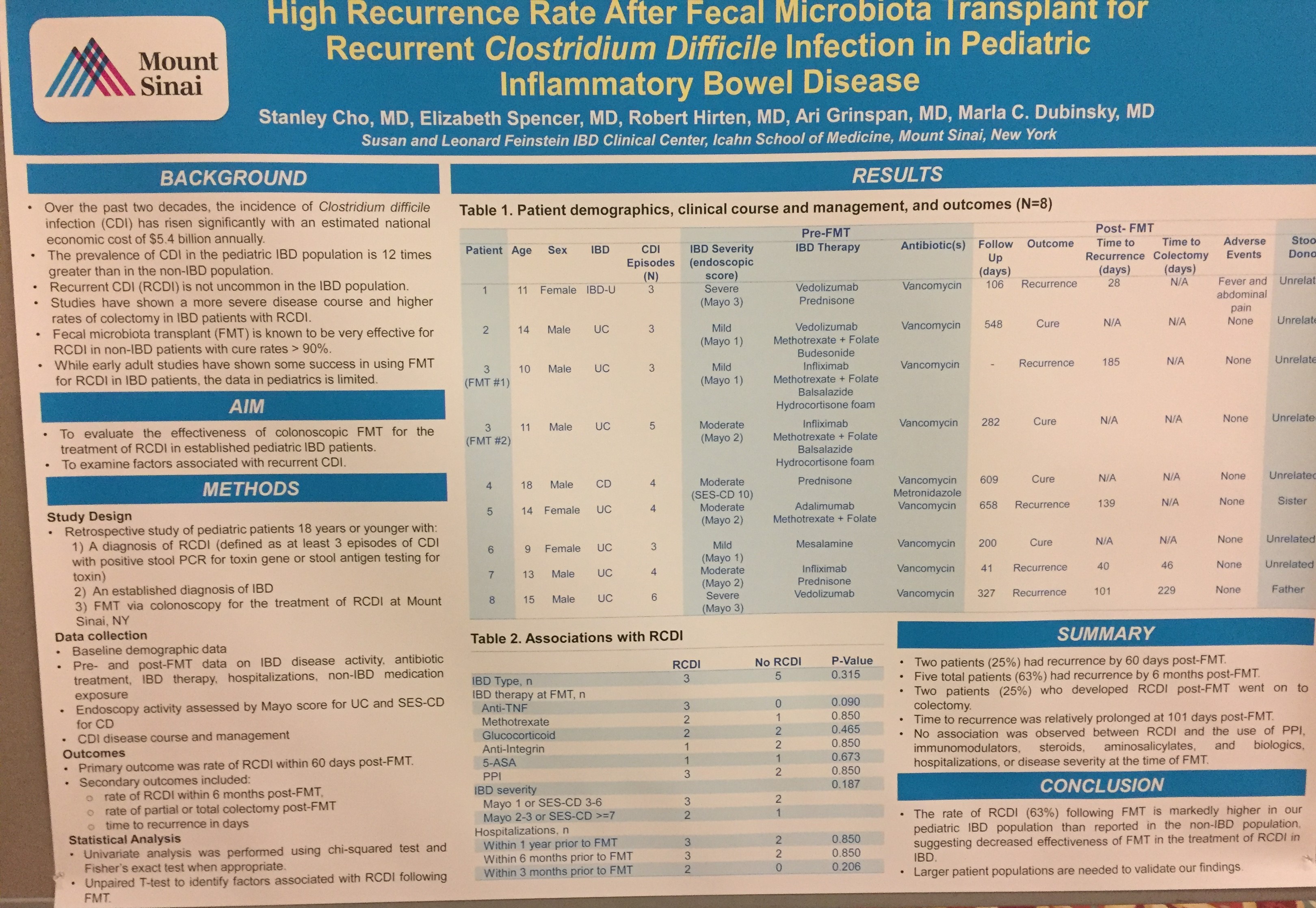
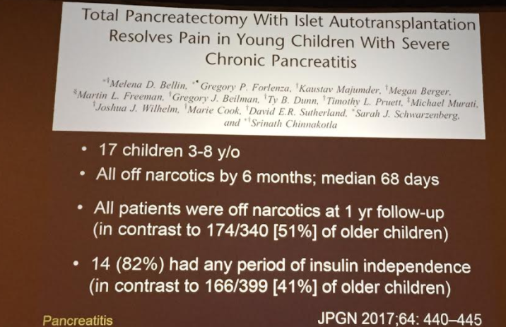
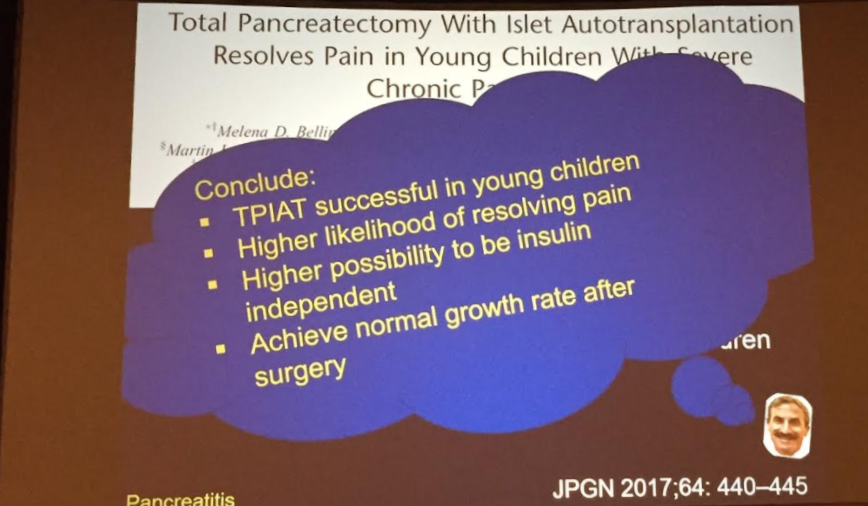
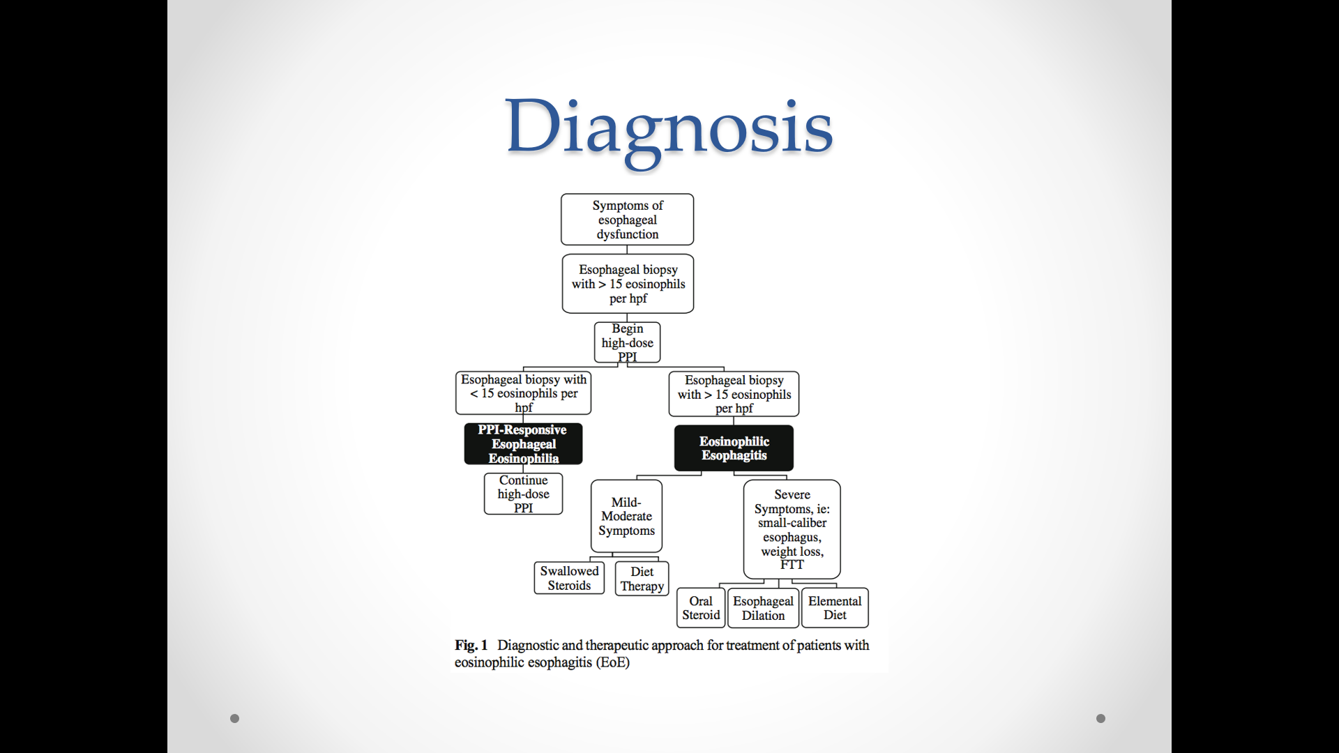
A prospective pediatric eosinophilic esophagitis (EoE) study (C Gutierrez-Junquera et al. JPGN 2018; 67: 210-6) examines the use of proton pump inhibitors (PPIs) for long-term management for this disorder.

After diagnosis of EoE, children received esomeproazole (1 mg/kg/dose BID).  For those with a response (<15 eos/hpf), they were maintained on 1 mg/kg/day for one year.

Key findings:

  • Of the initial cohort of 109, 72 (66%) had response to esomeprazole.
  • 57 of these responders were subsequently followed in this study.  At the lower daily esomeprazole dose, 70.1% (n=40) continued with <15 eos/hpf and 29.9% (n=17) had relapse.
  • Maintaining response was more common among those who achieved an initial response (with BID esomeprazole) of <5 eos/hpf compared to those who had achieved an initial response of 6-14 eos/hpf.  At 1 year, in those with who had a more complete response, 81% maintained eosinophil count <15/hpf compared with only 50% in those with a lesser initial response.
  • Adverse events with prolonged treatment were uncommon and included self-resolving diarrhea in three, headache in one and urticaria in one; the latter two adverse effects responded to change to lansoprazole

My takes: 

  1. PPI treatment is effective in probably 40-50% of individuals with EoE (though higher response in this study)
  2. Some individuals need higher doses of PPIs
  3. Due to the high response rate, this underscores the need to diagnose EoE prior to using PPIs or after they have been discontinued.

Related blog posts:

Eosinophilic Esophagitis -Three Subtypes

Eosinophilic oesophagitis endotype classification by molecular, clinical, and histopathological analyses: a cross-sectional study

T Shoda et al. The Lancet Gastroenterology & Hepatology, published online May 2, 2018. DOI: https://doi.org/10.1016/S2468-1253(18)30096-7

Abstract:

Background

Eosinophilic oesophagitis is understood in terms of quantifiable histological, endoscopic, and molecular features. Data are scant for inter-relations of these features and their potential to identify distinct disease endotypes. We aimed to identify clinical–pathological correlations between endoscopic and histological disease variables by transcription profiling of the oesophagus of patients with eosinophilic oesophagitis of varying severity and disease activity states.

Methods

We did a cross-sectional study across ten hospital sites in the USA associated with the Consortium of Eosinophilic Gastrointestinal Disease Researchers. We analysed oesophageal biopsy specimens taken from paediatric and adult patients with eosinophilic oesophagitis (discovery cohort), using the eosinophilic oesophagitis diagnostic panel (EDP), a set of 96 informative transcripts. Histological and endoscopic features were assessed by quantification of oesophageal eosinophils and use of the eosinophilic oesophagitis histology scoring system (HSS) and the eosinophilic oesophagitis endoscopic reference score (EREFS). Associations among the various histological, endoscopic, and molecular features were analysed by Spearman correlation. Results were replicated in a biologically independent, single-centre, validation cohort of patients with active eosinophilic oesophagitis.

Findings

The discovery cohort contained 185 samples and the validation cohort comprised 100 specimens. In the discovery cohort, EDP showed intersite consistency, significant correlation with oesophageal eosinophils (p<0·0001), and similar findings between paediatric and adult patients. Of eight HSS domains, basal zone hyperplasia correlated with the EDP (median Spearman ρ 0·47 [IQR 0·36–0·60]). Of five EREFS features, distal furrows correlated with the EDP (median Spearman ρ 0·42 [0·32–0·50]). By analysing active eosinophilic oesophagitis in the discovery cohort, the EDP identified three clusters associated with distinct endotypes (termed EoEe1–3) despite similar eosinophil levels. EoEe1 was associated with a normal-appearing oesophagus (risk ratio [RR] 3·27, 95% CI 1·04–10·27; p=0·0443), an inverse association with a history of oesophageal dilation (0·27, 0·09–0·82; p=0·0105) and showed relatively mild histological, endoscopic, and molecular changes. EoEe2 showed an inflammatory and steroid-refractory phenotype (RR 2·77, 95% CI 1·11–6·95; p=0·0376) and had the highest expression of inflammatory cytokines and steroid-responding genes. EoEe3 was associated with a narrow-calibre oesophagus (RR 7·98, 95% CI 1·84–34·64; p=0·0013) and adult onset (2·22, 1·19–4·12; p=0·0155), and showed the highest degree of endoscopic and histological severity and the lowest expression of epithelial differentiation genes. These endotypes were replicated in the validation cohort by clustering and with an eosinophilic oesophagitis endotype-prediction algorithm.

Interpretation

Our new disease classification stratifies patients with eosinophilic oesophagitis into subgroups with potential clinical and therapeutic significance and provides a framework for a precision medicine approach to eosinophilic oesophagitis.

Related blog entries:

Esophageal Diseases Special

Gastroenterology published a ‘special issue’ in January 2018 (volume 154; pages 263-451) which reviewed several esophageal diseases in-depth: gastroesophageal reflux disease (GERD), eosinophilic esophagitis (EoE), and esophageal cancer. For me, this issue served as a good review on GERD and EoE.

A couple of items that I picked up:

  • For both GERD and functional dyspepsia, “estimated prevalence values are approximately 20% for each.” (pg 269)
  • “15% of healthy individuals may have microscopic esophagitis” (pg 291)
  • For pH-impedance, the current view of non-acid reflux is unchanged: “unknown clinical relevance of non-acid reflux in the setting of aggressive acid suppression.” (pg 291)
  • Treatment algorithm for EoE (pg 353):
    • Induction treatment with any of the three approaches:  high dose topical corticosteroids, double dose proton pump inhibitor (PPI) or elimination diet “because no comparative studies have shown any of these to be superior to the others.”
    • Then, re-evaluation after 2-3 months (clinical, endoscopic, and histologic).  Responders should continue on therapy but maintenance treatment suggests low dose topical corticosteroid, lowering PPI to single dose, or continuing elimination diet.  For nonresponders, switching to one of the other two treatment approaches is recommended.
    • The algorithm indicates that followup evaluation of responders to insure ongoing response should be considered 1 year later
  • As for dilatation, the authors note that this does not control the underlying inflammation and thus should not be used as monotherapy. Also, “after dilatation, 75% of patients have considerable chest pain that may last several days.” (pg 354)

Unrelated twitter post below -IgG allergy testing is NOT a good idea:

#NASPGHAN 17 More Abstracts

This link for the NASPGHAN abstracts :NASPGHAN 2017 Scientific Abstracts

The following slides are from some of the abstract posters. This first poster (next 5 pics) showed that symptom association with meals is not predictive of aspiration among a selected group of children who underwent swallow study evaluations. In the figures, the blue bars are children who passed the swallow study whereas the red bars indicate the children who failed the swallow study.

This next slide demonstrated that a six food diet for EoE could be administered blenderized via a gastrostomy tube.

The next slide showed that irritable bowel syndrome was more frequent (overall hazard ratio of 1.52) following a urinary tract infection in the first year of life.

The next pictures are from a poster discussing high rates of recurrent C difficile infection following fecal microbial transplantation in pediatric patients with inflammatory bowel disease (mainly ulcerative colitis).  An inference from this study would be that many cases of C difficile that were attributed as causing symptoms could in fact have been from a flare up of their IBD.  More details about the diagnosis of C difficile (based on PCR or ELISA) would be helpful

The next poster provides data from CHOP experience with Ustekinumab.  Overall, in this highly-selected (refrcactory) population the long term improvement was low; while one-third had steroid-free remission at week 8, this was not maintained at week 16 and week 24.  In addition, among the 22 patients, one developed transverse myelitis.

This study that follows (next two pics) documented the relative safety of liver biopsies (mainly percutaneous without interventional radiology) in the post-transplant period.  The two most serious adverse events, cholangitis and bile leak, helped identify biliary strictures.

The following collaborative study examined the neurocognitive status of children with Alagille syndrome.  Overall, this study shows that children with Alagille syndrome are at increased risk of low IQ compared to children with other cholestatic diseases.


#NASPGHAN17 Annual Meeting Notes (Part 2): Year in Review

This blog entry has abbreviated/summarized this presentation. Though not intentional, some important material is likely to have been omitted; in addition, transcription errors are possible as well.

This first slide shows the growth in NASPGHAN membership:

Year in Review

Melvin Heyman  Editor, JPGN

This lecture reviewed a number of influential studies that have been published in the past year.  After brief review of the study, Dr. Heyman summarized the key take-home point.


Diet and Stress in Pediatric Eosinophilic Esophagitis

When it comes to eosinophilic esophagitis (EoE), I sometimes worry that some treatments are worse than the disease, depending on the severity of the EoE. A recent study (C Case et al. JPGN 2017; 65: 281-84) indicates that dietary therapy is often stressful for families.

This study examined children ages 2-18 during an annual American Partnership for Eosinophilic Diseases (APFED.org) patient education conference. What I found most interesting was Table 3. “Stress associated with eosinophilc esophagitis.”

Some of the data:

  • In response to ‘how stressful do you find the following since your child’s diagnosis of EoE?’ Family structure at mealtimes: Not stressful 13.5%, somewhat stressful 21.6%, moderate stressful 16.2%, significant stressful 32.4%, and severe stressful 16.2%
  • In response to ‘how stressful do you find the following since your child’s diagnosis of EoE? Buying and cooking separate foods/meals for your child: Not stressful 2.6%, somewhat stressful 21.1%, moderate stressful 21.1%, significant stressful 31.6%, and severe stressful 23.7%
  • In response to ‘how stressful do you find the following since your child’s diagnosis of EoE? Financial strain due to cost of food: Not stressful 10.5%, somewhat stressful 21.1%, moderate stressful 18.4%, significant stressful 23.7%, and severe stressful 36.3%
  • What is your current stress level in response to your child’s EoE? Not stressful 2.6%, somewhat stressful 15.8%, moderate stressful 36.8%, significant stressful 42.1%, and severe stressful 2.6%
  • 62% of respondents indicated that child’s EoE has affected marital relationship.

In addition, the study documented that “half of youth were affected by worry, anger, and sadness related to specialized diets.”  As this study relied on participants at an APFED meeting, this could skew the EoE population to be more severely affected.

My take: This study shows the emotional burden that dietary treatment of EoE places on families.

Related blog posts:

Berry College

An Allergy-Immunology View of GI Diseases

Recently, one of our allergy-immunology colleagues, Dr. Kiran Patel, from Emory presented an update on GI Diseases from an allergist viewpoint at one of our GI clinical education meetings. With his permission, many of the slides are noted below.  The slides present a good deal of information, though a lot of nuance and further details were provided by Dr. Patel.

Next few slides discuss typical GI food allergies.  It is not surprising that a lot of allergies manifest with GI symptoms given the amount of immune cells in the intestines and frequent interactions with foods and antigens.

This next slide points out that four of the most common food allergens (cow’s milk, egg, soy, and wheat) are frequently outgrown, whereas with peanuts, tree nuts, fish, and shellfish, it is uncommon to outgrow these allergies..

The next slide discusses potential evaluation.  While the slide states that the positive predictive value of skin prick tests and serum-based IgE tests may be as high as 50%; in fact, when broad panels of allergy tests are ordered, the positive predictive value can be quite low.

Related blog posts:

Dr. Patel did discuss the LEAP study and the LEAP-ON study which overall indicate that early antigen introduction is likely to reduce food allergies. Related blog posts:


The next few slides review Food Protein-Induced Enterocolitis Syndrome. Related blog posts:

The next few slides discuss eosinophilic esophagitis (EoE).  Allergy testing has not been very helpful in most patients with EoE. Related blog posts:

The last part of Dr. Patel’s talk focused on GI disease (eg. inflammatory bowel disease presentation) of primary immune deficiencies.  In the bottom slide, the diseases that often present with GI symptoms are boxed.

Disclaimer: These blog posts are for educational purposes only. Specific dosing of medications (along with potential adverse effects) and changes in diet should be confirmed by prescribing physician.  This content is not a substitute for medical advice, diagnosis or treatment provided by a qualified healthcare provider. Always seek the advice of your physician or other qualified health provider with any questions you may have regarding a condition.

Is there a link between Eosinophilic Esophagitis and Celiac Disease?

Probably most pediatric gastroenterologists have seen patients who underwent endoscopy for celiac disease and found out that the patient had both celiac disease and esophageal eosinophilia.  Whether the esophageal eosinophilia should be classified as eosinophilic esophagitis (EoE) is based in part on whether one concludes that the EoE is a separate disorder and unrelated to the celiac disease.

One useful retrospective study on this topic (S Hommeida et al. JPGN 2017; 65: 58-63) examines the association between celiac disease and EoE.   Key findings:

  • Among a cohort of 10,201 children seen at the Mayo clinic, 595 were considered to have EoE and 546 had celiac disease.
  • Only 10 patients had both celiac disease and EoE.
  • The risk of EoE was not increased in children with celiac disease compared to those without celiac disease (odds ratio 0.29).  The prevalence of EoE in children with celiac disease was 1.8% whereas the prevalence among all children undergoing endoscopy was 5.8%.
  • 4 of 10 children treated only with GFD clinically improved (no followup histology)

Limitations:

  • The diagnosis of EoE was not clear in this study.  As noted in the associated editorial (pg 1-2), “the use of a high-dose proton pump inhibitor at the time of initial diagnosis is not mentioned.”
  • Overall, the number of patients with both EoE and celiac disease was small.  Thus, a much larger study could be necessary to prove the lack of an association.

My take: This study suggests that there is not an association between EoE and celiac disease. Some patients with both disorders will respond to a gluten free diet, whereas some will require additional treatment directed at EoE.

Related study: T Wallach et al. JPGN 2017; 65: 64-8. This retrospective study showed poor adherence to biopsy guidelines in EoE and celiac disease.  Among 9171 children, 8% were biopsied in accordance with 2007 AGA EoE consensus recommendations and 35% in accordance with  2006 AGA celiac guidelines.  Higher detection rates were observed among patients who had higher adherence to diagnostic guidelines. With both diseases, obtaining sufficient number of biopsies is key; and with celiac disease, obtaining biopsies from duodenal bulb as well as distal duodenum is recommended.

Chattahoochee River, Sandy Springs

Dilatation for Eosinophilic Esophagitis -Pediatric Data

The most recent data in adults has indicated that dilatation for eosinophilic esophagitis (EoE) likely does not have increased risk compare to esophageal dilatation for other causes.  A recent pediatric retrospective study (C Menard-Katcher et al. JPGN 2017; 64: 701-6) reaches a similar conclusion.

In this study over a 5-year period, there were 68 dilatations among 40 patients with EoE.

Dilatation was considered complete if a diameter of 15 mm (45 French) was reached or if a deep rent in the mucosa was evident; small (<0.5 cm) shallow rents were “not considered criteria for cessation of dilations.”

Methods:

  • In their institution, areas of narrowing >5 cm in length were typically treated with Maloney dilators and shorter narrowings were managed with balloon dilators (through the scope).
  • For Maloney bougie dilators, often dilations started at 24 French; typically 30 French if scope could traverse narrowing.
  • For balloon, often dilations started at 10 mm.  Fluoroscopy was often used at initial dilation (12 of 19).
  • 17 of 40 required more than one dilation in the study period

Some of the key findings:

  • Approximately 5% of their EoE patients needed dilations.
  • Patients with EoE who needed dilations were older than EoE patients who did not need this: 13.8 vs 8.2 years
  • Postoperative chest pain was most common adverse event, affecting 15% of dilations. In this small series, there were no perforations.
  • At this institution, half of the patients had dilation at their diagnostic endoscopy before starting EoE-specific therapy. However, as noted in their commentary, medical management may obviate the need for dilations.
  • Medical management consisted of “swallowed steroids (62%), dietary therapy (12%) or both (24%).”

My take: Overall, this study indicates that dilations are fairly safe in the EoE population. That being said, in my view, all dilations carry a small but significant risk.

Related blog posts:

Musee d’Orsay, Naissance de Venus, Alexandre Cabanel, 1863