New Era in Cholestatic Liver Diseases

H Sutton, RJ Sokol, BM Kamath. Hepatology 2025; 82: 985-995. Open Access! IBAT inhibitors in pediatric cholestatic liver diseases: Transformation on the horizon?

This review article is one of many in the same issue (#4) of Hepatology.

Key points:

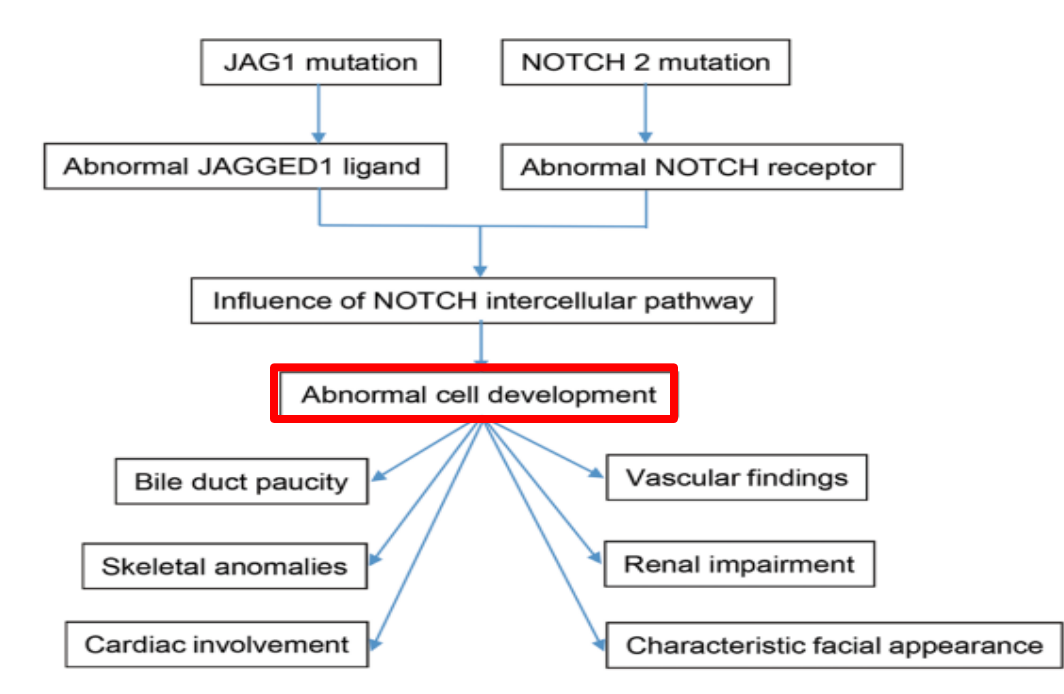
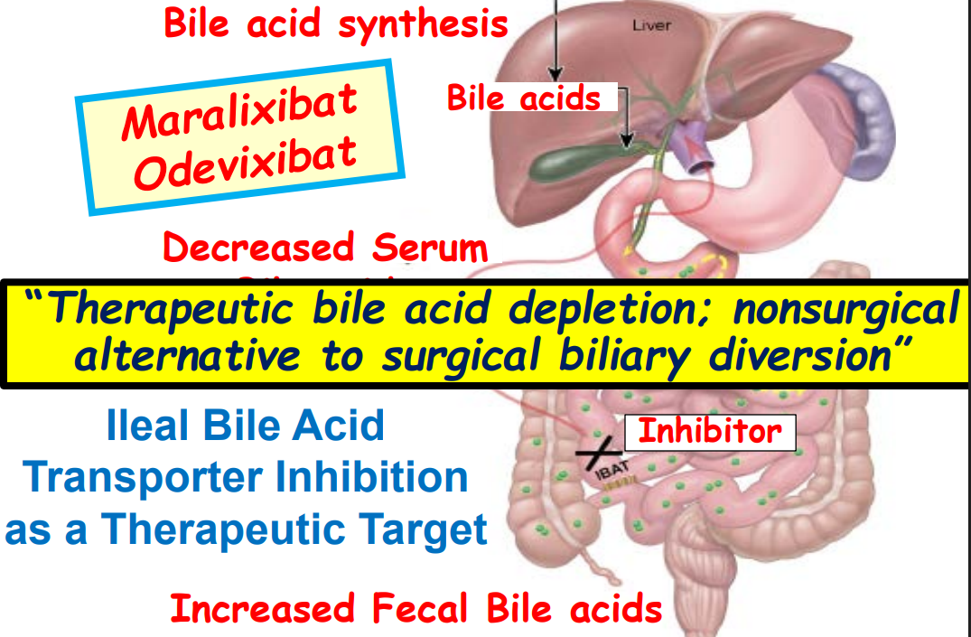
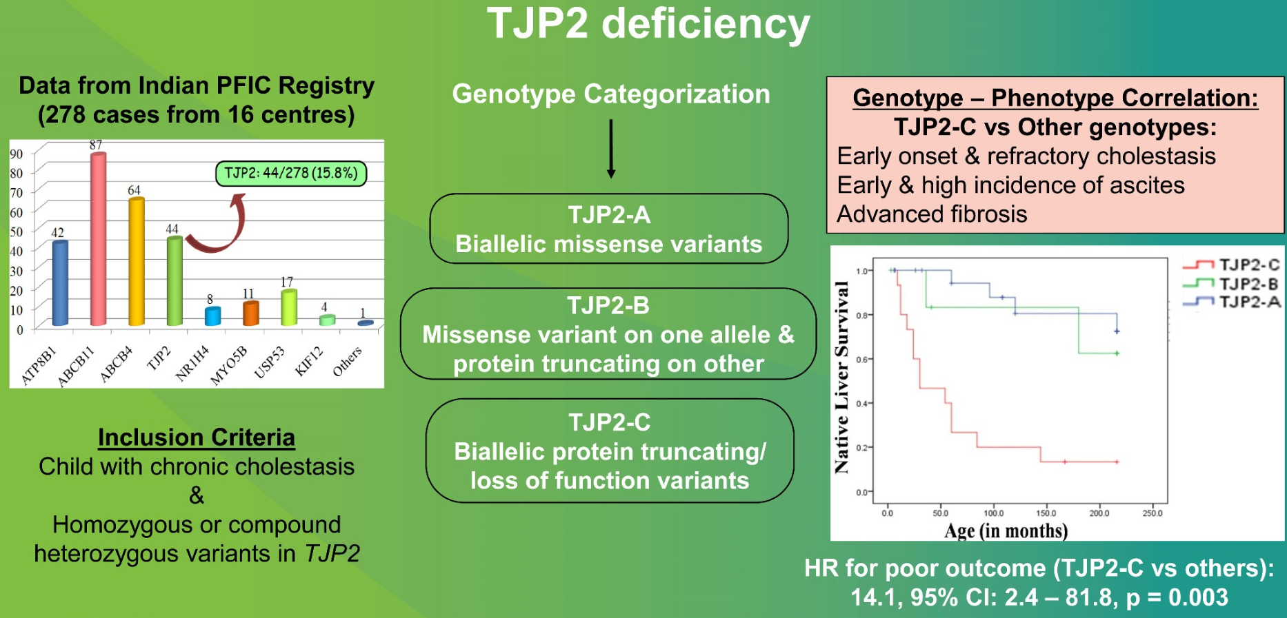
  • “In the last few years, a novel class of agents, intestinal bile acid transporter (Ileal bile acid transporter (IBAT); also known as apical sodium-dependent bile acid transporter [ASBT]) inhibitors, has emerged and gained approval from the FDA… the pivotal studies on which these approvals were granted were all performed in rare pediatric cholestatic diseases, namely Alagille syndrome (ALGS) and progressive familial intrahepatic cholestasis (PFIC).3 Additional expansion of these approvals will possibly follow as there are ongoing trials of IBAT inhibitors in primary biliary cirrhosis, primary sclerosing cholangitis, and biliary atresia.”
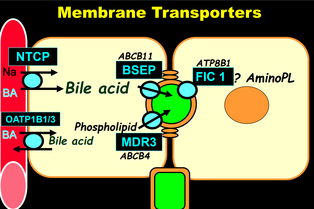
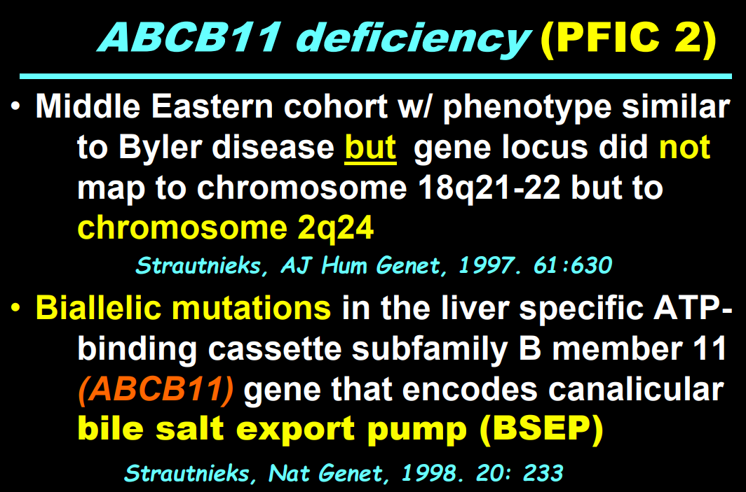
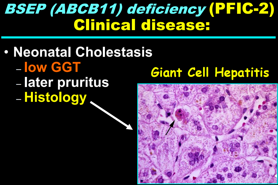
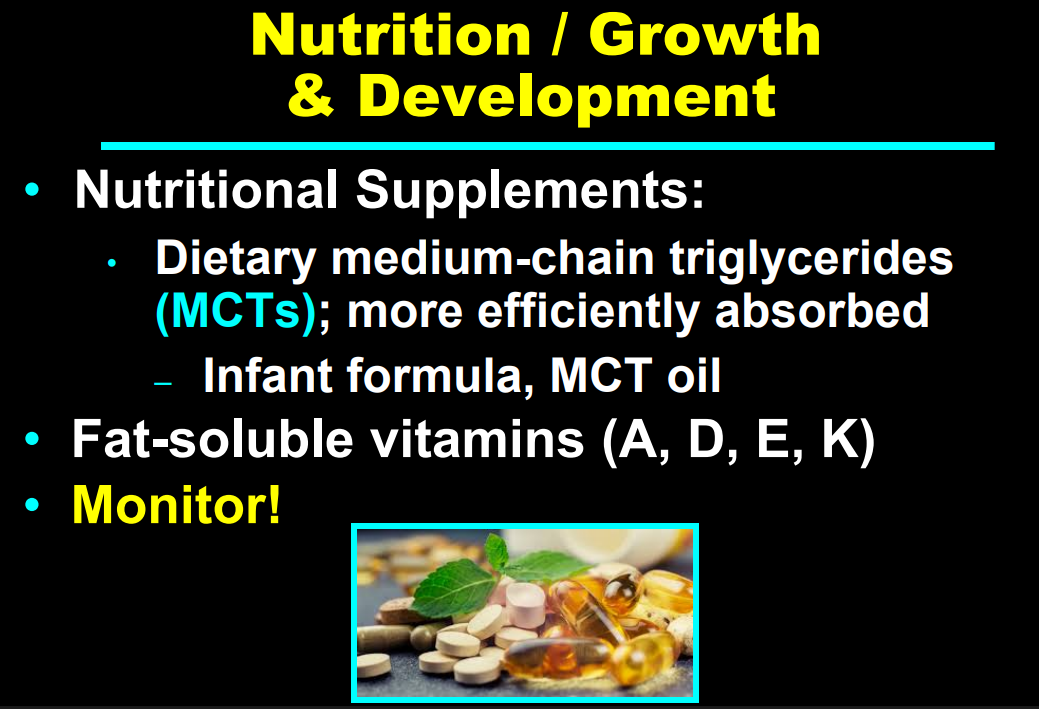
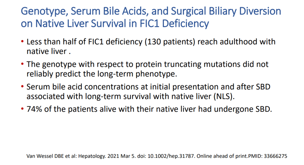
  • “The role of bile acids in promoting hepatic injury in cholestasis is perhaps best illustrated in human infants with ABCB11 (bile salt export pump; BSEP) disease or PFIC type 2…The response to IBAT inhibition in this disease further supports the notion that retained bile acids are a key factor leading to progressive liver injury and cholestatic symptoms including pruritus, fat-soluble vitamin deficiencies, and growth failure.4
  • These medications may improve liver histology and not just reduce pruritic symptoms: “Using the MDR2−/− mouse cholangiopathy model, Miethke et al22 demonstrated that ASBT inhibition led to a reduction in both serum and intrahepatic bile acid concentrations by 98% and 65%, respectively. These reductions in bile acid concentrations were associated with improved liver biochemistry and a reduction in peri-portal inflammation and fibrosis on histology. The histopathologic improvements seen in these treated MDR2−/− are important to highlight, as they support the rationale of this therapeutic approach: that lowering serum bile acid (sBA) with IBAT inhibition leads to a reduction in intrahepatic bile acid accumulation and toxicity, improvements in liver inflammation and fibrosis, and ultimately improved liver disease biology.”
  • Numerous clinical trials are listed in Table 1 (completed trials) and Table 2 (ongoing).
  • Physiology: “Bile acids are key regulators of their own enterohepatic circulation, predominately through activation of the farnesoid X receptor (FXR)…the fecal elimination of bile acids in IBAT inhibitor–treated patients appears to far exceed the rate of synthesis of new bile acids in the liver; thus, IBAT inhibitors reduce the total bile acid pool size and the bile acid load presented to the liver.22,34,39
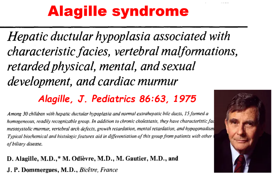
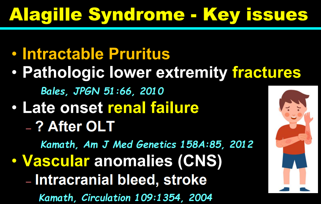
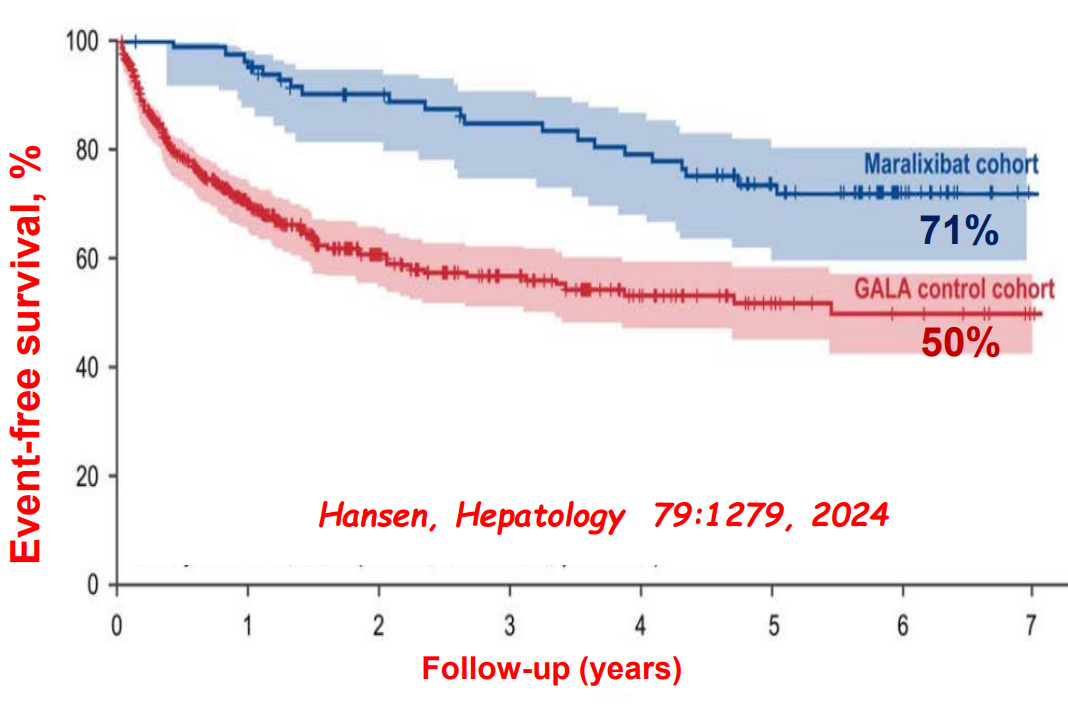
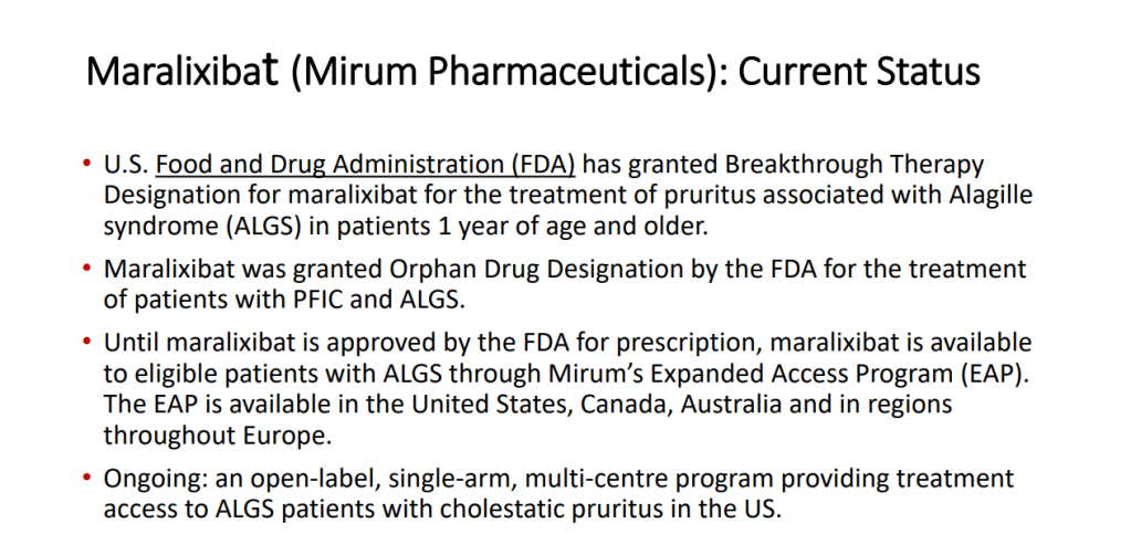
  • Alagille syndrome (ALGS): Key trials are summarized including the ICONIC trial with maralixibat and the ASSERT trial with odevixibat.
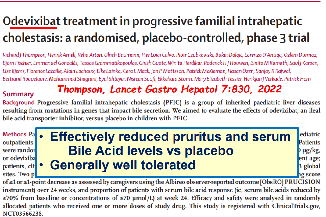
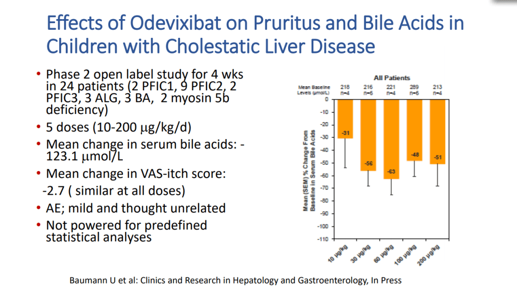
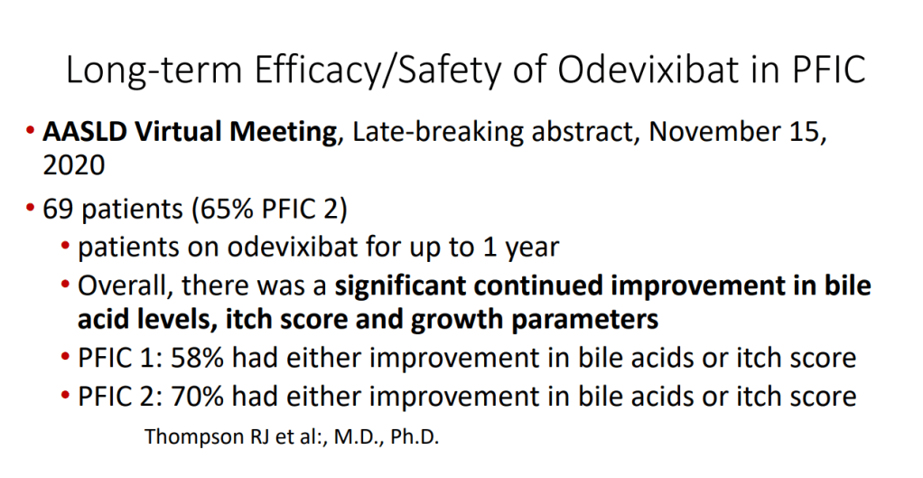
  • PFIC (Type 1 and 2) Trials: Key trials are summarized including the MARCH-PFIC trial with maralixibat and the PEDFIC1 & PEDFIC 2 trialswith odevixibat.
  • Safety: These medications are well-tolerated with self-limiting diarrhea and abdominal pain especially at the initiation of these medications. Liver blood test abnormalities have been noted in up to 20%. “This is an interesting finding, and the underlying etiology is unknown. Maralixibat is largely luminally restricted and so, without systemic absorption, a direct hepatotoxic effect is unlikely. It may reflect an alteration in the speciation of the bile acid pool with increasing bile acid synthesis or alterations in the gut-liver axis signaling. More importantly, it is not known if there are any clinical consequences to the increase in ALT.”
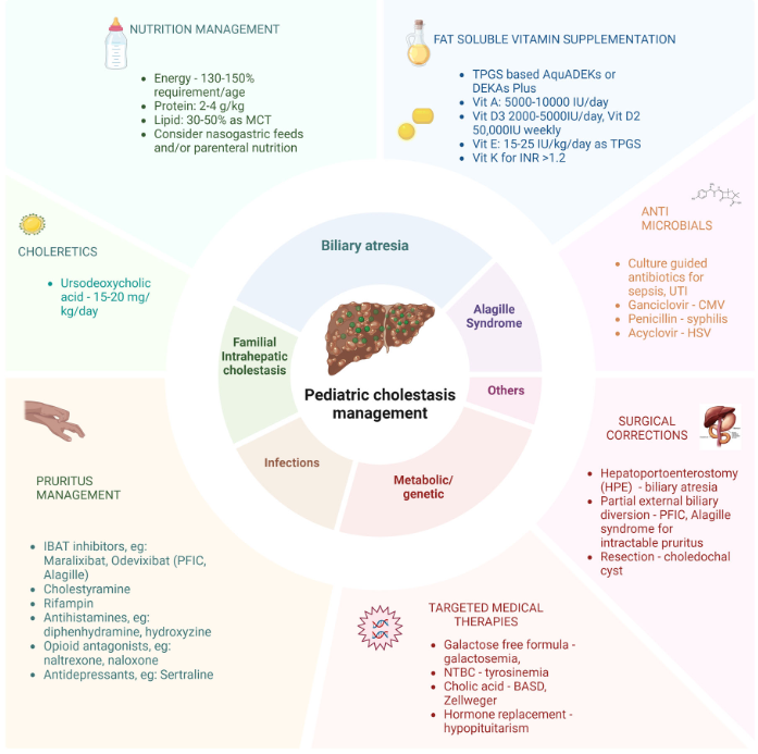
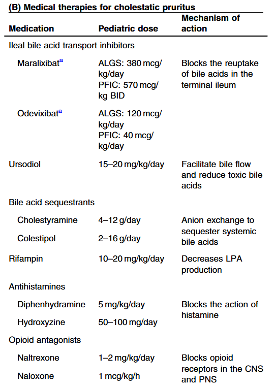
  • Cost: The authors note that ursodeoxycholic acid and antihistamines are frequently used for management of pruritus. They also not that “from a cost standpoint, it seems appropriate to offer rifampin before IBAT inhibitors in the treatment of cholestatic pruritus.”
  • Conclusions: “The clinical trial data are encouraging. As more physicians gain experience prescribing IBAT inhibitors, we will continue to learn how to best apply them to our patient populations. Like any new drug, there are still several unknowns. One of these unknowns is the potential for loss of efficacy…The short-term to medium-term clinical effects of IBAT inhibitors are clear, but we have not yet begun to see the long-term benefits. Whether durable reductions in oncogenic and fibrogenic bile acids reduce rates of HCC or slow the progression of (or reverse) portal hypertension remains to be seen.”

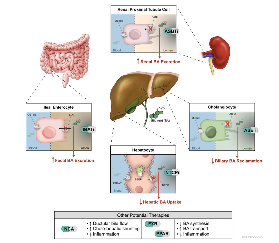
Related article: M Trauner, SJ Karpen, PA Dawson. Hepatology 2025; 82: 855-876. Open Access! Benefits and challenges to therapeutic targeting of bile acid circulation in cholestatic liver disease

“Recent advances in understanding bile acid (BA) transport in the liver… This has led to new treatments targeting BA transport and signaling. These include inhibitors of BA transport systems in the intestine and kidney (IBAT/ASBT inhibitors) and liver (NTCP inhibitors), as well as receptor agonists that modify BA synthesis and transport genes. BA analogs like norucholic acid also show promise. This review discusses the molecular and clinical basis for these therapies, particularly for cholestatic liver disorders.

Principal therapeutic targets within the entero-nephro-hepatic circulation of BAs in cholestasis.

My take (borrowed from Trauner et al): “We have arrived at a new era in the treatment of cholestatic disorders. This has been made possible by incorporating findings from discoveries into the molecular pathogenesis of cholestasis and adaptive processes that direct rational therapeutics to improve patients’ lives.”

Related blog posts:

Dr. William Balistreri: Whatever Happened to Neonatal Hepatitis (Part 2)

Recently Dr. Balistreri gave our group an excellent lecture. I have taken some notes and shared some slides. There may be inadvertent omissions and mistakes in my notes.

Key Points:

  • Producing enough bile acids and recycling bile acids in enterohepatic circulation is crucial for bile acid flow. In addition, there are ‘good’ bile acids like cholic acid that have trophic properties and ‘bad’ bile acids like lithocholic acid that cause liver toxicity
  • In addition to defects in the metabolic pathway of bile acids, discoveries identified defects in the membrane transporters (eg. FIC1, BSEP, MDR3), trafficking proteins (eg. MYO5B, VPS33B), nuclear control receptors (eg. FXR), and tight junction proteins (eg. TJP2). Tight junction protein defects are associated with bile leakage from bile canaliculus
  • Alagille syndrome, a disorder of embryogenesis, related to JAG1-NOTCH2 signaling pathways affects organs throughout the body
  • Many of these genetic mutations are now being identified in adults with unexplained liver diseases (eg. intrahepatic cholestasis of pregnancy and cryptogenic cirrhosis)
  • Cholestasis panels and whole exome sequencing are important tools
  • Ileal bile acid transporter (IBAT) inhibitors have emerged as important therapies for conditions like Alagille which were previously treated with biliary diversion

Cholestasis Evaluation:

See blog post: Identifying Biliary Atresia in Infants: New Guidelines

Baby with Carbamoyl-phosphate synthetase 1 (CPS1) deficiency (urea cycle defect)

My take: This lecture really shows how the field of pediatric liver disease has been a puzzle. Now one can see how almost all of the pieces of the puzzle work together.

Related blog posts:

Advancements in Pediatric Cholestatic Liver Disease Management

KR Mysore et al. J Pediatr Gastroenterol Nutr. 2025;80:549–558. Recent advances in the management of pediatric cholestatic liver diseases

This is a useful review summarizing advances in the management of cholestatic diseases.

Treatment with IBAT inhibitors:

“Improvement in both pruritus and serum BAs/bilirubin levels has been associated with improved event‐free survival and 6‐year transplant‐free survival in ALGS patients treated with maralixibat. Additionally, this class of medication improved overall growth of the patient by improving mean height and weight Z scores that may be related to reduced impact of high serum bile acid levels on the growth axis although further studies are needed to better define the mechanism responsible for this out-come. This finding suggests these parameters could be used as surrogate end‐points for disease severity in diseases like ALGS or PFIC, where the time course to develop the need for LT commonly occurs over many years.”

Related blog posts:

Disclaimer: This blog, gutsandgrowth, assumes no responsibility for any use or operation of any method, product, instruction, concept or idea contained in the material herein or for any injury or damage to persons or property (whether products liability, negligence or otherwise) resulting from such use or operation. These blog posts are for educational purposes only. Specific dosing of medications (along with potential adverse effects) should be confirmed by prescribing physician.  Because of rapid advances in the medical sciences, the gutsandgrowth blog cautions that independent verification should be made of diagnosis and drug dosages. The reader is solely responsible for the conduct of any suggested test or procedure.  This content is not a substitute for medical advice, diagnosis or treatment provided by a qualified healthcare provider. Always seek the advice of your physician or other qualified health provider with any questions you may have regarding a condition.

Autoimmune Diseases in Patients with Primary Sclerosing Cholangitis Plus One

A Lundberg Bave et al. Hepatology 2024; 80: 527-535. Autoimmune diseases in primary sclerosing cholangitis and their first-degree relatives

Methods: Using National Swedish registries, the authors evaluated a matched cohort study, 1378 individuals with PSC and 13,549 general population comparators and their first-degree relatives.

Key findings:

  • After excluding inflammatory bowel disease and autoimmune hepatitis, the prevalence of autoimmune disease was 18% in PSC and 11% in comparators, OR: 1.77
  • Highest odds were seen for celiac disease [OR: 4.3], sarcoidosis [OR: 2.74], diabetes type 1 [OR: 2.91], and autoimmune skin disease [OR: 2.15]
  • First-degree relatives of individuals with PSC had higher odds of developing IBD [OR: 3.25], autoimmune hepatitis [OR: 5.94], and any autoimmune disease than relatives of the comparators [OR: 1.34] 

My take: Keep an eye out for other autoimmune diseases in patients (& their 1st-degree relatives) with PSC.

Related blog posts:

Briefly noted: BB Lai et al. Hepatology 2024; 80: 511-526. Genotype correlates with clinical course and outcome of children with tight junction protein 2 (TJP2) deficiency–related cholestasis Key finding: “Patients with the TJP2-C genotype carrying PPTMs [predicted protein-truncating mutation] in both alleles had a rapidly progressive course, leading to early decompensation and death if they did not receive timely liver transplantation.”

IQ and Pediatric Chronic Liver Disease

DH Leung et al. JPGN2022 – Volume 74 – Issue 1 – p 96-103. Neurodevelopmental Outcomes in Children With Inherited Liver Disease and Native Liver

In this longitudinal study, the authors evaluated Full Scale Intelligence Quotient (FSIQ) in children with chronic liver disease (mean age 7.6 yrs). Key finding:

  • Patients with Alagille syndrome (ALGS) are at increased risk of lower FSIQ (with 29% <85), whereas our data suggest A1AT and PFIC are not

Related blog posts:

Liver Shorts: PFIC/FIC1, Best Tenofovir, Ascites Practice Guidance

DBE van Wessel et al. Hepatology 2021; 74: 892-906. Open Access: Impact of Genotype, Serum Bile Acids, and Surgical Biliary Diversion on Native Liver Survival in FIC1 Deficiency

This huge collaborative study with 130 patients provides a great deal of information about familial intrahepatic cholestasis type 1 (FIC1). Key findings:

  • Survival analysis showed an overall native liver survival (NLS) of 44% at age 18 years. NLS was comparable among FIC1-A, FIC1-B, and FIC1-C (% NLS at age 10 years: 67%, 41%, and 59%, respectively; P = 0.12)
  • The number of predicted protein truncating mutations did not correlate with natural history or prognosis

H Toyoda et al. Hepatology 2021; 74: 656-666. Treatment and Renal Outcomes Up to 96 Weeks After Tenofovir Alafenamide Switch From Tenofovir Disoproxil Fumarate in Routine Practice

In this study, the researchers 834 patients with CHB previously treated with TDF for ≥12 months who were switched to TAF in routine practice at 13 US and Asian center. Key findings:

  • “Overall, we observed continued improvement in virologic response, ALT normalization, and no significant changes in eGFR following switch to TAF from TDF.”
  • HBV DNA suppression increased from 88% to 92% at 48 weeks post-switch, and then 95% at 96 weeks postswitch
  • Improved renal function: “By week 96, 21% (55/267) of patients with CKD stage 2 at switch improved to stage 1 and 35% (30/85) of CKD stage 3-5 patients improved to stage 2 and 1.2% (1/85) to stage 1.”

SW Biggins et al. Hepatology 2021; 74: 1014-1048. Open Access. Diagnosis, Evaluation, and Management of Ascites, Spontaneous Bacterial Peritonitis and Hepatorenal Syndrome: 2021 Practice Guidance by the American Association for the Study of Liver Diseases

This practice guidance (with 276 references) is an update from similar guidelines published in 2012.

Key Points For Children:

  • Children with cirrhosis and ascites should be referred for evaluation for LT
  • Children undergoing LVP should receive 25% albumin infusion of 0.5-1.0 g/kg, or 6-8 g per liter of ascites removed.
  • Diagnostic paracentesis should be performed in children with ascites and fever, abdominal pain, or clinical deterioration. The risks and benefits of this procedure for use in all children with new ascites but without these symptoms have not been defined.
Atlanta Beltline Murals

Aspen Webinar 2021 Part 3-IBAT Inhibitors

This blog entry has abbreviated/summarized this presentation. Though not intentional, some important material is likely to have been omitted; in addition, transcription errors are possible as well. Another great lecture from Dr. Suchy.

IBAT Inhibitors Frederick Suchy

Key  points:

  • IBAT inhibitors block intestinal absorption of bile acids/disrupt enterohepatic circulation; this leads to augmented bile acid excretion in stools
  • IBAT inhibitors may reduce liver damage in the setting of cholestasis/accumulation of toxic bile acids
  • Potential diseases for IBAT inhibitors include Alagille syndrome and PFIC
  • Van Wessel et al (J Hepatol 2020; 73: 84-93) correlated survival with PFIC1/PFIC2 with bile acid levels and showed improvement in survival in those with surgical biliary diversion
  • Goals for IBAT inhibitor trials: improvement in pruritus, bile acids, reduced ALT, hepatic fibrosis, HCC and need for liver transplantation
  • Marixibat is available for use as an FDA approved breakthrough medication for Alagille and PFIC2 in pediatric patients older than 1 year
  • Odexibat is designated as an orphan drug for Alagille, PFIC, PBC, and biliary atresia
  • Safety appears good with IBAT inhibitors. Fat soluble vitamin monitoring is needed
This image has an empty alt attribute; its file name is image-132.png
This image has an empty alt attribute; its file name is image-134.png
This image has an empty alt attribute; its file name is image-136.png
This image has an empty alt attribute; its file name is image-138.png
This image has an empty alt attribute; its file name is image-140.png
This image has an empty alt attribute; its file name is image-142.png
This image has an empty alt attribute; its file name is image-144.png
This image has an empty alt attribute; its file name is image-146.png
This image has an empty alt attribute; its file name is image-148.png

Related blog posts:

Case report: Alejandro Velez Lopez

3 yo presented with fatigue and jaundice, 3 weeks after COVID-19 infection. She was not taking any medications.  Labs:  ALT 939, AST 1321, T bili 5.5, D bili 0.9, INR 2, Plts 174, Hgb 12.8, LDH 1297. remained positive for SARS-CoV2 by PCR. Acetaminophen -no exposure.  Evaluation: LKM 1:1280. Neg ANA, NL Ferritin, NL sIL2r, Other viral studies negative, NL IgG. Developed encephalopathy with NH4 317, INR peaked at 2.8.  Treated with steroids, rifaximin and lactulose.  Liver biopsy showed sub-massive necrosis and fibrosis (indicative of  autoimmune hepatitis, likely triggered or exacerbated by COVID-19).  Patient responded to medical therapy and did not require liver transplantation.

Hypothyroidism with ATP8B1 Deficiency

A recent study (L Li et al. J Pediatr 2015; 157: 1334-9) indicated that hypothyroidism may be another extrahepatic feature of patients with ATP8B1 deficiency; this mutation’s main manifestation has been intrahepatic cholestasis in either progressive familial intrahepatic cholestasis (PFIC type 1) or benign recurrent intrahepatic cholestasis (BRIC type 1).

In this study, 3/13 were hypothyroid and an additional 2/13 had subclinical hypothyroidism.  These patients were compared with a cohort of children with ABCB11 deficiency (PFIC type 2 or BRIC type 2) in which 0/19 had hypothyroidism.

Related blog posts:

Clinical Features of Byler Disease

A recent article (Morris AL, et al. JPGN 2015; 60: 460-6) provides a detailed analysis of six cases of Byler disease during their first two years of life.  These cases were strictly defined and defined by homozygous c.923G>T mutation of ATP8b1.

Presenting features:

  • 2 with newborn direct hyperbilirubinemia
  • 2 with complications of coagulopathy. “Bleeding diathesis is a particular issue in the Amish community where home delivery is common and vitamin K may not be administered perinatally.”
  • 1 with failure to thrive and rickets
  • 1 was a sibling identified with newborn genetic testing

Key features:

  • Intensive fat-soluble vitamin supplementation was needed. “Vitamin K deficiency can be lethal.”
  • Poor growth was frequent (Figure 2): “growth trajectories were generally at the low end of percentiles and did not reflect parental size.” It was “typically responsive to supplementation with medium-chain triglyceride-based formula. and/or use of 30 cal per ounce formulae.”
  • Elevated serum bile acids and low normal GGT (Υ-glutamyltranspeptidase)
  • Diarrhea was commonly reported
  • Intractable pruritus in 4 of 6 children which developed between 6-12 months of age;  in two patients rifampin therapy was effective.
  • Partial external biliary diversion was used in 4 children during 2nd year of life; there was a “generally favorable response to PEBD.”
  • There were not issues noted with portal hypertension

Bottomline: This report shares some practical experience with this rare disorder.

Related blog posts:

BRIC, PFIC, and nasobiliary drainage

Case reports, when effective, help clinicians understand meaningful differences in disease presentation; in addition, they highlight practical treatment approaches.  An excellent example of one such case report is the following:

  • Zellos A et al.  JPGN 2012; 55: 88-90

These authors present a case with unique features that highlight some of the clinical problems with benign recurrent intrahepatic cholestasis (BRIC) and progressive familial intrahepatic cholestasis (PFIC).  BRIC1 and PFIC1 are associated with mutations in ATP8B1; BRIC2 and PFIC2 are associated with mutations in ABCB11.  The primary difference between BRIC and PFIC is the phenotypic expression.  In BRIC, individuals have episodes of cholestasis; in PFIC, progressive chronic liver disease develops in the first months of life.  PFIC2/ABCB11 mutations cause defective bile salt export pump (BSEP) at the bile-canniculus membrane.

Both ATP8B1 and ABCB11 intrahepatic cholestasis conditions present in a similar fashion with low GGT values.  In this case report, a 5-year-old presented with jaundice, acholic stools and dark urine.  His laboratory values revealed an ALT of 60 U/L, direct bilirubin of 7.6 mg/dL and gamma-glutamyl transpeptidase (GGT) of 10 U/L.  Initially, after exclusion of other liver conditions (eg. NL MRCP, copper studies, α-1 antitrypsin, autoimmune serology, infectious etiologies), the authors suspected ‘a clinicopathologic intergrade between BRIC and PFIC’ likely due to ATP8B1 as there was BSEP expression on liver biopsy immunostaining.  After sequencing did not demonstrate any ATP8B1 mutations, the authors identified two heterozygote mutations in ABCB11.

From a treatment standpoint, once nasobiliary drainage (NBD) was in place, the patient quickly improved.  This occurred after >6 weeks of failure with urosdeoxycholic acid/conservative measures.  As a precaution, the authors cultured the bile once a week and instituted antibiotic treatment when positive cultures were identified.

One other point alluded to by the authors is that the natural history of BRIC2 is poorly described.  Whether this disorder is truly ‘benign’ as the name suggests is unclear.  In patients with similar mutations who develop PFIC2, there is a high risk of hepatocellular carcinoma (HCC).

Intrahepatic Cholestasis Genes/Disorder (Clin Liver Dis 2006; 10: 27-53.)

Gene: Disorder (protein)
ABCB11: PFIC 2, BRIC 2 (BSEP)
ABCB4: PFIC 3, ICP (MDR3)
CFTR: CF (CFTR)
ATP8B1: PFIC1 -Byler’s (FIC1), BRIC, GFC -Greenland Familial
CLDN1: NISCH (Claudin 1) -neonatal sclerosing cholangitis/icthyosis
VPS33B: ARC syndrome (Vascular protein sorting 33) -arthrogryposis-renal dysfn-cholestasis, low GGT
AKR1D1: BAS: Bile acid synthetic defect: neonatal cholestasis with giant cell hepatitis
(5β-reductase)
HSD3B7: BAS (C27-3β-HSD)
CYP7BI: BAS (CYP7BI)
TJP2: (ZO-2) FHC: Familial hypercholanemia (tight junction protein)
BAAT: FHC (BAAT)
EPHX1: FHC (epoxide hydrolase)
JAG1: Alagille (JAG1) JAG1 is transmembrane cell-surface protein important in regulating cell fate during embryogenesis
PKHD1: ARPKD (fibrocystin -important in ciliary function and tubulogenesis)
PRKCSH: ADPLD (hepatocystin)
ABCC2: Dubin-Johnson syndrome (MRP2)
CIRH1A: NAIC -N Amer Indian childhood cirrhosis (Cirhin)

Additional references for BRIC/low GGT PFIC:

  • -JPGN 2010; 51: 494.  Use of biliary diversion –helpful in 18 PFIC2 cases with long-term f/u.
  • -Liver Transplantation 2010; 16: 856.  6 patients developed recurrent low gamma-glutamyl transpeptidase cholestasis, that mimics BSEP disease, following transplantation. All had documented genetic defects in ABCB11 that were predicted to lead to a congenital absence of BSEP protein.
  • -NEJM 2009; 361: 1359. Recurrence of BSEP deficiency p OLT due to antibodies against BSEP
  • -Hepatology 2010; 51: 1645. n=62 children & clinical course.
  • -Gastroenterol 2008; 134: 1203. Severe BSEP –82 different mutations in 109 families. (n=132 patients)
  • -JPGN 2008; 46: 241. Excellent review. FIC1 caused by mutations in ATP8B1, PFIC 2 caused by mutations in ABCB11 which encodes BSEP – bile salt export pump. Increased risk of HCC in PFIC2 especially.
  • -J Pediatr 2007; 150: 556.  Increase risk of HCC in PFIC2.
  • -Hepatology 2006; 44: 478-486. Cases of pediatric HCC in PFIC-2
  • -Gastroenterol 2006; 130: 908. Review of canalicular transport defects.
  • -Hepatology 2005; 42: 222. summary of cholestasis workshop
  • -Gastroenterol 2004; 126: 322. Review of bile salt transporters.
  • -JPGN 2002; 34: 7A. FTT, diarrhea persist p biliary diversion or transplant.

PFIC3 -High GGT

  • -Gastroenterol 2003; 124: 1037-42. MDR3 mutations causing cholelithiasis, cholestasis, biliary cirrhosis, & pregnancy cholestasis.
  • -Gastroenterol 2001; 120: 1448-1458. n=31 cases. MDR3 mutations. ABCB4 gene
  • -Gastroenterol 2001; 120: 1459-67. Gallbladder stones & chronic cholestasis in 6 MDR3+ pts. Avg age of presentation: 2.9yrs. Avg age of Tx: 7.5yrs.  Sx/S : high ggt cholestasis, pruritus, intrahepatic cholestasis of pregnancy in heterozygotes (& c contraception)
  • -Hepatology 1996; 23: 904-8. MDR3 gene assoc c PFIC