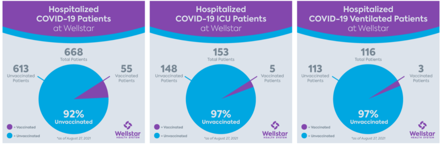
I am a pediatric gastroenterologist at GI Care for Kids (previously called CCDHC) in Atlanta, Georgia. The goal of my blog is to share some of my reading in my field more broadly. In addition, I wanted to provide my voice to a wide range of topics that often have inaccurate or incomplete information.
Before starting this blog in 2011, I would tear out articles from journals and/or keep notes in a palm pilot. This blog helps provide an updated source of information that is easy to access and search, along with links to useful multimedia sources.
I was born and raised in Chattanooga. After graduating from the University of Virginia, I attended Baylor College of Medicine. I completed residency and fellowship training at the University of Cincinnati at the Children’s Hospital Medical Center. I received funding from the National Institutes of Health for molecular biology research of the gastrointestinal tract.
During my fellowship, I had the opportunity to work with some of the most amazing pediatric gastroenterologists and mentors. Some of these individuals included Mitchell Cohen, William Balistreri, James Heubi, Jorge Bezerra, Colin Rudolph, John Bucuvalas, and Michael Farrell. I am grateful for their teaching and their friendship. During my training with their help, I received a nationwide award for the best research by a GI fellow.
I have authored numerous publications/presentations including original research, case reports, review articles, and textbook chapters on various pediatric gastrointestinal problems. In addition, I have been recognized by Atlanta Magazine as a "Top Doctor" in my field multiple times.
Currently, I am the vice chair of the section of nutrition for the Georgia Chapter of the American Academy of Pediatrics. In addition, I am an adjunct Associate Clinical Professor of Pediatrics at Emory University School of Medicine. Other society memberships have included the North American Society for Pediatric Gastroenterology Hepatology and Nutrition (NASPGHAN), American Academy of Pediatrics, the Food Allergy Network, the American Gastroenterology Association, the American Association for the Study of Liver Diseases, and the Crohn’s and Colitis Foundation.
As part of a national pediatric GI organization called NASPGHAN (and its affiliated website GIKids), I have helped develop educational materials on a wide-range of gastrointestinal and liver diseases which are used across the country. Also, I have been an invited speaker for national campaigns to improve the evaluation and treatment of gastroesophageal reflux disease, celiac disease, eosinophilic esophagitis, hepatitis C, and inflammatory bowel disease (IBD). Some information on these topics has been posted at my work website, www.gicareforkids.com, which has links to multiple other useful resources.
I am fortunate to work at GI Care For Kids. Our group has 17 terrific physicians with a wide range of subspecialization, including liver diseases, feeding disorders, eosinophilic diseases, inflammatory bowel disease, cystic fibrosis, DiGeorge/22q, celiac disease, and motility disorders. Many of our physicians are recognized nationally for their achievements. Our group of physicians have worked closely together for many years. None of the physicians in our group have ever left to join other groups. I have also worked with the same nurse (Bernadette) since I moved to Atlanta in 1997.
For many families, more practical matters about our office include the following:
– 14 office/satellite locations
– physicians who speak Spanish
– cutting edge research
– on-site nutritionists
– on-site psychology support for abdominal pain and feeding disorders
– participation in ImproveCareNow to better the outcomes for children with inflammatory bowel disease
– office endoscopy suite (lower costs and easier scheduling)
– office infusion center (lower costs and easier for families)
– easy access to nursing advice (each physician has at least one nurse)
I am married and have two sons (both adults). I like to read, walk/hike, bike, swim, and play tennis with my free time.
I do not have any financial relationships with pharmaceutical companies or other financial relationships to disclose. I have helped enroll patients in industry-sponsored research studies.
A long time ago I heard a joke from a mentor about how can you tell if a person is an optimist. An optimist is a person who finds a pile of manure under the tree on Christmas morning and declares: ‘Oh boy, I’m getting a pony.’
Researchers who are trying to identify oral treatments for celiac disease are probably true optimists. Yet, despite my skepticism, a recent study (D Schuppan et al. NEJM 2021; 385: 35-45. A Randomized Trial of a Transglutaminase 2 Inhibitor for Celiac Disease) provides the best proof yet that an oral treatment may be helpful.
In this 6-week randomized, double-blind, placebo-controlled study with 159 participants, treatment with ZED1227, a selective oral transglutaminiase 2 inhibitor reduced histologic injury compared to placebo; all patients were receiving a diet with 3 grams of daily gluten. Key findings:
Treatment with ZED1227 at all three dose levels attenuated gluten-induced duodenal mucosal injury. The estimated difference from placebo in the change in the mean ratio of villus height to crypt depth from baseline to week 6 was 0.44 (95% confidence interval [CI], 0.15 to 0.73) in the 10-mg group (P=0.001), 0.49 (95% CI, 0.20 to 0.77) in the 50-mg group (P<0.001), and 0.48 (95% CI, 0.20 to 0.77) in the 100-mg group (P<0.001)
The estimated differences from placebo in the change in intraepithelial lymphocyte density were −2.7 cells per 100 epithelial cells (95% CI, −7.6 to 2.2) in the 10-mg group, −4.2 cells per 100 epithelial cells (95% CI, −8.9 to 0.6) in the 50-mg group, and −9.6 cells per 100 epithelial cells (95% CI, −14.4 to −4.8) in the 100-mg group
Adverse events were similar to placebo; 3 (8%) patients in the 100 mg group developed a rash
The need for a treatment besides a gluten-free diet is significant; among adults, 40-50% do not achieve mucosal healing/recovery despite GFD institution; in addition, the diet is difficult and costly.
My take: I think it is still a long journey to find an effective & safe oral treatment for celiac disease.
F Wang et al JPGN 2021; 73: 17-22. Glucocorticoids Improve Enteral Feeding Tolerance in Pediatric Short Bowel Syndrome With Chronic Intestinal Inflammation In this retrospective study with 15 patients who had histologically-confirmed chronic intestinal inflammation, glucocorticoids (budesonide or prednisone) were associated with clinical improvement. Key findings: 7 of the 15 patients gained enteral autonomy. 6 of 7 of those had eosinophilic infiltrates as part of their histologic findings. 11 of 15 had a reduction in parenteral calories.
L Norsa et al JPGN 2021; 73: 48-53. Scoring Endoscopy in Pediatric Inflammatory Bowel Disease: A Way to Improve Quality This study showed very poor agreement between 11 pediatric GIs in evaluating videos of 15 endoscopies (see below). Key finding: Intraclass correlation was 0.298 (95% confidence interval [CI]: 0.13–0.55) for ulcerative colitis (UC) and 0.266 (0.11–0.52) for Crohn disease (CD). My take: This study indicates either a need for rigorous training of endoscopists and/or need for AI review of endoscopy.
NY Times 7/2/21: Full text: For Surprise Medical Bills, It’s the Beginning of the End Key issues for regulators: define a standard price for out-of-network care, determine what hospitals and doctors will need to do to notify patients they are not in their insurance network, and establish a complaint system for consumers who believe they were illegally billed.
This single-center retrospective study with 43 patients (over 11 year timeframe) identified etiology and outcomes for neonatal acute liver failure (NALF).
Key findings:
Etiologies included viral infection (23%), gestational alloimmune liver disease with neonatal hemochromatosis (GALD-NH) (21%), cardiac-associated ischemia (16%), other ischemia (14%), genetic etiologies (9%), Trisomy 21-associated myelodysplasia (TAM) (7%), hemophagocytic lymphohistiocytosis (HLH) (2%), and not identified (7%)
Infants with viral etiologies had the highest alanine aminotransferase (ALT) at presentation (1179 IU/L, interquartile range [IQR] 683–1585 IU/L) in contrast to low levels in GALD-NH (23 IU/L, IQR 18–64 IU/L)
Across all etiologies, only 33% were alive at 1 year
Among laboratory values at presentation, alpha-fetoprotein (AFP) was significantly higher in patients that survived with their native liver (P = 0.04)
Figure 4 provides a helpful management algorithm for NALF. Figure 4 is similar to the slide below (shared by lead author).
-Consider empiric acyclovir in those with INR >/= 2.0 in the first 30 days of life
-In those with normal to low elevation of aminotransferases, consider empiric IVIG while undergoing workup. Part of workup should include either MRI or lip biopsy for GALD
-In those with moderate to severe elevation of aminotransferases, workup should include assessment for viral, HLH and genetic etiologies. Fulminant viral hepatitis or HLH likely with Ferritin levels >10,000. Hypoglycemia and hyperammonemia is suggestive of metabolic/mitochondrial disorder
-Liver biopsy may be needed if etiologies not identified
Disclaimer: This blog, gutsandgrowth, assumes no responsibility for any use or operation of any method, product, instruction, concept or idea contained in the material herein or for any injury or damage to persons or property (whether products liability, negligence or otherwise) resulting from such use or operation. These blog posts are for educational purposes only. Specific dosing of medications (along with potential adverse effects) should be confirmed by prescribing physician. Because of rapid advances in the medical sciences, the gutsandgrowth blog cautions that independent verification should be made of diagnosis and drug dosages. The reader is solely responsible for the conduct of any suggested test or procedure. This content is not a substitute for medical advice, diagnosis or treatment provided by a qualified healthcare provider. Always seek the advice of your physician or other qualified health provider with any questions you may have regarding a condition.
65% of respondents (n=238 of 2300 responded to survey) perform repeat endoscopy to assess for endoscopic remission in pediatric IBD as part of routine clinical practice (usually 9 to 12 months later)
“Symptoms are not sufficient to follow IBD patients” was reported by 82% of those who repeat endoscopy
“I perform endoscopy based on clinical, biomarker, and/or imaging trends” was reported by 81% of those who do not repeat endoscopy
In those inclined to do repeat endoscopy (n=134 total), the authors state there was a significant difference based on years in practice but this is difficult to discern based on the data presented in Table 1; the numbers in both groups are much greater than the number of total patients in each group. They state in the repeat endoscopy group (n=134), the practitioner experience was n=58 (1-5 yrs), n=43 (6-10 yrs), n= 34 (11-15 yrs), and 70 (>15 yrs) and the “no repeat group” (n=67 total) was n=43 (1-5 yrs), n=33 (6-10 yrs), n=21 (11-15 yrs), and n=37 (>15 yrs). Apparently, according to the discussion, those in practice more than 15 years were less likely to recommend a ‘treat-to-target’ endoscopy.
There is also a discrepancy in the report with regard to ImproveCareNow participation, stated to be 63% in the abstract and 71% in the results section
Discussion: I would propose that the first part of a journal club start with these two lines from the discussion: “As the paradigm of clinical endpoints has evolved in the management of IBD, there has been a shift from using clinical symptoms to drive major therapeutic decisions to using endoscopic assessment. This lag time to adopt new practices in medicine has been highlighted in research demonstrating the slow adoption of new clinical practices by physicians, possibly related to the difficulty with “unlearning” common practices and shifting to new ones.” As an aside, 77% of the survey respondents were in an academic practice; it would be fun to see how the section chief views this assertion.
While the majority of survey respondents supported repeat endoscopy in all patients, the discussion point above is making a different distinction (“drive major therapeutic decisions”). I think a much higher proportion of practitioners would endorse endoscopy prior to major therapeutic decisions. However, with regard to supporting more widespread routine followup in all of those in clinical remission, the discussion references data from a single retrospective pediatric cohort study with 104 patients (Inflamm Bowel Dis 2017; 23: 1447-1453), that 30% of patients in clinical remission had active disease on endoscopy.
My take: As alluded to in the conclusion, long-term data from prospective studies are needed to determine the benefit (or lack of benefit) of followup endoscopy, especially in patients with combined clinical/biomarker remission.
Background: “The incidence of EoE during OIT has been estimated at 2.7%.” (AJ Lucendo et al. Ann Allergy Asthma Immunol 2014; 113: 624-629)
Methods: Twenty adults with IgE-mediated peanut allergy were randomly assigned to groups given peanut OIT (n = 15) or placebo (n = 5) in this prospective study. Serial gastrointestinal biopsies were collected at baseline (n = 21, 0 weeks), following dose escalation (n = 10, 52 weeks), and during the maintenance phase (n = 11, 104 weeks)
Key findings:
At baseline: 3 of the 21 subjects (14%) had esophageal peak eosinophil counts ≥15 eos/hpf and all subjects had dilated intercellular spaces (DIS)
At 52 weeks: OIT induced or exacerbated esophageal eosinophilia (EoE) at 52 weeks with peak eosinophil counts ≥15 eos/hpf in 4 of 7 patients [57%] who did not have EoE at baseline. EoE did not develop in patients receiving placebo
At 104 weeks: In 4 of 6 participants (67%), OIT-induced EoE and gastrointestinal eosinophilia resolved by the end of the maintenance phase
One patient developed a clinical diagnosis of EoE.
The discussion notes overlap between EoE and IgE-mediated food allergy. The risk of EoE in patients with IgE-mediated food allergy is 118 times that of the general population (4.7% vs 0.04%) (J Allergy Clin Immunol Pract 2017; 5: 369-375). Also, the authors note that in this study all of the peanut allergic subjects had evidence of epithelial barrier dsyfunction.
My take: This small study shows, that for most adult patients, the development of EoE during OIT is often transitory.
A series of articles details the 2021 AGA Guidelines for Crohn’s disease (CD) including a clinical practice guideline (pg 2496-2508), a clinical decision support tool (2509-2510), a spotlight summary (pg 2511), a technical review (2512-2557), and a review of the recommendations (pg 2557-2262). I will highlight the first article.
For me the most important of their recommendations was #7:
In adult outpatients with moderate to severe CD, the AGA suggests early introduction with a biologic with or without an immunomodulator rather than delaying their use until after failure of 5-aminosalicylates and/or corticosteroids.
Other points:
For moderate to severe CD, the authors support the use of anti-TNF therapy in combination with immunomodulators, support the use of ustekinumab and vedolizumab, and support the use of SC/IM (but not oral) methotrexate.
The authors are AGAINST the use of mesalamine/sulfasalazine products as well as corticosteroids for maintenance of remission for CD
Methods: This article describes the development a computed-tomography enterography (CTE)–based radiomic model (RM). This retrospective multicenter study included 167 CD patients who underwent preoperative CTE and bowel resection. 1454 radiomic features were extracted from venous-phase CTE and a machine learning–based RM was developed based on the reproducible features using logistic regression. The RM was validated in an independent external test cohort recruited from 3 centers.
Key findings:
In the training cohort, the area under the ROC curve (AUC) of RM for distinguishing moderate–severe from none–mild intestinal fibrosis was 0.888.
In the test cohort, the RM had an AUC of 0.816.
RM was more accurate than visual interpretations by either radiologist (radiologist 1, AUC = 0.554; radiologist 2, AUC = 0.598; both, P < .001) in the test cohort
My take: This CT approach with RM allowed for accurate characterization of intestinal fibrosis in CD. The images look pretty cool too.
Methods: Overall in this phase 1 randomized, open-label study in patients with either ulcerative colitis or Crohn’s disease, 66 and 65 patients were randomized to CT-P13 SC (every 2 weeks) and CT-P13 IV, respectively
Key findings: The pharmacokinetic noninferiority of CT-P13 SC to CT-P13 IV, and the comparable efficacy, safety, and immunogenicity profiles
These findings are in agreement with similar studies performed in patients with Rheumatoid Arthritis.
My take: If confirmed with additional studies, it is likely that SC infliximab treatment will be a useful alternative to intravenous infliximab. This is similar to data presented with vedolizumab which is currently administered intravenously.