Unknown's avatar

About gutsandgrowth

I am a pediatric gastroenterologist at GI Care for Kids (previously called CCDHC) in Atlanta, Georgia. The goal of my blog is to share some of my reading in my field more broadly. In addition, I wanted to provide my voice to a wide range of topics that often have inaccurate or incomplete information. Before starting this blog in 2011, I would tear out articles from journals and/or keep notes in a palm pilot. This blog helps provide an updated source of information that is easy to access and search, along with links to useful multimedia sources. I was born and raised in Chattanooga. After graduating from the University of Virginia, I attended Baylor College of Medicine. I completed residency and fellowship training at the University of Cincinnati at the Children’s Hospital Medical Center. I received funding from the National Institutes of Health for molecular biology research of the gastrointestinal tract. During my fellowship, I had the opportunity to work with some of the most amazing pediatric gastroenterologists and mentors. Some of these individuals included Mitchell Cohen, William Balistreri, James Heubi, Jorge Bezerra, Colin Rudolph, John Bucuvalas, and Michael Farrell. I am grateful for their teaching and their friendship. During my training with their help, I received a nationwide award for the best research by a GI fellow. I have authored numerous publications/presentations including original research, case reports, review articles, and textbook chapters on various pediatric gastrointestinal problems. In addition, I have been recognized by Atlanta Magazine as a "Top Doctor" in my field multiple times. Currently, I am the vice chair of the section of nutrition for the Georgia Chapter of the American Academy of Pediatrics. In addition, I am an adjunct Associate Clinical Professor of Pediatrics at Emory University School of Medicine. Other society memberships have included the North American Society for Pediatric Gastroenterology Hepatology and Nutrition (NASPGHAN), American Academy of Pediatrics, the Food Allergy Network, the American Gastroenterology Association, the American Association for the Study of Liver Diseases, and the Crohn’s and Colitis Foundation. As part of a national pediatric GI organization called NASPGHAN (and its affiliated website GIKids), I have helped develop educational materials on a wide-range of gastrointestinal and liver diseases which are used across the country. Also, I have been an invited speaker for national campaigns to improve the evaluation and treatment of gastroesophageal reflux disease, celiac disease, eosinophilic esophagitis, hepatitis C, and inflammatory bowel disease (IBD). Some information on these topics has been posted at my work website, www.gicareforkids.com, which has links to multiple other useful resources. I am fortunate to work at GI Care For Kids. Our group has 17 terrific physicians with a wide range of subspecialization, including liver diseases, feeding disorders, eosinophilic diseases, inflammatory bowel disease, cystic fibrosis, DiGeorge/22q, celiac disease, and motility disorders. Many of our physicians are recognized nationally for their achievements. Our group of physicians have worked closely together for many years. None of the physicians in our group have ever left to join other groups. I have also worked with the same nurse (Bernadette) since I moved to Atlanta in 1997. For many families, more practical matters about our office include the following: – 14 office/satellite locations – physicians who speak Spanish – cutting edge research – on-site nutritionists – on-site psychology support for abdominal pain and feeding disorders – participation in ImproveCareNow to better the outcomes for children with inflammatory bowel disease – office endoscopy suite (lower costs and easier scheduling) – office infusion center (lower costs and easier for families) – easy access to nursing advice (each physician has at least one nurse) I am married and have two sons (both adults). I like to read, walk/hike, bike, swim, and play tennis with my free time. I do not have any financial relationships with pharmaceutical companies or other financial relationships to disclose. I have helped enroll patients in industry-sponsored research studies.

NEJM: Functional Dyspepsia

A recent NEJM had a concise review of functional dyspepsia (Talley NJ, Ford AC. NEJM 2015; 373: 1853-63).

With regard to functional dyspepsia in adults, the authors note that using the Rome III criteria, the global prevalence is between 5% and 11%.

While symptoms do not reliably distinguish organic and functional dyspepsia, they note that “with a relatively low rate of identification of organic disease, it is neither desirable nor realistic to perform this test [upper gastrointestinal endoscopy] in all patients with dyspepsia.”

Their review suggests several criteria to consider to help determine who needs endoscopy including age >55 yrs, GI bleeding, dysphagia, persistent vomiting, unintentional weight loss, family history of gastric or esophageal cancer, and iron-deficiency anemia.

With regard to workup, they suggest testing for H pylori non invasively with either breath testing or stool antigen testing.  The review covers treatment approaches including acid suppression (“effect is modest”), antidepressants (“tricyclic antidepressants…should be preferred over selective serotonin-reuptake inhibitors”), prokinetic agents, psychological treatments, and complementary approaches.  Figure 3 provides a helpful algorithm.

With regard to prognosis, “approximately 15 to 20% of people with functional dyspepsia have persistent symptoms and 50% have resolution of symptoms; in the remaining 30 to 35% of patients symptoms will fluctuate and meet the criteria for another functional gastrointestinal disorder.”

Briefly noted: “Acute Anxiety and Anxiety Disorders are Associated with Impaired Gastric Accommodation in Patients with Functional Dyspepsia” HG Ly et al. Clin Gastroenterol Hepatol 2015; 13: 1584-91.

Related blog posts:

Sunrise in Sandy Springs

Sunrise in Sandy Springs

Why Does Enteral Nutrition Work for Crohn’s Disease? Is it due to the Microbiome?

A recent study (K Gerasimidis et al. Am J Gastroenterol advance online publication 3 November 2015; doi: 10.1038/ajg.2015.357. Full Text: Extensive Modulation of the Fecal Metagenome in Children With Crohn’s Disease During Exclusive Enteral Nutrition) finds that treatment with Exclusive Enteral Nutrition further reduces microbiome diversity compared to healthy controls. This was an unexpected finding as the authors state: “we would expect EEN treatment to normalize the perceived ‘dysbiotic’ microbiota toward a healthier state.”

Reference from KT Park’s twitter feed. Here’s the abstract:

OBJECTIVES:

Exploring associations between the gut microbiota and colonic inflammation and assessing sequential changes during exclusive enteral nutrition (EEN) may offer clues into the microbial origins of Crohn’s disease (CD).

METHODS:

Fecal samples (n=117) were collected from 23 CD and 21 healthy children. From CD children fecal samples were collected before, during EEN, and when patients returned to their habitual diets. Microbiota composition and functional capacity were characterized using sequencing of the 16S rRNA gene and shotgun metagenomics.

RESULTS:

Microbial diversity was lower in CD than controls before EEN (P=0.006); differences were observed in 36 genera, 141 operational taxonomic units (OTUs), and 44 oligotypes. During EEN, the microbial diversity of CD children further decreased, and the community structure became even more dissimilar than that of controls. Every 10 days on EEN, 0.6 genus diversity equivalents were lost; 34 genera decreased and one increased during EEN. Fecal calprotectin correlated with 35 OTUs, 14 of which accounted for 78% of its variation. OTUs that correlated positively or negatively with calprotectin decreased during EEN. The microbiota of CD patients had a broader functional capacity than healthy controls, but diversity decreased with EEN. Genes involved in membrane transport, sulfur reduction, and nutrient biosynthesis differed between patients and controls. The abundance of genes involved in biotin (P=0.005) and thiamine biosynthesis decreased (P=0.017), whereas those involved in spermidine/putrescine biosynthesis (P=0.031), or the shikimate pathway (P=0.058), increased during EEN.

CONCLUSIONS:

Disease improvement following treatment with EEN is associated with extensive modulation of the gut microbiome.

My take: We really don’t know why EEN works and we have a lot to learn about a ‘healthy’ microbiome.

Related blog posts:

New Hepatitis B Treatment Guidelines

Link to full article: Updated Hepatitis B Treatment Guidelines from AASLD

With regard to pediatrics:

9A. The AASLD suggests antiviral therapy in HBeAg-positive children (ages 2 to <18 years) with both elevated ALT and measurable HBV DNA levels, with the goal of achieving sustained HBeAg seroconversion.

“Most studies required ALT elevation (>1.3 times ULN) for at least 6 months with HBV DNA elevations for inclusion. Given that HBV DNA levels are typically very high during childhood (>106 IU/mL), there is no basis for a recommendation for a lower-limit value with respect to treatment. However, if a level <104 IU/mL is observed, therapy might be deferred until other causes of liver disease and spontaneous HBeAg seroconversion are excluded.”

“Duration of treatment with oral antivirals that has been studied is 1-4 years. It may be prudent to use HBeAg seroconversion as a therapeutic endpoint when oral antivirals are used, continuing treatment for an additional 12 months of consolidation, as recommended in adults. It is currently unknown whether a longer duration of consolidation would reduce rates of virological relapse.”

“Children who stop antiviral therapy should be monitored every 3 months for at least 1 year for recurrent viremia, ALT flares, and clinical decompensation.”

9B. The AASLD recommends against use of antiviral therapy in HBeAg-positive children (ages 2 to <18 years) with persistently normal ALT, regardless of HBV DNA level.

Another nice summary of current treatment recommendations: P Martin et al. Clin Gastroenterol Hepatol 2015; 13: 2071-87.  Table 5 lists recommendations for treatment of HBeAg-positive.

  • The main group needing treatment (entecavir, tenofovir, or PEGinterferon alfa-2a) are those with HBV DNA >2000 IU/mL and elevated ALT.  Table 6 lists recommendations for those with HBeAg-negative.  Main group needing treatment are the same (HBV DNA >2000 IU/mL and elevated ALT).
  • With both groups (HBe-Ag negative and positive), “consider liver biopsy or transient elastography” if elevated HBV DNA >2000 and normal ALT.  If histologic disease present, consider treatment.
  • One point the authors make about therapy regards duration: “Historically, HBeAg seroconversion was considered a durable response, and discontinuation of antiviral therapy was recommended after a period of consolidation therapy of 6-12 months from the time of HBeAg seroconversion. However, patients who discontinue therapy …can experience recurrent viremia and ALT flares.  Thus, long-term therapy is justified.”
  • For HBeAg negative patients who have compensated liver disease, loss of HBsAg for 6-12 months may be discontinued from therapy.

Hepatitis C Prevalence Underestimated

A recent study (BR Edlin et al. Hepatology 2015; 62: 1353-63) provides data suggesting that Hepatitis C virus (HCV) infection has been underestimated.

The number most commonly used is derived from the 2003-2010 National Health and Nutrition Examination Survey (NHANES) which showed 3.6 million in the U.S. with antibodies to HCV and 2.7 million currently infected.

The authors performed a systemic review and note that ~1 million people were excluded from this survey including a large number at high risk for HCV: ~500,000 incarcerated people, and 220,000 homeless people.

Based on their analysis, they conclude that “the number of US residents who have been infected with hepatitis C is unknown but is probably at least 4.6 million…and of these, at least 3.5 million… are currently infected.”

Related blog posts:

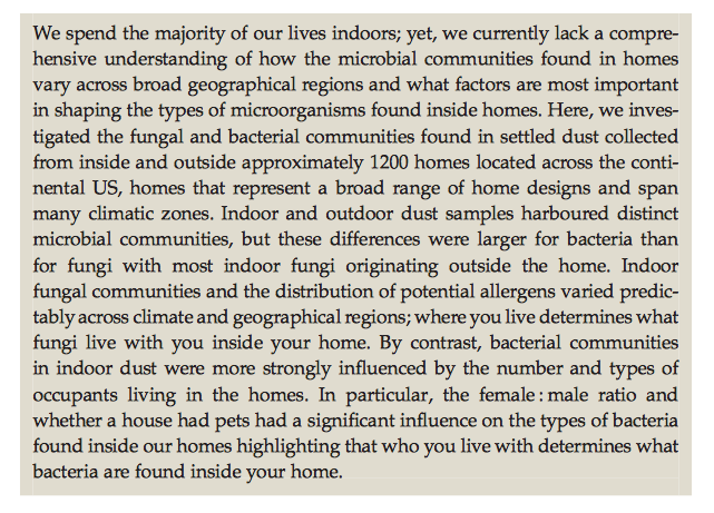
The Ecology of Microscopic Life in Household Dust

An interesting article about the fungi and bacterial in our households –has implications for our microbiome and for our predilection for allergies: full text: The Ecology of Microscopic Life in Household Dust

Barbera ́n A et al. The ecology of microscopic life in household dust. Proc. R. Soc. B 282: 20151139. http://dx.doi.org/10.1098/rspb.2015.1139 (reference from KT Park’s twitter feed)

Screen Shot 2015-11-22 at 9.07.00 AM

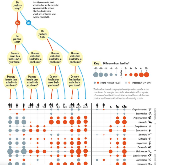
This article was summarized in by Mark Fischetti in Scientific American: full text: Men and Women Alter a Home’s Bacteria Differently

Screen Shot 2015-11-22 at 9.15.14 AM

“To biopsy or not to biopsy” –that is the question (for Celiac disease)

First off -thanks to Ben Gold for the following reference and the blog title as well.

  • CM Trovato et al. Am J Gastroenterol 2015; 110: 1485-89.

In this retrospective study (alluded to in a previous post:Celiac Update September 2015 | gutsandgrowth), the researchers examined whether “biopsy-sparing” protocols for symptomatic children with high titers of serum anti-transglutaminase (anti-TTG) antibody levels (>10 times upper limit of normal [ULN]) would be suitable for asymptomatic patients.

Background: In 2012, the European Society for Pediatric Gastroenterology Hepatology and Nutrition (ESPGHAN) published guidelines that it is possible to omit endoscopic biopsies for celiac disease if patients older than 2 years of age had high anti-TTG titers (>10 times ULN), positivity for EMA, compatible HLA DQ2 or HLA DQ8 and were symptomatic.

Findings:

  • Among 196 patients, the 40 who were asymptomatic had severe Marsh lesions (3a, 3b, or 3c) in 92% compared with 91% of 156 who were symptomatic. In both groups, the remaining patients had either Marsh 1 or 2 lesions.
  • 94.4% of patients had improved serology during followup along with symptomatic improvement (in those with symptoms)

Bottomline:  Whether symptomatic or not, those with high antiTTG titers who meet all of the other ESPGHAN criteria have a very high probability of celiac disease.

Briefly noted: K Marild et al. Am J Gastroenterol 2015; 110: 1475-84. This study, based on a large prospective Norwegian cohort (72,921 children) that frequent infections (>10) in the first 18 months of life increased the risk of celiac disease with an adjusted odds ratio of 1.32 (highest infection quartile compared to lowest infection quartile).  However, alternative explanations, including surveillance bias and reverse causation, cannot be excluded.

Related blog posts:


The Science Behind IBS Dietary Interventions

A succinct review (BE Lacy. Clin Gastroenterol Hepatol 2015; 13: 1899-1906) reviews the topic of dietary interventions for irritable bowel syndrome (IBS).

Here are some of the points:

  • “True food allergies are present in 1% to 4% of the US population, but are not more prevalent in IBS patients.”
  • One study found that “more than 1 in 4 patients with self-reported NCGS [nonceliac gluten sensitivity] actually fulfill the diagnosis.”  In other words, most patients with self-reported NCGS do not have NCGS.
  • “The prevalence of lactase deficiency is similar, or slightly higher, in IBS patients compared with healthy subjects; however, the self-reporting of symptoms attributed to lactose intolerance is not reliable.”
  • Potential mechanisms of food triggering GI symptoms were discussed, including intestinal permeability, visceral hypersensitivity, small intestine bacterial overgrowth, and gut microbiome.

Another article which covers the same topic: PR Gibson et al. Gastroenterol 2015; 148: 1158-74.


Which Diet is Best for Irritable Bowel Syndrome?

As noted in this blog previously, there has been increasing evidence that a low FODMAPs (fermentable oligo-, di-, monosaccharides and polyols) diet is an effective option for irritable bowel syndrome (IBS) in adults and children. Now, a study (L Bohn et al. Gastroenterol 2015; 149: 1399-1407) directly compares a low FODMAPs diet with an IBS diet in a multicenter, parallel, single-blind study of 75 patients (adults) with Rome III criteria for IBS.

The comparison IBS diet recommended regular meal patterns, avoidance of large meals, reduced intake of fat and reduced insoluble fibers, caffeine, and gas-producing foods, such as beans, cabbage and onions.  In addition, this diet recommended avoidance of spicy foods, coffee, alcohol, soft drinks, and sweeteners that end with “-ol.” This diet has been recommended by the British Dietetic Association and by the National Institute for Health and Care Excellence (NICE).  NICE Guidelines for IBS

Key findings:

  • 67 patients completed the study.  The severity of IBS symptoms improved in both groups (P<.0001) without a difference between the two groups
  • 19 (50%) of low FODMAPs had reductions in IBS severity scores of >50 compared with baseline and 17 patients (46%) in the ‘traditional’ IBS diet group had this degree of improvement.

My take: Diet changes often result in symptom improvement in IBS.  Both of these diets can be recommended in patients with IBS.

Atlanta Sky

Atlanta Sky

The Story Behind a 30 Year Esophagitis Study

A recent retrospective study ( SS Baker et al. JPGN 2015; 61: 538-40) reported on changes in esophagitis over a 30 year period at one center.  While the authors focus on the fluctuating percentage of esophagitis noted during three periods, in my opinion, they miss the opportunity to discuss more relevant findings.

Specifically, the authors note the following:

  • From 1980-88 (n=186 over 8 years) that 26.9% had esophagitis and 4.8% had >15 eos/hpf.  Normal pathology in the esophagus was noted in 73.1%.
  • From 2000-2002 (n=321 over 2 years), 41.2% had esophagitis and 8.5% had >15 eos/hpf.  Normal pathology in the esophagus was noted in 58.8%.
  • In the most recent period, 2011, (n=675 over 1 year), 31%* had esophagitis and 12.7% had >15 eos/hpf.  Normal pathology in the esophagus was noted in 69%.     *erroneously reported as 33%

What is baffling to me are the following:

  • Why the authors assert that there has been a fluctuating prevalence.  In absolute terms, the increase in cases is marked, though one can argue that in earlier periods there may have been many undiagnosed cases.
  • Why the authors do not comment on the tremendous increase in the use of endoscopy in their discussion.  In the first period, they were averaging ~23/year, the second period ~95/year and in the most recent period, they performed 675 in one year.

My take: This study shows that esophageal eosinophilia has been present for a long time and that identification of cases has increased considerably over 32 years.  In addition, the use of endoscopy has increased markedly, yet the yield of abnormal findings remains similar.

Briefly noted: C Menard-Katcher et al. JPGN 2015; 61: 541-46.  This retrospective study of 22 children showed that 55% had esophageal strictures identified by esophagram but not endoscopy.

Related blog posts:

Bamboo

Eosinophilic Esophagitis Review -NEJM

Good review:  Glenn T. Furuta, M.D., and David A. Katzka, M.D. N Engl J Med 2015; 373:1640-1648

A couple pointers from this review:

  • Estimated prevalence of eosinophilic esophagitis (EoE) 0.4% in Western countries.  Symptoms are often underestimated due to patient ‘accommodation’ which includes eating slowly/carefully, drinking a lot of liquids and avoiding items more prone to become lodged (meats, pills, breads)
  • Pathogenesis: “Birth by cesarean section, premature delivery, antibiotic exposure during infancy, food allergy, lack of breast-feeding, and living in an area of lower population density have all been associated with eosinophilic esophagitis.”
  • Impaired barrier function and enhanced the activity play a role in pathogenesis
  • Food allergy is a non-IgE-mediated process.  Omalizumab, an anti-IgE biologic, is ineffective in EoE and EoE can develop in IgE-null mice
  • Male predominance (3:1) suggests that there is a genetic component.
Esophagus with ringed appearance, furrowing, and loss of vascular markings

Esophagus with ringed appearance, furrowing, and loss of vascular markings

Another useful reference on Eosinophilic Gastritis in Children: Am J Gastroenterol 2014; 109; 1277-85.  This article provides data on clinical and histologic remission with eosinophilic gastritis (>70 eos/hpf), n=30 children.  “Response to dietary restriction was high” (82% clinical, 78% histologic response) Thanks to Seth Marcus for this reference.

Related blog posts: