Prospective Outcome Data for Infants with Gastroschisis

A recent study (BS Fullerton et al. J Pediatr 2017; 188: 192-7) reports outcome data from 4420 neonates with gastroschisis from 175 North Amercan centers.

This study, using prospectively-collected data from the Vermont Oxford Network, was restricted to infants with birth weights >1500 g and gestational age >28 weeks.

Key findings from this cohort:

  • Survival was 97.8%
  • Length of stay (LOS) 37 day median
  • Sepsis, confirmed with either positive blood culture or CSF culture, “was the only independent predictor of mortality.”
  • In addition to gastroschisis repair, abdominal surgery was needed in 22.3%
  • At discharge, 57.0% were <10% weight for age; whereas, only 37.2% were born small for gestational age.
  • There were no outcome differences based on mode of delivery (eg. vaginal vs. cesarean)
  • Other congenital anomalies were noted in many infants, with 5.8% had an intestinal atresia: 4.6% jejunal/ileal, 1.9% colonic, 0.4% duodenal atresia.

My take: This contemporary study shows excellent survival of neonates with gastroschisis.  Sepsis, need for additional surgery, and poor growth remain important challenges.

Related blog posts:

Patient with FeedingTubeAwareness.org Tshirt

Expert 2017 Opinion: Miralax is (Still) First Choice Laxative for Children

IJN Koppen et al. Journal of Pediatric Gastroenterology & Nutrition: October 2017 – Volume 65 – Issue 4 – p 361–363

Abstract:

 According to international guidelines, polyethylene glycol (PEG) is the laxative of first choice in the treatment of functional constipation in children, both for disimpaction and for maintenance treatment. PEG acts as an osmotic laxative and its efficacy is dose dependent. PEG is highly effective, has a good safety profile, and is well tolerated by children. Only minor adverse events have been reported. Overall the use of PEG in children has been reported to be safe, although in patients predisposed to water and electrolyte imbalances monitoring of serum electrolytes should be considered.

Because this topic is of great importance to the families that are seen by pediatric gastroenterologists (and pediatricians), I wanted to review this brief article which describes the efficacy and safety of polyethylene glycol (aka miralax).

Key Points:

  • Polyethylene glycol (PEG) is the most widely used laxative in children and adults
  • It works by interacting “with water molecules by forming hydrogen bonds, in a ratio of 100 water molecules per 1 PEG molecule, which leads to an additional increase in colonic water content.” It is minimally absorbed.
  • Studies have demonstrated that PEG is better or noninferior to all of the following: lactulose, milk of magnesia, mineral oil, and flixweed (a medicinal herb)

Safety:

  • Only minor adverse events have been reported in studies.  In randomized, placebo-controlled trials, adverse events “did not occur more frequently in patients receiving PEG compared to patients receiving placebo.”
  • The main safety issue has been when it has been administered via nasogastric administration; improper placement can lead to severe pulmonary complications.  In addition, PEG should be used “cautiously in children with swallowing problems…because of risk of aspiration.”

Combatting Myths: 

  • The authors assert that there has never been reports of physical or psychological dependence.  Weaning from PEG is to prevent relapse of constipation.
  • There is no evidence to support loss of efficacy.
  • The phenomenon of “lazy bowel syndrome” in which there is worsened colonic function has not been described due to PEG; though, patients with underlying motility problems have had these problems misattributed to PEG use.
  • Despite anecdotal reports of tremors, tics, and obsessive-compulsive behavior in children taking PEG, there has been no evidence of a causal relationship.  “These events …are still under investigation, but the FDA has decided that no action is necessary.”  The authors note that the co-occurrence of neuro-behavioral problems and constipation is well-recognized in children with and without laxative use.

Clinical Pearl: Stimulant Laxatives After Repaired Anorectal Malformations:

  • “In children with constipation after repaired anorectal malformations, …stimulant laxatives (eg. senna) should be the laxative of choice.” (J Pediatr Surg 2017; 52: 84-8)

My take (borrowed from the authors): “PEG has rapidly become the treatment of first choice for children with functional constipation.”

Related blog posts:


How Many Kids with Reflux Actually Have Reflux?

A terrific recent retrospective study (LB Mahoney, S Nurko, R Rosen. J Pediatr 2017; 189: 86-91) examined how often children with reflux symptoms actually have reflux.

This study reviewed 45 children ≥5 years (mean age 11.8 years) who had undergone both upper endoscopy and impedance pH study (off PPI therapy). Inclusion criteria: no erosive esophagitis. Common symptoms included heartburn, abdominal pain, chest pain, and regurgitation.

Definitions:

  • Nonerosive reflux disease –had abnormal esophageal acid exposure
  • Reflux hypersensitivity -had normal acid exposure but had a positive symptom association to acid or nonacid reflux
  • Functional heartburn -had normal acid exposure and negative symptom association

Key findings:

  • 44% had functional heartburn, 29% with reflux hypersensitivity (27% acid, 2% nonacid), 27% had nonerosive reflux disease (NERD)
  • Response to a proton pump inhibitor (PPI) was not predictive of reflux phenotype: 58% with NERD, 67% with reflux hypersensitivity, and 55% with functional heartburn. Response to PPI was stated as “at least some symptomatic improvement with PPI use.”  There was not a difference in PPI response among those who received a dose <1 mg/kg and those ≥1 mg/kg.
  • Microscopic esophagitis was present in 17% in NERD, 25% with reflux hypersensitivity, and in 20% of functional heartburn

While this study has limitations, including referral bias, it is likely that these patients are typical for many pediatric gastroenterologists. The authors note that typical patients were “patients who underwent a PPI trial but continued to have persistent symptoms.”

My take: In a pediatric gastroenterology setting, the most common reason for “reflux” is actually functional heartburn.  Thus, in those with persistent symptoms, evaluation with endoscopy and pH probe is worthwhile, especially as there has been more attention to potential risks of PPI therapy.

Related blog posts:

View from Asheville, NC

Diet and Stress in Pediatric Eosinophilic Esophagitis

When it comes to eosinophilic esophagitis (EoE), I sometimes worry that some treatments are worse than the disease, depending on the severity of the EoE. A recent study (C Case et al. JPGN 2017; 65: 281-84) indicates that dietary therapy is often stressful for families.

This study examined children ages 2-18 during an annual American Partnership for Eosinophilic Diseases (APFED.org) patient education conference. What I found most interesting was Table 3. “Stress associated with eosinophilc esophagitis.”

Some of the data:

  • In response to ‘how stressful do you find the following since your child’s diagnosis of EoE?’ Family structure at mealtimes: Not stressful 13.5%, somewhat stressful 21.6%, moderate stressful 16.2%, significant stressful 32.4%, and severe stressful 16.2%
  • In response to ‘how stressful do you find the following since your child’s diagnosis of EoE? Buying and cooking separate foods/meals for your child: Not stressful 2.6%, somewhat stressful 21.1%, moderate stressful 21.1%, significant stressful 31.6%, and severe stressful 23.7%
  • In response to ‘how stressful do you find the following since your child’s diagnosis of EoE? Financial strain due to cost of food: Not stressful 10.5%, somewhat stressful 21.1%, moderate stressful 18.4%, significant stressful 23.7%, and severe stressful 36.3%
  • What is your current stress level in response to your child’s EoE? Not stressful 2.6%, somewhat stressful 15.8%, moderate stressful 36.8%, significant stressful 42.1%, and severe stressful 2.6%
  • 62% of respondents indicated that child’s EoE has affected marital relationship.

In addition, the study documented that “half of youth were affected by worry, anger, and sadness related to specialized diets.”  As this study relied on participants at an APFED meeting, this could skew the EoE population to be more severely affected.

My take: This study shows the emotional burden that dietary treatment of EoE places on families.

Related blog posts:

Berry College

Top Anti-TNF for Ulcerative Colitis

A recent retrospective cohort study (S Singh et al. Clin Gastroenterol Hepatol 2017; 15: 1218-25) compared infliximab and adalimumab in a nationwide Danish cohort of adults with ulcerative colitis (UC) from 2005-2014. The authors used propensity score and selected 171 patients who received infliximab (IFX) from a total of 1580 and 104 patients who received adalimumab (ADA) among a total of 139.

Key findings:

  • Patients who received ADA had higher hospitalization rates (HR 1.84) and a trend toward higher UC-related hospitalization (HR 1.71, CI 0.95-3.07) compared to IFX
  • Risk of abdominal surgery was not significantly higher in ADA patients (HR 1.35) compared to IFX
  • Serious infections were higher in ADA group, HR of 5.11 of needing hospitalization due to infections

There have been no randomized clinical trials  to determine if a specific anti-TNF agent is superior to another. In an associated editorial (MT Osterman, GR Lichtenstein, 1197-99), the authors note that while we don’t know which agent is superior, by comparing similar trials (ACT 1 & 2 for IFX and ULTRA 1 & 2 for ADA), “raw week 8 induction rates of clinical remission, clinical response, and mucosal healing are approximately 16%, 18%, and 19%, respectively, higher for infliximab”..”less dramatic differences favoring infliximab (approximately 9%, 13%, and 15%, respectively) are seen during maintenance at 1 year.”

My take: Due to the lack of randomized head-to-head studies, we do not know with certainty which anti-TNF is best for UC.  However, the data we have from retrospective cohort studies and from using raw data from prospective studies suggests that infliximab is effective in more patients.

Related blog posts:

University of Virginia Rotunda Pctures


Disclaimer: These blog posts are for educational purposes only. Specific dosing of medications (along with potential adverse effects) should be confirmed by prescribing physician.  This content is not a substitute for medical advice, diagnosis or treatment provided by a qualified healthcare provider. Always seek the advice of your physician or other qualified health provider with any questions you may have regarding a condition.

Briefly: Notable Recent IBD Publications

Vermeire S et al. Lancet 2017; 389: 266-75.  The “FITZROY ” study examined clinical remission in patients with moderate-to-severe Crohn’s disease treated with filgotinib, a orally administered selective JAK inhibitor.  This agent is 30 times more selective fo rJAK1 over JAK3. This study enrolled 174 patients in a phase II study. Key findings:

  • Among patients naive to anti-TNF agents, clinical remission (based on CDAI <150 at week 10) noted in 47% of filgotinib-treated compared with 23% of placebo group (P=.0077)
  • Among patients naive to anti-TNF agents, clinical response was noted in 67% of filgotinib-treated compared with 44% in the placebo group.

H Singh et al. Gastroenterol 2017; 153: 430-8.  Using the large Manitoba Epidemiology Database with 1.3 million population (2005-2014), the authors found that individuals with IBD had a 4.8 fold increase risk of Clostridium difficile infection.

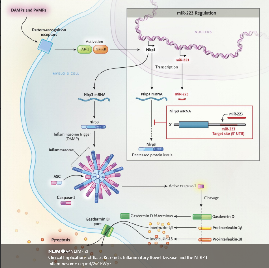
T-D Kanneganti. NEJM 2017; 377: 694-6. This review examined the NLRP3 Inflammasome.  Neudecker et al (J Exp Med 2017; 214: 1737-52) identified microRNA miR-223 which functions “to suppress the Nlrp3 inflammasone during acute colitis.” Other useful points in this review of basic research:

  • “The majority of the immune cells in the body are located in the intestine, where they are spatially separated from more than 10 trillion microorganisms by a layer of mucus and a layer of epithelial cells.  Deterioration of this physical barrier …underlies inflammatory bowel disease.”
  • miR-223 is increased in the inflamed colon. “During inflammation, the expression of miR-223 is also upregulated..and the molecule binds to its complementary sequence in a regulatory part of Nlrp3 mRNA…lead[ing] to decreased Nlrp3 expression and the consequent dampening of interleukin-1β maturation and associated inflammation.”

Intestinal Failure -Concise Review

A recent review (CP Duggan, T Jaksic. NEJM 2017; 377: 666-75) concisely reviews recent advances in pediatric intestinal failure.  Most of the review has been covered elsewhere in this blog.  A couple of key points:

Outcomes of intestinal failure:

  • The authors note that a 2012 study identified a 25% mortality rate of infants enrolled between 2000-2004.  “More recent advances have resulted in substantially improved survival rates (>90%).”

Epidemiology of intestinal failure:

  • Using a definition of needing parenteral nutrition for more than 42 days after bowel resection or a residual small-bowel length of less than 25% of normal (for gestational age), intestinal failure was identified in 24.5 cases per 100,000 live births
  • Among infants with birth weight <1500 g, the incidence is 7 per 1000 live births.
  • Frequent causes: necrotizing enterocolitis, gastroschisis

Adaptation of Intestine:

  • Improved chances of attaining enteral autonomy if longer residual small bowel, younger age at time of intestinal resection, preservation of ileocecal valve, absence of severe liver disease, diagnosis of necrotizing enterocolitis, and normal motility.

Parenteral nutrition:

  • Lower rates of liver disease noted with routine restriction of soy-based fat emulsions to 1 g per kilogram
  • Fish oil preparations (with n-3 fatty acids): switching to fish oil preparation “reduces biochemical measures of cholestasis.”
  • Newer preparations of fat emulsions: Smoflipid, Clinolipid are FDA-approved for adults.  Smoflipid, in small studies, is associated with lower conjugated bilirubin compared with soy-based lipids.

Enteral nutrition:

  • “Prompt initiation of enteral feeding after bowel resection has been reported to improve the rate of enteral autonomy….little justification for prolonged ‘gut rest'”
  • The authors note that human milk is often chosen for enteral nutrition and when unavailable, amino acid based formulas are typically chosen due to “more favorable outcomes than protein hydrolysates.”
  • Chronic diarrhea is improved with drip feedings, though bolus feeds may have trophic effects.  “In our experience, a combined approach (e.g. continuous feeding at night and bolus feeding during the day) is feasible.”
  • Oral motor stimulation is important.  Thus, try to give oral human milk feeds when feasible.

Medical Therapies:

  • Acid blockers: used for hyperacidity after massive resection
  • Loperamide
  • Bile acid sequestrants (eg. cholestyramine)
  • NOT evaluated in intestinal failure: octreotide, racecadotril, crofelemer
  • Motility agents
  • Antibiotics for bacterial overgrowth.  “Cyclical use (1 week per month) of broad-spectrum antibiotics…is the mainstay of therapy…at many centers.”
  • Probiotics: “No evidence of benefit in small studies; risk of sepsis”
  • Pancreatic enzymes: rarely used. Indicated if pancreatic atrophy or exocrine insufficiency
  • Growth factors: Teduglutide -licensed for adults, studies in children are ongoing

Surgical Therapies:

  • Central lines
  • Gastrostomy Tubes
  • STEP procedure or possibly lengthening procedure (Bianchi).  STEP procedure is less technically difficult.

Previous related blog entries:

The Non-Biopsy Diagnosis of Pediatric Celiac Disease

In some corners, experts have suggested the need for a followup intestinal biopsy to assure that celiac disease is responding to a gluten-free diet (Related blog post: Are followup biopsies necessary in celiac disease? Look beyond the headlines).  Meanwhile, many are looking at performing zero biopsies –at diagnosis or later. A recent study (J Wolf et al. Gastroenterol 2017; 153: 410-9) involved a prospective enrollment of 898 children undergoing duodenal biopsy to confirm or rule out celiac disease (CD).  Patients had tissue transglutaminase IgA (TTG IgA), total IgA and deamidated gliadin IgG (DGL-IgG) measured.

Key findings:

  • 592 had CD and 345 did not have CD.  24 did not have a final diagnosis.
  • In examining non-IgA deficient patients that had either TTG IgA >10 times ULN or normal (<1 times ULN), the positive predictive value for CD was 0.988 and the negative predictive value was 0.934.
  • In examining patients with both TTG IgA elevation (>10 times ULN) and DGP-IgG (>10 times ULN) or normal labs, the positive predictive value (PPV) for CD was also 0.988 and the negative predictive value (NPV) was 0.958.  The authors estimated that the PPV and NPV would remain >0.95 even at disease prevalence rates as low as 4%.
  • In this high prevalence population, the authors note that only 23% would have required an endoscopy to confirm or exclude CD; however, they note that in another study using consecutive serologic data, a much higher proportion (57%) needed biopsies due to serology that was <10 times ULN.
  • The authors note that HLA status genotyping, which has been recommended as needed in non-biopsy diagnosis, is not helpful.  Compatible HLA status was noted in all 277 cases of TTG-IgA >10 times ULN and was deemed “unnecessary” by the study authors in those with high titers.

The ESPGHAN guidelines for non-biopsy diagnosis indicate that a repeat serologic study should be performed to exclude a sample mix-up and to only forego biopsies in symptomatic patients.

My take: This study show\ed that individuals with high celiac serology titers have celiac disease >98% of the time.  This information should be discussed with families in determining whether endoscopic biopsy is needed.  Among those who pursue a non-biopsy approach, some individuals could have competing etiologies for their symptoms; thus, a low threshold for evaluation is needed in those who do not respond to a gluten free diet

Related blog posts:

Disclaimer: These blog posts are for educational purposes only. Specific dosing of medications (along with potential adverse effects) should be confirmed by prescribing physician.  This content is not a substitute for medical advice, diagnosis or treatment provided by a qualified healthcare provider. Always seek the advice of your physician or other qualified health provider with any questions you may have regarding a condition.

Rotunda View at UVa


Followup Biopsies in Pediatric Celiac Disease?

Last December, this blog site discussed a widely-reported, provocative study suggesting that “1 in 5 pediatric celiac disease patients on gluten-free diet sustain persistent intestinal damage” (Are followup biopsies necessary for Celiac disease? Look beyond the headlines).

Apparently a great number of celiac disease (CD) experts took exception to the authors’ conclusion that “neither the presence of symptoms nor positive serology were predictive of a patient’s histology at the time of repeat biopsy. These findings suggest a revisitation of monitoring and management criteria of celiac disease in childhood.” 

A recent commentary (S Koletzko et al. JPGN 2017; 65: 267-9) from the CD working group of ESPGHAN critiques the limitations of the study. Limitations included the following:

  • Selection bias -70% of patients in this retrospective study had repeat endoscopy were symptomatic
  • Retrospective study
  • Including children with admitted non-adherence to gluten-free diet (GFD)
  • Including ~25% of children who were rebiopsied within 17 months of starting GFD
  • Lack of standardized tTG testing
  • Lack of blinding of pathologist

Their conclusions:

  • “We do not think that the data..give sufficient support…that routine biopsies should be performed in all children at diagnosis and after the initiation of GFD.”
  • “The CD working group of ESPGHAN strongly advises against regular rebiopsy in children on a GFD…Re-endoscopy should be reserved for symptomatic patients–in particular when seronegative.”

The authors of the initial study (MM Leonard, A Fasano. JPGN 2017; 65: 270-1) were given an opportunity to defend their conclusions. Their commentary was much less provocative in my view, and titled: “Zero, One, or Two Endoscopies to Diagnosis and Monitor Pediatric Celiac Disease? The Jury is Still Out”

They note that there is a lack of data to know whether there are “no real clinical consequences” of persistent enteropathy, as stated by S Koletzko et al.  They state that until further research is completed a “personalized approach to follow-up care is needed.”  It is encouraging that they have started a prospective study to address the limitations of their retrospective study.

My commentary:

  • For patients with CD who are strictly-adherent, asymptomatic and with normal serology, repeat endoscopy is of questionable benefit.  If there are abnormalities in the histology, what is the appropriate intervention?
  • There has been a study (Gastroenterology 2010; 139: 763) which showed that mortality was NOT worsened in undiagnosed CD (identified by review of serology) in Olmstead County. In this population, the main long-term detrimental effect was reduced bone density. My inference is that for CD patients who are asymptomatic, particularly those with normalized serology, they are unlikely to have easily-identifiable adverse effects noted, even if their histology is abnormal.

My take (unchanged from last year): I think it is premature to recommend routine followup biopsies in asymptomatic patients with normal serology.

Prague Castle -clever door knocker