Is a High Protein Diet Beneficial and Safe?

There has been a lot of hype about the benefits of a high protein diet. In a recent substack article (8/31/25) , Eric Topol reviews the data on this (for adults).

Here’s the link:Our Preoccupation With Protein Intake

Key points:

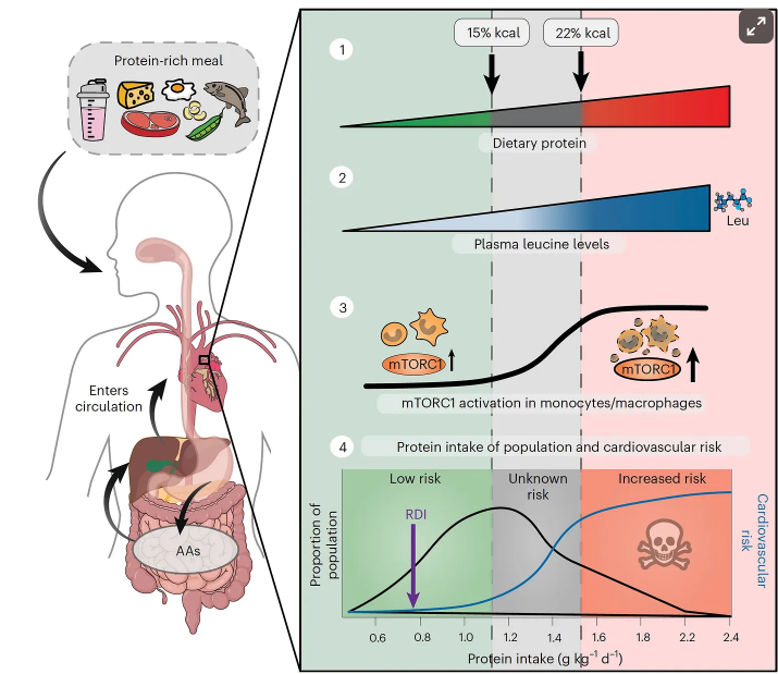
  • “The pervasive call for higher protein intake stems from the assertion that people are not getting adequate amounts in their diet, namely the 0.8 g/kg/day recommend by the National Academy of Medicine and the World Health Organization….
  • Regarding the need to increase protein intake 2-3 fold per day, Stuart Phillips, a leading expert on protein, energy, and building muscle mass, who is a professor at McMaster University in Canada, said “It’s baloney. But there’s a generation, particularly young men, and now an increasing number of young women, who are absolutely brainwashed by what they hear online”…there are no data to support more than 1.6 g/kg/day of protein intake.
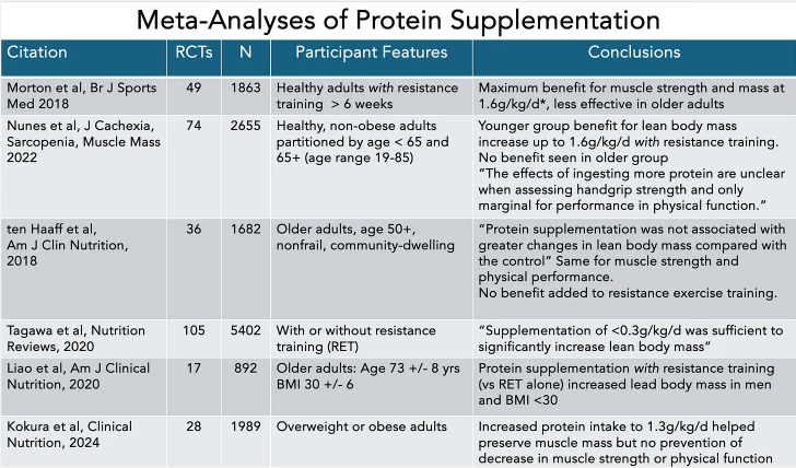
Safety concerns:

  • “There are many observational studies that have raised the safety concerns for high-protein intake, particularly derived from animal protein, for increased risk of type 2 diabetes, cardiovascular disease, and higher all-cause mortality. A prospective study of ~44,000 women in Sweden followed for 15.7 years found an association of high-protein diet with heightened cardiovascular risk.”
  • A “high protein intake is dangerous for people with kidney disease, present in 1 of 7 adults, but 9 of 10 people with reduced kidney function are unaware of it.”

My take (borrowed from Dr. Topol): “The body of evidence about protein does not provide support [for] very high protein intake, certainly not in excess of 1.6 g/kg/day…there is no way to store protein in the body…Resistance training is the principal driver for building muscle mass and strength, not high protein intake.” While this article focuses on adults, the premise is similar in children; though, on a per kilogram basis, children need modestly higher amounts. (Reference: JL Hudson et al. Nutrients. 2021 May 5;13(5):1554. Dietary Protein Requirements in Children: Methods for Consideration)

Related blog posts:

How Often Esophageal Coins Pass Into the Stomach

P Quitadomo et al. Am J Gastroenterol 2025; 120: 1388-1390. “Insert-Coin”: A Prospective Study of Coin Ingestion in Children of Southern Italy

Thanks to Ben Gold for this reference.

This prospective study from Naples, Italy examined children 0-14 yrs of age with a coin ingestion (n=807). Children with coins in the proximal esophagus underwent endoscopic removal within 4 hours whereas those with middle to lower esophageal coins had re-evaluation after 12 hours before removal.

Key findings:

  • 52 of 807 (6.4%) had a coin retained in the esophagus, the remainder were in the stomach or beyond
  • 20 of 52 (38%) were located in the middle to lower esophagus (10 in each)
  • 13 of 20 (65%) coins in the middle to lower esophagus had spontaneous gastric passage
  • The mean age of patients with gastric passage (72 months) was higher than those without passage (48 months)

My take: Only 6% of patients in this study who had a coin ingestion had esophageal retention of the coin. In addition, one-fourth of those with esophageal coins had spontaneous passage into the stomach. This occurred only with the mid-distal esophageal coins; in this subset it occurred in 65%. Thus, in those with mid-distal esophageal coins, watchful waiting for ~12 hrs may be beneficial for patients. The ultimate primary prevention of this problem may occur with more widespread adoption of electronic payments.

Related blog posts:

Agnes Northrup of Louis Tiffany Studio,
Tiffany Garden Landscape Window (1912)
at The Metropolitan Museum of Art
Agnes Northrup of Louis Tiffany Studio,
Autumn Landscape  (1923) at The Metropolitan Museum of Art

“Are Marathons and Extreme Running Linked to Colon Cancer?”

NY Times 8/19/25: Are Marathons and Extreme Running Linked to Colon Cancer?

An excerpt:

A small, preliminary study found that marathoners were much more likely to have precancerous growths. Experts aren’t sure why…

Dr. Cannon, an oncologist with Inova Schar Cancer in Fairfax, Va., launched a study, recruiting 100 marathon and ultramarathon runners aged 35 to 50 to undergo a colonoscopy.

The results were staggering. Almost half the participants had polyps, and 15 percent had advanced adenomas likely to become cancerous. The rate of advanced adenomas was much higher than that seen among adults in their late 40s in the general population, which ranges from 4.5 percent to 6 percent, according to recent studies.

The research was presented at an American Society of Clinical Oncology conference but has not yet been published in a medical journal…

Dr. David Rubin, chief of gastroenterology and director of the Inflammatory Bowel Disease Center at the University of Chicago, said the study was important but limited. It lacked a control arm consisting of similar young adults who were not long-distance runners, he noted, and the family histories of colon cancer among the marathoners were not entirely known…

Runners often develop gastrointestinal symptoms that they dismiss as benign — so-called runner’s trots, for example. The symptoms can be caused by ischemic colitis, a condition that develops when blood flow to the colon is temporarily reduced as it is redirected to muscles in other parts of body (like a runner’s legs).

My take: While this is a small study, it indicates that extreme runners could have an increased risk of colonic polyps and cancer. If there are symptoms (especially rectal bleeding and weight loss), a low threshold for further evaluation is needed.

Related blog posts:

View of Statue of Liberty from Governor’s Island

IBD Management in Pregnancy: Global Consensus

U Mahadevan et al. Clinical Gastroenterology and Hepatology 2025 (published ahead of print). Open Access! Global Consensus Statement on the Management of Pregnancy in Inflammatory Bowel Disease

Addendum -updated reference: U Mahadevan et al. Clinical Gastroenterology and Hepatology 2025; 23: S1-S60. Open Access! Global Consensus Statement on the Management of Pregnancy in Inflammatory Bowel Disease

This is a 60 page open access article. Table 1 lists 34 “GRADE” statements and Table 2 lists 35 consensus statements. This article is also jointly published in the following:

  • Gut
  • Am J Gastroenterol
  • Inflammatory Bowel Diseases
  • Journal of Crohn’s and Colitis
  • Aliment Pharmacol Ther

For Moms:

For Babies:

My take: This is a useful reference –mainly helpful for gastroenterologists rather than pediatric providers.

Related blog posts:

The Best Way to Judge Pediatric Poo

J Orozco et al. Am J Gastroenterol 2025; 120: 1381-1387. Comparison of the Bristol Stool Scale and Modified Version for Children: Use by Providers vs Children

Thanks to Ben Gold for this reference.

Background: The modified Bristol Stool Form Scale for Children (mBSFS-C) removes #3 and #5 from the Bristol Stool Form Scale (BSFS), leaving only one normal image and shortening the options from seven to five.

Methods: Pediatric gastroenterology providers  (21 faculty, 11 fellows, 3 nurse practitioners)  and 200 children/families rated the same 35 stool photographs, reflecting diverse stool forms, using both scales. The order of photograph presentation and scale use were randomized.

Modified Bristol Stool Scale
Bristol Stool Chart

Key findings:

  •  Of 1,225 provider ratings using the mBSFS-C, 90.0% agreed with the provider’s modal ratings vs 77.8% using the BSFS.
  • Of 7,000 child ratings using the mBSFS-C, 84.6% agreed with the children’s modal ratings vs 71.8% using the BSFS.
  • Using providers’ modal ratings as the reference, all mBSFS-C photograph modal ratings matched between children and providers (35/35 photographs) whereas only 86% (30/35 photographs) matched with the BSFS.

Discussion:

  • “Unique and new in this study is the direct head-to-head comparison of the 2 scales (BSFS, mBSFS-C) when used by pediatric gastroenterology providers and children. Both the BSFS and mBSFS-C demonstrated excellent reliability…modal rating agreement was significantly poorer for the BSFS than for the mBSFS-C.”
  • “Almost 20% of the time expert raters using the BSFS (vs. 8% with the mBSFS-C) deemed a stool to be a different clinical delineation than that selected by the majority of their peers.”

Related editorial: Peter Lu, The American Journal of Gastroenterology 120(6):p 1267, June 2025. Is It Time to Scale Down the Bristol?

My take: The modified BSFS is easier and better. This study indicates it should be widely used for children but probably for adults too. As Dr. Lu’s editorial notes, “aren’t adults just big children?”

Related blog posts:

“The Broken Promises of Profit-Driven Medicine”

N Tomes. NEJM 2025; 393: 521-524. A Gilded Age for Patients? The Broken Promises of Profit-Driven Medicine

This article (one of several in a series) describes the development of the current corporate-centered health care system over the past 100 years and the drawbacks.

An excerpt:

Between the 1920s and the 1960s, the American medical profession adopted a new doctor-controlled business model of care delivery, dependent on continual investment in new drugs, technologies, and procedures. That model created the profit opportunities that enticed corporate stakeholders to invest in health care in the 1970s and 1980s. But as the corporate presence increased, physicians lost control of their business model; the “tail” of financialization began wagging the “dog” of medical practice. That shift coincided with corporate cooptation of the language of consumerism to justify these changes as in patients’ best interests. In the process, physicians and patients lost economic autonomy over health care choices…

Insurance plans let doctors and hospitals set prices, facilitating more investment in the capital-intensive technologies equated with higher-quality care. But that dynamic also created an upward spiral of costs…These problems led to a second transformative movement: the federal government’s entry into the health insurance business, with the creation of Medicare and Medicaid, which also let doctors and hospitals set prices. This… aimed to relieve Americans of the duty of financing the medical system themselves, bringing in third parties to help. Enrollees in the new plans got more coverage but only on the terms set by those parties; uninsured people had to manage on their own. Meanwhile, the cycle of rising costs continued unabated…

Medical entrepreneurs believed that applying market discipline to health care would produce the right combination of innovation, efficiency, and cost–benefit balance to ensure better care for patients while profiting investors…

While adopting some consumer-industry practices, corporate health care players strove to avoid others, such as direct price competition. Policy efforts to rein in costs stressed the need for price transparency: consumer–patients needed to “shop” more critically for the cheapest care. In this spirit, political conservatives promoted “consumer-driven” health care. But such schemes foundered on the reality that many insurance plans gave patients little flexibility to price-shop for care…

Many powerful health care industry stakeholders still believe allowing corporate interests freer rein will produce that “golden age for patients.” The health care economy’s fragility suggests otherwise…When they [hospitals] pivoted from profitable surgeries to unprofitable Covid care, they needed massive infusions of government funding to survive.  Similarly, recent shortages of essential but low-profit drugs, including chemotherapy agents and insulin, reveal the limits of the pharmaceutical industry’s profit-oriented approach to essential drug production…

No market solution has arisen for the most critical determinant of poor health and health care outcomes in the United States: extreme income inequality. Countless studies indicate that poverty is the most important health risk factor that Americans face…little profit can be made by preventing or treating poverty-induced illnesses.

U.S. health care needs a new business model…only more collective physician and patient action will help medicine find a more equitable, sustainable model…Many people will suffer if the system collapses completely, but perhaps a more sustainable health care system can be built from the rubble.

My take: This analysis of the U.S. healthcare system, like many others, is analogous to getting the license plate of the truck that ran you over. At this time, there is little prospect of stopping the truck.

Related blog posts:

Stone Mountain, Ga from the Cherokee Trail

New Data on the Reliability of the No-Biopsy Diagnosis in Pediatric Celiac Disease

Chang D, Wong M, Camila Cardenas M, et al. Pediatrics. 2025;156(3):e2025070897. Positive Predictive Value of Tissue Transglutaminase IgA for Celiac Disease.

Background:For a no-biopsy celiac diagnosis, “the [ESPGHAN] criteria were revised in 2020, to specify that only a highly positive tTG IgA greater than or equal to 10× ULN and a positive EMA on a second blood sample are sufficient to diagnose celiac disease, obviating the need for HLA testing or symptoms.” NASPGHAN “has yet to adopt similar serologic criteria for diagnosing celiac disease. A clinical report in 2016 continued to recommend a confirmatory biopsy in all suspected cases of celiac disease. This recommendation cited concerns of interassay variability.”

Methods: This was a retrospective, multicenter North American cohort study of children (<18 years) with an elevated tTG IgA within 6 months of an esophagogastroduodenoscopy between January 2016 and December 2021. Biopsy-confirmed celiac disease was determined by the presence of intraepithelial lymphocytosis and villous atrophy. The primary outcomes were the PPV of an elevated tTG IgA and tTG IgA greater than or equal to 10 times the upper limit of normal (10× ULN).

The study identified 4019 children with the following characteristics: 63.3% female; 9% type 1 diabetes, and 2% Down syndrome

Key findings:

  • Histologic findings in the entire cohort were consistent with celiac disease for 3321 children (PPV = 82.6%)
  • Among the 1739/4019 (43.2%) children with tTG IgA greater than or equal
    to 10× ULN, 1651 had biopsy-confirmed celiac disease (PPV10× = 94.9%). Five percent (88/1739) of children did not have histologic findings of celiac disease, including 41/1739 (2%) with normal histology
  • EMA only marginally improved specificity as 76% of children without celiac disease and tTG IgA greater than or equal to 10× ULN had a positive EMA, albeit on the same sample. There was variations across providers, practices, and countries, which resulted in EMA not being performed in all cases or tested on the same blood sample as the tTG IgA
  • Assays performed worse in children with type 1 diabetes (PPV10× 89%) than that in those without T1DM (95.7%)
  • Of those with esophageal biopsies, 175/2980 (6%) had at least 15 eos/hpf in at least 1 esophageal biopsy
  • Of those with gastric biopsies, 912/3534 (25.8%) had histologic gastritis, including 1.4% (51/3534) with evidence of Helicobacter infection (n (49 cases of H pylori and 2 cases of H heilmannii)

My take: This study indicates that the no-biopsy diagnostic approach for celiac disease may need to be reconsidered. Though, it is likely that the no-biopsy approach would have had a higher PPV if the guidelines were actually followed (eg. separate EMA confirmatory sample). The values in this study show a much lower PPV than prior studies (96% in those without T1DM). In addition, among the 4% without celiac disease (potential celiac disease), about half of them would likely progress to celiac disease and could potentially benefit from a gluten free diet. Thus, up to 2%of patients, based on this study, would probably be harmed by being placed on a gluten free diet.

Another point to reiterate based on this study, diagnostic confirmation by a specialist prior to dietary changes is recommended. Increasingly, patients are referred after starting a gluten free diet.

Related blog posts:

Brooklyn Botanic Gardens

Can Smartphone Use Increase the Risk of Hemorrhoids?

There are many reasons NOT to use a phone while you are on the toilet. Now a small study indicates that smartphone use is associated with longer time on the toilet and increased rate of endoscopically-detected hemorrhoids.

An excerpt from the NBC summary:

“The longer you sit on the toilet, the worse it is for you,” said Dr. Trisha Pasricha, director of the Beth Israel Deaconess Medical Center’s Institute for Gut-Brain Research Institute in Boston. Pasricha is also an author of the study, which was published Wednesday in PLOS One

Pasricha and colleagues surveyed 125 adults just before they were about to have a routine colonoscopy to screen for colorectal cancer. Eighty-three (66%) of the participants admitted to using their phones in the bathroom — mostly to catch up on news of the day and scroll through social media.

Gastroenterologists performing the colonoscopies looked for evidence of inflamed veins, or hemorrhoids. People who said they took their phone into the bathroom were 46% more likely to have hemorrhoids compared to the others…

The experts agreed that business on the toilet should take no longer than 5 minutes. More than 37% of study participants who used a smartphone in the bathroom stayed for longer than that, compared to 7% of people who kept their phones out of the bathroom.

My take: This study has a number of limitations; so, a definite link between smartphones and hemorrhoids has not been established. For example, there could be reverse causation. In this case, individuals who expected to be on the toilet longer may be more likely to use their smartphones rather than the smartphones making them stay longer. Nevertheless, I think multitasking on the toilet is generally not a good idea.

Brooklyn Bridge, August 2025

Is Lactated Ringer’s Better Than Normal Saline For Routine Use?

L McIntyre et al. NEJM 2025; 393: 660-670. A Crossover Trial of Hospital-Wide Lactated Ringer’s Solution versus Normal Saline

Background: “A single-center, multiple-crossover trial involving noncritically ill patients in the emergency department found that balanced crystalloid fluids were associated with a lower incidence of major adverse kidney events at 30 days than normal saline. A systematic review of 13 randomized, controlled trials comparing balanced crystalloids with normal saline in a total of 35,884 critically ill participants showed no significant difference in mortality (17.4% with balanced crystalloids and 18.2% with saline; relative risk, 0.96; 95% confidence interval [CI], 0.91 to 1.01) or in the incidence of the use of renal replacement therapy (5.6% and 6.0%, respectively; relative risk, 0.95; 95% CI, 0.81 to 1.11) in trials with a low risk of bias.4 However, the investigators in that analysis and those in another patient-level meta-analysis involving a Bayesian approach concluded that there is a high probability that balanced crystalloids are associated with lower in-hospital mortality and a lower incidence of the use of renal replacement therapy than normal saline.5

Methods of th “FLUID” trial: 3 hospitals used lactated ringer’s (LR) and 4 hospitals used normal saline throughout hospital setting for 12 weeks. Then after a 1-2 week washout period, the hospitals switched to the other fluid for 12 weeks.

Key finding:

Discussion: “A limitation of this trial was the inability to recruit the total of 16 hospitals as originally planned owing to the Covid-19 pandemic. Hence, the trial had less power to detect differences that were small — but important to patients — at the level of the hospital or health care system…Our findings align with those of recent meta-analyses of randomized, controlled trials that suggest a small but clinically relevant reduction in mortality with balanced crystalloids as compared with normal saline.”

My take: This study did not show a significant difference in death or readmission at 90 days. Yet, lactated ringer’s is probably just a bit better fluid for most adult patients. In the pediatric population, more studies are needed.

Related blog posts:


Disclaimer
: This blog, gutsandgrowth, assumes no responsibility for any use or operation of any method, product, instruction, concept or idea contained in the material herein or for any injury or damage to persons or property (whether products liability, negligence or otherwise) resulting from such use or operation. These blog posts are for educational purposes only. Specific dosing of medications (along with potential adverse effects) should be confirmed by prescribing physician.  Because of rapid advances in the medical sciences, the gutsandgrowth blog cautions that independent verification should be made of diagnosis and drug dosages. The reader is solely responsible for the conduct of any suggested test or procedure.  This content is not a substitute for medical advice, diagnosis or treatment provided by a qualified healthcare provider. Always seek the advice of your physician or other qualified health provider with any questions you may have regarding a condition

Expert Advice for GI Manifestations of Hypermobile Ehlers-Danlos Syndrome Including Association with POTS and Mast Cell Activation Syndrome (MCAS)

Q Aziz et al. Clinical Gastroenterology and Hepatology, Volume 23, Issue 8, 1291 – 1302. Open Access! AGA Clinical Practice Update on GI Manifestations and Autonomic or Immune Dysfunction in Hypermobile Ehlers-Danlos Syndrome: Expert Review

This review and practice update includes 16 “best practice advice” statements. Here are nine of them:

  • #1: Clinicians should be aware of the observed associations between hEDS or HSDs and POTS and/or MCAS and their overlapping gastrointestinal (GI) manifestations; while theoretical explanations exist, experimental evidence of the biological mechanisms that explain relationships is limited and evolving.
  • #2: Testing for POTS/MCAS should be targeted to patients presenting with clinical manifestations of POTS/MCAS, but universal testing for POTS/MCAS in all patients with hEDS/HSDs is not supported by the current evidence.
  • #3: Gastroenterologists seeing patients with DGBI should inquire about joint hypermobility and strongly consider incorporating the Beighton score for assessing joint hypermobility into their practice as a screening tool; if the screen is positive, gastroenterologists may consider applying 2017 diagnostic criteria to diagnose hEDS (https://www.ehlers-danlos.com/wp-content/uploads/2017/05/hEDS-Dx-Criteria-checklist-1.pdf) or offer appropriate referral to a specialist where resources are available.
  • #4: Testing for POTS through postural vital signs (eg, symptomatic increase in heart rate of 30 beats/min [40 beats/min for 12-19 yo] or more with 10 minutes of standing during an active stand or head-up tilt table test in the absence of orthostasis) and referral to specialty practices (eg, cardiology or neurology) for autonomic testing should be considered in patients with hEDS/HSDs and refractory GI symptoms who also report orthostatic intolerance after exclusion of medication side effects and appropriate lifestyle or behavioral modifications (eg, adequate hydration and physical exercise) have been attempted but is not required for all patients with hEDS/HSDs who report GI symptoms alone.
  • #5: In patients presenting to gastroenterology providers, testing for mast cell disorders including MCAS should be considered in patients with hEDS/HSDs and DGBI who also present with episodic symptoms that suggest a more generalized mast cell disorder (eg, visceral and somatic pain, pruritus, flushing, sweating, urticaria, angioedema, wheezing, tachycardia, abdominal cramping, vomiting, nausea, diarrhea, urogynecological and neurological complaints) involving 2 or more physiological systems (eg, cutaneous, GI, cardiac, respiratory, and neuropsychiatric), but current data do not support the use of these tests for routine evaluation of GI symptoms in all patients with hEDS/HSDs without clinical or laboratory evidence of a primary or secondary mast cell disorder.
  • #6: If MCAS is suspected, diagnostic testing with serum tryptase levels collected at baseline and 1–4 hours following symptom flares may be considered by the gastroenterologist; increases of 20% above baseline plus 2 ng/mL are necessary to demonstrate evidence of mast cell activation.
  • #12: Medical management of GI symptoms in hEDS/HSDs and POTS/MCAS should focus on treating the most prominent GI symptoms and abnormal GI function test results. In addition to general DGBIs and GI motility disorder treatment, management should also include treating any symptoms attributable to POTS and/or MCAS.
  • #13: Treatment of POTS may include increasing fluid and salt intake, exercise training, and use of compression garments. Special pharmacological treatments for volume expansion, heart rate control, and vasoconstriction with integrated care from multiple specialties (eg, cardiology, neurology) should be considered in patients who do not respond to conservative lifestyle measures.
  • #14: When MCAS is suspected, patients can benefit from treatment with histamine receptor antagonists and/or mast cell stabilizers, in addition to avoiding triggers such as certain foods, alcohol, strong smells, temperature changes, mechanical stimuli (eg, friction), emotional distress (eg, pollen, mold), or specific medications (eg, opioids, nonsteroidal anti-inflammatory agents, iodinated contrast).

Background: “Clinical gastroenterologists are encountering an increasing number of patients with chronic GI symptoms who also appear to experience comorbid hEDS/HSDs, POTS, and/or MCAS.15,16 Recognizing and treating GI symptoms in patients with hEDS/HSDs and comorbid POTS or MCAS present major challenges for clinicians, who often feel under equipped to address their needs.”

The article provides guidance on measuring hypermobility (Beighton Scoring System), Diagnosis/classification of mast cell activation (Table 1) and treatments for these disorders (Table 2)

My take: This is a useful reference for the overlap of DGBIs with hypermobile Ehlers-Danlos, POTS and Mast cell Activation. Nevertheless, the relationship between these disorders is unclear. In fact, there have been some studies indicating that joint mobility is not associated with an increase in functional GI disorders. Some of the association may be related to a surveillance bias.

Related blog posts: