D Turner et al. JPGN 2024; 79:315–324. Ustekinumab in paediatric patients with moderately to severely active Crohn’s disease: UniStar study long-term extension results
Dosing: “Patients were randomised 1:1 and stratified by body weight (<40 or ≥40 kg) to receive a single induction dose of lower- or higher-dose IV ustekinumab (lower dose: 3 mg/kg [<40 kg] and 130 mg [≥40 kg]; higher dose: 9 mg/kg [<40 kg] and 390 mg [≥40 kg]). Doses specified as higher were selected to deliver ustekinumab exposure comparable to a reference adult population with CD.7, 12 At Week 8, patients received a single SC maintenance dose of ustekinumab (2 mg/kg [<40 kg]; 90 mg [≥40 kg]).”
Key findings:
Of the 34 patients who entered the LTE, 25 patients with evaluable data completed Week 48, and 41.2% (14/34) achieved clinical remission at Week 48
Efficacy and PK through 1 year in ustekinumab-treated paediatric patients were comparable to those previously reported in adults. No new safety or immunogenicity signals were reported through 4 years of ustekinumab treatment.
My take (borrowed in part from authors): “Overall, long-term data support the SC dose regimens of 90 mg as maintenance therapy for the treatment of CD for a paediatric population with ≥40 kg body weight. A phase 3 study of ustekinumab (ClinicalTrials.gov Identifier: NCT04673357) is ongoing to further evaluate dose regimens for paediatric patients <40 kg and ≥40 kg.” This type of data is essential to support the use of advanced therapies like ustekinumab until they receive specific regulatory approval for children (often 8-10 years after approval in adults).
KA Hommel et al. JPGN 2024; 78:1273–1278. Pilot and feasibility of the SMART IBD mobile app to improve self-management in pediatric inflammatory bowel disease
The Self‐Management Assistance with Recommended Treatment (SMART) IBD app –Key findings:
Patients rated the app quality as good and accessed the app adequately overall, with some pages being used often.
Medication adherence increased over the course of the study and was associated with sleep duration, mood, and stool consistency and blood content.
My take: IBD Management apps could be quite helpful, especially for teens and young adults.
S Hsiang et al. Inflammatory Bowel Diseases, Volume 30, Issue 8, August 2024, Pages 1284–1294, https://doi.org/10.1093/ibd/izad166. Safety, Effectiveness, and Treatment Persistence of Subcutaneous Vedolizumab in IBD: A Multicenter Study From the United Kingdom
Methods: IBD patients (n=563) on IV vedolizumab across 11 UK sites agreed to transition to SC injections or otherwise continued IV treatment
Key findings:
Data from 563 patients, demonstrated no differences in disease activity, remission rates, and quality of life between the SC and IV groups at all time points
Drug persistence at week 52 was similar (81.1% vs 81.2%; P = .98)
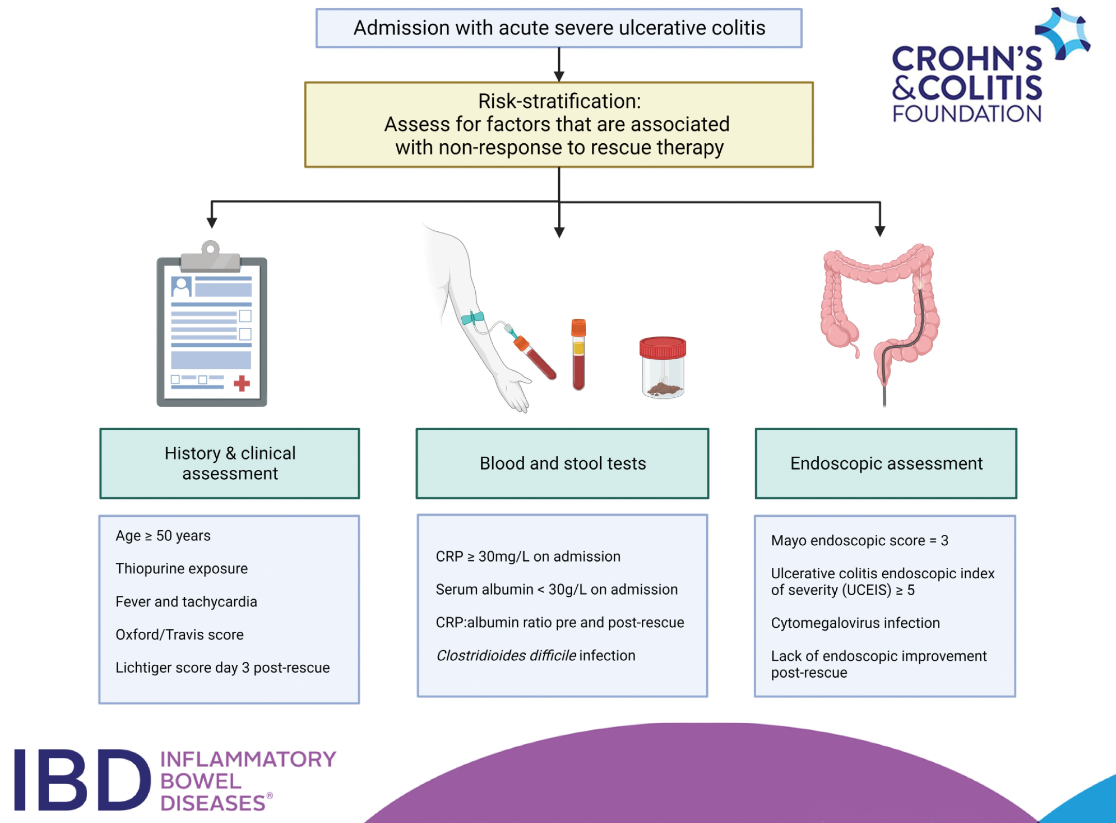
CFD Li Wai Suen, et al. Inflammatory Bowel Diseases, Volume 30, Issue 8, August 2024, Pages 1389–1405, https://doi.org/10.1093/ibd/izad183. Factors Associated With Response to Rescue Therapy in Acute Severe Ulcerative Colitis
This systematic review identified 101 completed studies were eligible for inclusion.
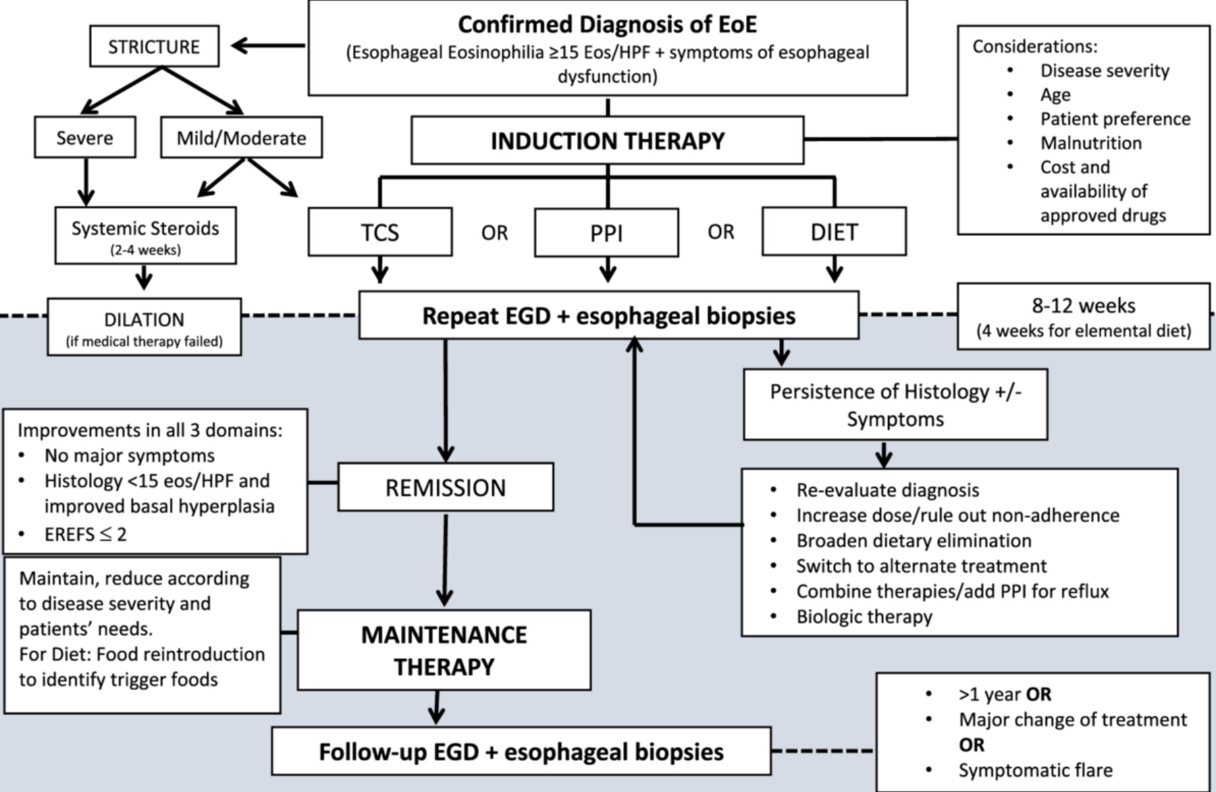
This report makes 52 statements and 44 recommendations. Overall this is a helpful report but there are many statements and recommendations that have NO value for pediatric gastroenterologists (except for those trapped in a cave for the last 10 years). Here are a couple examples of that:
“ESPGHAN EGID WG recommends that pediatricians should be aware of the higher incidence of EoE in relatives.”
“ESPGHAN EGID WG recommends that a high index of suspicion for EoE must be maintained in children with concomitant atopic disease.”
“ESPGHAN EGID WG recommends the peak value of 15 eos/HPF as the cut‐off value in esophageal biopsy specimens, for the histological diagnosis of EoE in an appropriate clinical context”
Some helpful recommendations:
“ESPGHAN EGID WG recommends against using available allergy tests to predict dietary triggers of EoE.” This is not new information but helpful to have clearly stated in guidelines.”
“ESPGHAN EGID WG recommends maintenance therapy to all patients after achieving histological remission” Commentary: “There are no prospective data on the best duration of maintenance therapy in pediatric EoE…[In a large study of adults} sustained untreated combined remission was seen in only 1.3% of patients who discontinued treatment.”
“ESPGHAN EGID WG suggests endoscopic and histological re‐evaluation after 1‐3 years during the maintenance phase in cases of stable clinical remission”
“ESPGHAN EGID WG recommends that dupilumab can be used in selected cases of children over1 year old weighing >15 kg with EoE refractory to conventional treatment and in those with concomitant atopic burden with approved indications for biologics”
“ESPGHAN EGID WG suggests that a short course of systemic steroids be considered as an alternative to dilation in the presence of moderate to severe esophageal strictures with severe symptoms.” “Treatment with short term systemic steroids can significantly reduce the need for mechanical esophageal dilation in moderate to severe strictures associated with pediatric EoE”
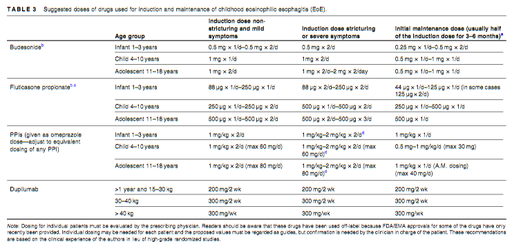
Suggested drug dosing is noted in Table 3 (see below)
Disclaimer: This blog, gutsandgrowth, assumes no responsibility for any use or operation of any method, product, instruction, concept or idea contained in the material herein or for any injury or damage to persons or property (whether products liability, negligence or otherwise) resulting from such use or operation. These blog posts are for educational purposes only. Specific dosing of medications (along with potential adverse effects) should be confirmed by prescribing physician. Because of rapid advances in the medical sciences, the gutsandgrowth blog cautions that independent verification should be made of diagnosis and drug dosages. The reader is solely responsible for the conduct of any suggested test or procedure. This content is not a substitute for medical advice, diagnosis or treatment provided by a qualified healthcare provider. Always seek the advice of your physician or other qualified health provider with any questions you may have regarding a condition.
I signed up for this webinar mainly to hear my partner Jose Garza’s presentation (who presented last), though all the speakers were good. I took a couple screenshots on my phone during the presentations. The webinar is available/archived at NASPGHAN website.
Dr. El-Chammas’ presentation gave a quick review on normal swallowing physiology, modalities for evaluation (eg. VSS, FEES) and showed some cool slides particularly with regard to pharyngeal manometry.
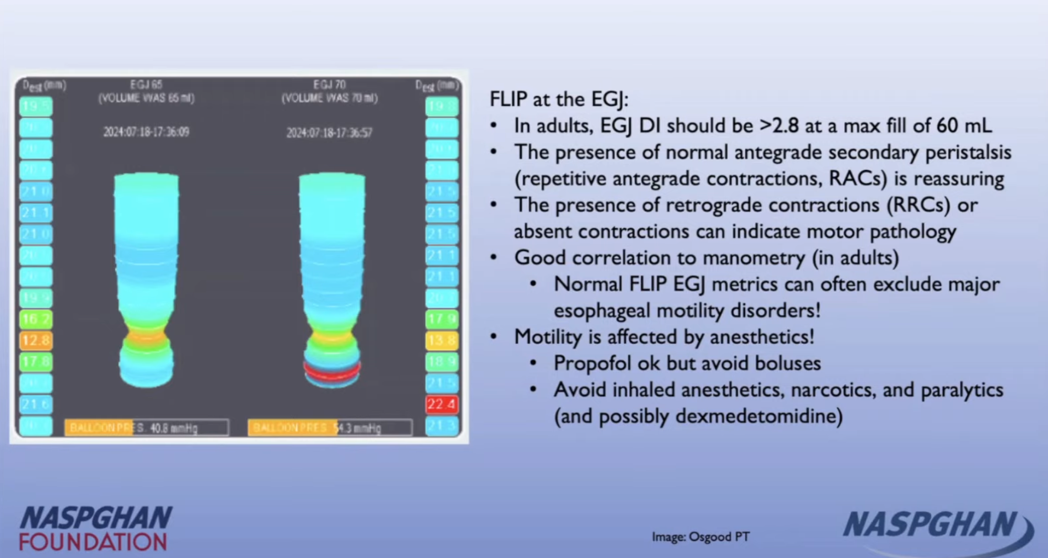
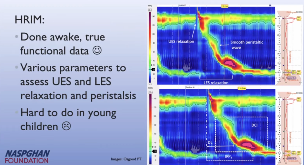
Dr. Osgood reviewed the etiologies/workup for dysphagia including helpful slides on esophagrams, FLIP and manometry.
Manometry typical of Type 2 Achalasia
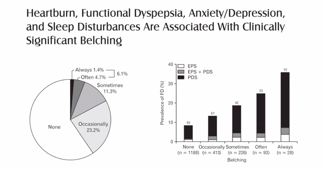
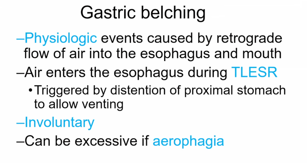
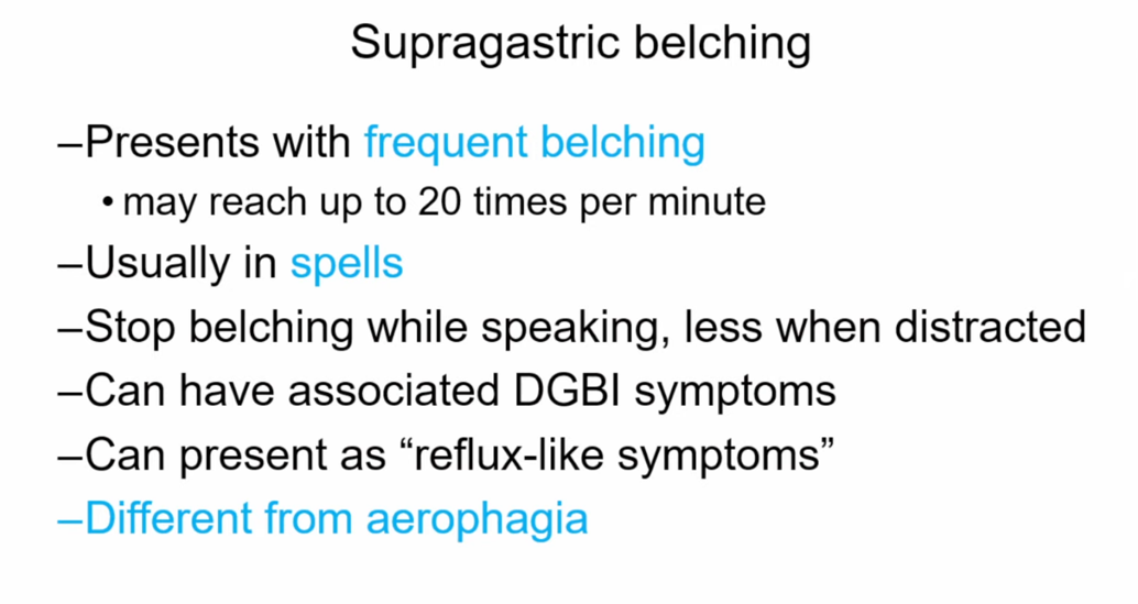
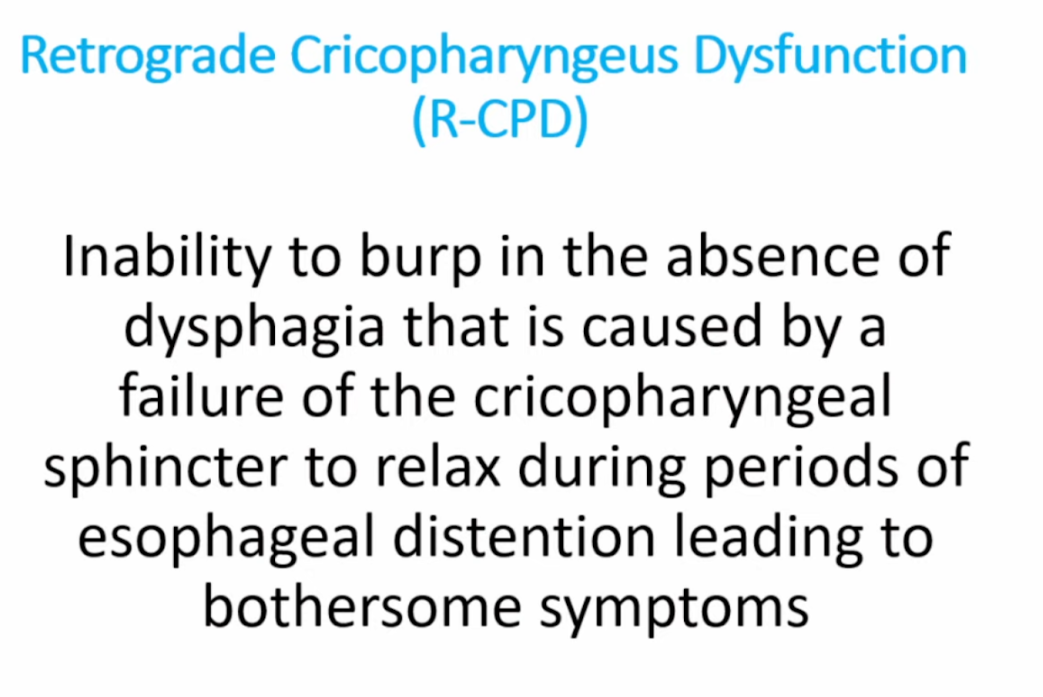
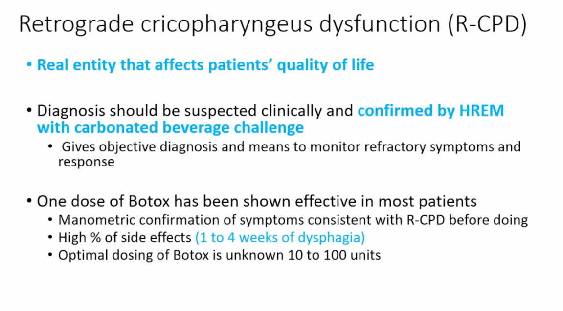
Dr. Garza provided insightful information on gastric vs supragastric belching. Supragastric belching can be treated with diaphragmatic breathing and cognitive behavioral therapy. Supragastric belching has shown poor response to pharmacologic therapy. He also explained the physiology behind the inability to burp.
Important to distinguish reason for belching as this affect managementThis study shows that with gastric belching the air works its way from the stomach up and with supragastric belching air is swallowed and expelled from the esophagus
My take: Our motility colleagues have some cool toys. When the treatments are as good as the toys, being a motility specialist will be even more fun.
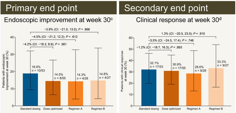
Methods: ENTERPRET was a phase 4, open-label, randomized, controlled trial (n=278) that included patients with moderate to severe UC who had high drug clearance at week 5 (serum concentration, <50 μg/mL) and nonresponse to standard vedolizumab treatment at week 6. At week 6, eligible patients were randomized 1:1 to receive standard dosing (300 mg every 8 weeks) or dose-optimized vedolizumab (600 mg at week 6, then 300 mg every 4 weeks; or 600 mg at week 6, then 600 mg every 4 weeks [based on week 5 serum concentration]).
After an initial clinical response of 47.5% (132/275) at week 6, 108 patients were subsequently randomized into either standard (n = 53) or dose-optimized vedolizumab (n = 55). The majority (86%) with nonresponse at week 6 had high drug clearance
Key findings at week 30:
Endoscopic improvement: 10 patients (18.9%) with standard vedolizumab vs 8 patients (14.5%) with dose-optimized vedolizumab
Clinical remission: Five patients (9.4%) with standard vedolizumab vs 5 patients (9.1%) with dose-optimized vedolizumab
Clinical response: 17 (32.1%) with standard vedolizumab vs 17 (30.9%) patients (30.9%) with dose-optimized vedolizumab
In their discussion, the authors note the gain of clinical response of ~16% in both groups is consistent with other studies showing “8% to 16% increase in the proportion of patients who had a clinical response between 6 and 14 weeks.” Thus, duration of treatment may be more important that serum drug level.
My take (borrowed in part from authors): “In patients with early nonresponse and high drug clearance, vedolizumab dose optimization is probably not required.” At week 30, both standard dosing and dose-optimized groups had a significant response despite lack of response at 6 weeks. So, it may be best to give a longer treatment course before reaching a conclusion on vedolizumab’s efficacy.
c Mayo endoscopic subscore </= 1. d Reduction in total Mayo score of >/=3 points and >/= 30% from baseline with an accompanying decrease in rectal bleeding subscore of >/=1 or absolute rectal subscore of </= 1
To examine the tongue, the authors recommend the following:
Assessment of tongue movement and coordination with a clean, gloved finger in the mouth to test the suck reflex and to palpate the hard and soft palate
Appearance of lingual frenulum, including the inability of the infant to extend tongue over the lower alveolar ridge or lift tongue midway to the palate, or a heart-shaped tongue on extension
Key Recommendations:
Ankyloglossia is a variation of a normal oral structure. Symptomatic ankyloglossia is defined as a restrictive lingual frenulum that causes problems with breastfeeding that are not improved with lactation support. Infants with ankyloglossia and normal feeding patterns need no intervention. Frenotomy for other problems or to prevent issues such as speech articulation or obstructive sleep apnea in the future is not evidence based.18
Posterior ankyloglossia is a poorly defined term, lacking agreement from experts, and should not be used as a reason to perform surgical intervention on an infant.
Labial and buccal frenae are normal oral structures unrelated to breastfeeding mechanics and do not require surgical intervention to improve breastfeeding. Sucking blisters are a normal finding in newborn infants, and as such, are not suggestive of pathology.
Suboptimal breastfeeding is a complex issue and every nursing dyad with painful or ineffective feeding should have a complete breastfeeding assessment before any treatment is offered.65,66 Here, multidisciplinary communication and management between lactation specialists, feeding therapists, surgeons, and pediatricians are paramount for the best outcome for the family.
Newborn infants with possible symptomatic ankyloglossia need close monitoring, support of breastfeeding while in the hospital, early postdischarge follow-up, and monitoring of weight gain in their medical home.
Surgical intervention for symptomatic ankyloglossia, versus laser, can reasonably be offered after other causes of breastfeeding problems have been evaluated and treated. Frenotomy may decrease maternal nipple pain.6,17,67 Although the evidence is not strong, addressing pain is important for successful continued breastfeeding.
Frenotomy should be performed by a trained professional, either the medical home provider or another to whom the medical home refers the patient. The performing professional should be experienced in the medical care of newborns and older infants and should maintain needed privileges for the procedure. As with any surgical procedure, before performing a frenotomy, the performing provider should take a “time out” to:
Obtain a signed consent
Discuss alternatives, risks, and benefits of the procedure
Discuss and provide pain control options
Document previous receipt of intramuscular vitamin K
Provide information on postsurgical care and follow-up
Attention to prevention of surgical complications, hemorrhage risk, pain mitigation, and evidence-based postsurgical care is recommended. Postoperative stretching exercises are not evidence-based and are not recommended.
Number of inpatient newborn ankyloglossia diagnoses in United States by year from Wei EX, Tunkel D, Boss E, Walsh J. Ankyloglossia: update on trends in diagnosis and management in the United States. 2012–2016. Otolaryngol Head Neck Surg. 2020;163(5):1029–1031.“(A) An example of anterior ankyloglossia with the lingual frenulum attaching at the tongue tip, limiting tongue mobility. (B) Posterior attachment of lingual frenulum. In the AAO-HNS consensus statement, consensus was not reached regarding the definition of posterior ankyloglossia. Some in the consensus group would describe this figure as an example of posterior ankyloglossia if there are objective findings of restricted tongue mobility caused by the lingual frenulum”
My take: Everyday I see infants with feeding problems that were attributed to being tongue-tied who do not improve after frenotomy. Most often, ankyloglossia does not need any intervention.
Related article: NY Times 7/2/24: Pediatricians Warn Against Overuse of Tongue-Tie Surgeries “The tongue procedures, which often cost several hundred dollars, should be done only to the small fraction of infants with severely tethered tongues, the report said.”
A recent episode of Bowel Sounds Podcast with my colleague Dr. Jose Garza was terrific. I scribbled a few notes afterwards but I usually listen while on a ride to an outreach clinic. So, there may be errors in omission, and in transcription.
Toddlers frequently have aerophagia and asymptomatic bloating
Nitrogen gas needs to be expelled from GI tract
Bloating symptoms can occur with and without distention
Bloating without other symptoms usually does not require an extensive workup. Bloating with diarrhea could indicate malabsorption
Bloating symptoms can worsen due to constipation
Chronic symptoms are usually reassuring; acute changes are more concerning
Intermittent distention is less concerning than persistent distention
Abdomino-phrenic dyssynergia occurs when the diaphragm pushes down on the abdomen and results in distention (see reference below)
Bloating symptoms frequently occur in those with only a small amount of excess air (less than 1 oz). There is often a degree of hypersensitivity.
Treatments for bloating could include diaphragmatic breathing, peppermint products, neuromodulators and sometimes diet. Diets can result in adverse effects including eating disorders, nutrient deficiencies, and cost
SIBO is over diagnosed in those without risk factors
pH-Impedance study, read manually, is a good test for belching: differentiates aerophagia, supragastric belching, and gastric belching
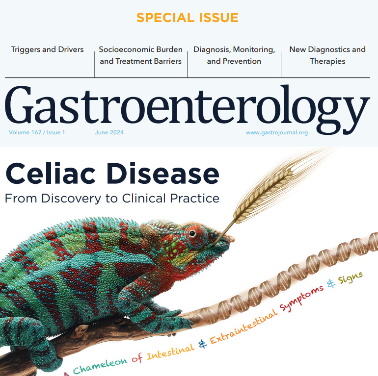
In June 2024 (special issue), Gastroenterology published an entire issue (193 pages) focused on celiac disease. There was a lot of useful information on almost every aspect of this disease. Below I have summarized some of the points –this is the last of 4 posts on this issue.
In addition to these articles, another good update on aspects of CeD was the Bowel Sounds podcast with Arun Singh. Link: Clinical Conundrums in Celiac Disease.
This article highlights the myriad presentations of CeD. Due to screening of at-risk groups. fewer patients are presenting with malabsorption and many patients at diagnosis are overweight or obese. In adults, 15-28% are overweight and 7-11% are obese.
Hematology: Among adults with iron-deficiency anemia, 1 in 31 have histologic evidence of CeD
Musculoskeletal/rheumatologic: osteopenia/osteoporosis/fractures, arthralgias/arthritis. Low bone mass with “z-scores less than -2 SD for age is present in 6-16% of children” with CeD
Growth in children: pubertal delay, short stature, growth failure
GI: stomach pain, diarrhea, bloating/distention, constipation, weight loss
Liver: elevated liver enzymes in up to 40% of patients with CeD at diagnosis. 1 in 20 patients with cryptogenic cirrhosis have CeD. In children, ~25% have “modest elevations in liver enzymes” at diagnosis. “Normalization …is generally seen within 6-12 months of a GFD.”
Endocrine: higher rates of type 1 diabetes and autoimmune thyroid disease
Asymptomatic: “10-27% of CeD patients are asymptomatic at diagnosis…Asymptomatic patients with CeD were taller, had lower TTG-IgA levels, and had less severe intestinal damage.” However, “patients reporting to be asymptomatic often record improvement” with GFD.
The authors detail how gluten exposure triggers symptoms. Gluten ingestion activates gluten-specific T cells and acute increases in interleukin-2 (IL-2). IL-2 is postulated to cause neuro-enteric symptoms and leads to release of serotonin. Serum IL-2 is a biomarker for acute gluten exposure.
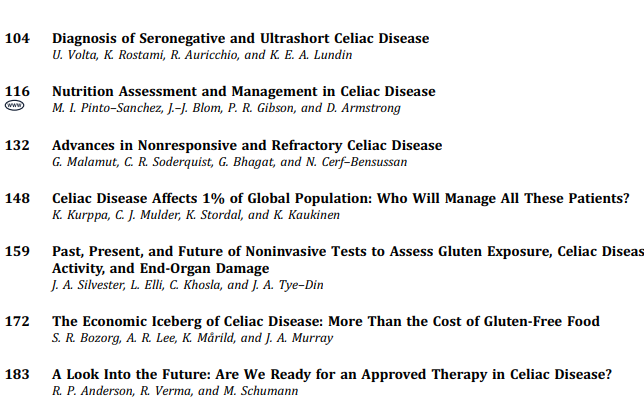
This article provides specific nutritional recommendations for patients with CeD including involvement of a dietitian. The authors note that some studies in children have found low iron, vitamin D, calcium, vitamin B12, vitamin B6, folate, zinc and magnesium in their GFD. They recommend checking for iron, ferritin, folate, vitamin B12, vitamin D, vitamin A, zinc, selenium, copper, and chromium at diagnosis.
The adoption of a GFD “has been associated with an increased risk of MASLD likely related to the nutritional composition of packaged gluten-free foods.” (see: Gluten-Free Diet Can Be Unhealthy)
30% of CeD adult patients experience persistent symptoms. The most common cause is continued gluten exposure (often inadvertent). Gluten contamination elimination diet can be implemented in this setting “though not feasible as a long-term solution.” (See: What To Do For Pediatric Patients with Non-Responsive Celiac Disease)
My take: More recent studies have indicated that more limited nutrient testing is appropriate in the pediatric population (see: How to Provide More Cost-Effective Celiac Care and Nutrient Deficiencies with Celiac Disease). Many nutrient deficiencies (in pediatrics) improve with institution of GFD and other nutrient deficiencies are similar in frequency as the general population.
“Seronegative villous flattening is a very rare condition” in children…and is “often attributable to other diagnosis (eg. inflammatory bowel disease…). The authors provide a diagnostic algorithm. In this setting, first step is to check HLA-DQ2/DQ8. A negative test “virtually rules out CeD.” A positive test indicates CeD is possible if other conditions are excluded. This includes autoimmune enteropathy, immune deficiencies, medication effects, infections, and other conditions. (See:Seronegative Villous Atrophy and@AmyOxentenkoMD: Celiac Disease and Mimics and How Many Cases of Celiac Disease Are We Missing?)
CeD confined to 1st portion of duodenum is considered ultra-short CeD. Algorithm for ultra-short CeD (Figure 2). The disease is supported by positive serology and positive HLA DQ2/DQ8. If serology and/or HLA DQ2/DQ8 are negative, it is very unlikely to be CeD and other diagnosis need to be identified (eg. peptic duodenitis).
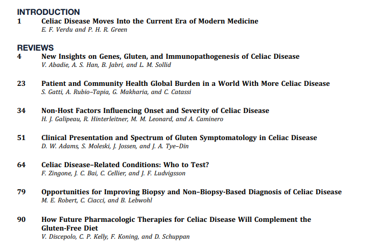
In June 2024 (special issue), Gastroenterology published an entire issue (193 pages) focused on celiac disease. There was a lot of useful information on almost every aspect of this disease. Below I have summarized some of the points.
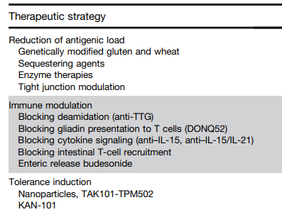
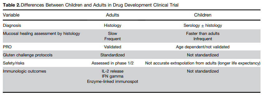
This article focuses on the emerging pharmacologic treatments
One of the treatment strategies has been to try to sequester gluten. This has included using enzymes to degrade dietary gluten. Other approaches include genetically modifying diet, tight junction modulation, immune modulation and tolerance induction.Some therapies being developed in adults may have unacceptable risk profiles for children. This could include blocking cytokine signaling (anti–IL-15, anti–IL-15/IL-21) and blocking intestinal T-cell recruitment
This article addresses nonresponsive CeD and refractory CeD. In the pediatric population, nonresponsive CeD is mainly related to persistent gluten exposure. Refractory CeD is rare in children.
In June 2024 (special issue), Gastroenterology published an entire issue (193 pages) focused on celiac disease. There was a lot of useful information on almost every aspect of this disease. Below I have summarized some of the points.
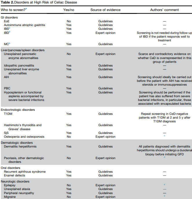
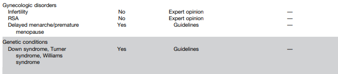
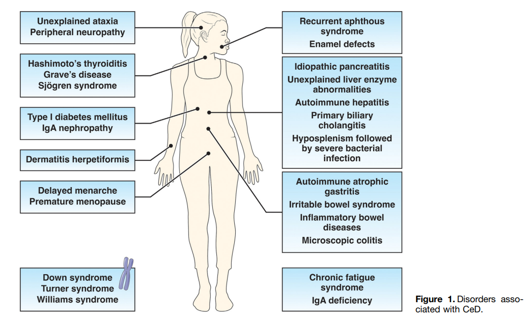
The authors detail disorders with increased risk for CeD and which merit screening (see Table 2 below). Some disorders that merit screening that are more obscure include idiopathic pancreatitis, autoimmune hepatitis, delayed menarche, and chronic fatigue.
They note that type 1 diabetes mellitus could require serial screening. “Because CeD can manifest at any time and with greater frequency during the initial 5 years, conducting additional screenings in CeD-negative T1DM patients 2 and 5 years after T1DM diagnosis and those who later develop gastrointestinal (GI) symptoms may be advisable.” In addition, it is important to recognize that CeD serology testing is less reliable in patients with T1DM.
They note that the worldwide prevalence is between 0.7% and 2.9%. In this issue, most authors estimate the prevalence to be about 1% with more than 50% undetected.
There are many places with higher rates. In U.S. “children in Colorado had a 2.5-fold higher risk compared to Washington State…similar regional differences were seen …in Sweden, Finland, and Germany.”
They note the burden before and after diagnosis. Before diagnosis/undetected, there can be persistent symptoms, complications (eg. osteoporosis, decreased fertility) and impaired quality of life. Afterwards, there are increased costs of a GFD and psycho-social burden of GFD.
In terms of generalized screening compared to case-finding, the authors note that given the number of at-risk groups, the case-finding approach could entail screening >50% of the population.
V Abadie et al.New Insights on Genes, Gluten, and Immunopathogenesis of Celiac Disease, reviews the intricate details of genetic, biochemical, and immunologic studies, which together have revealed mechanisms of gluten peptide modification and HLA binding, thereby enabling a maladapted anti-gluten immune response.
What I was most interested in was the mechanisms behind ‘potential’ celiac disease (PCeD) in which patients have autoimmunity (+serology) but normal histology. In potential CeD, the anti-CD4+ T-cell response is present but decoupled from tissue cytotoxicity. However, notably, IL-21, a cytokine produced by gluten-specific CD4+ T cells in active CeD, is not up-regulated in potential CeD…In addition, patients with potential CeD lack the presence of an epithelial stress response associated with IL-15, HSP70, and HSP27 upregulation in epithelial cells.” “The presence of epithelial stress is a crucial prerequisite for the development of tissue damage.”