FDA Approves Higher Dosage of Linaclotide for IBS-C and ByHeart Formula Recall Due to Botulinism

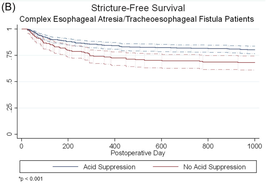
A Brooks. HCPLive 11/5/25: FDA Approves Linaclotide (Linzess) Capsules for Pediatric IBS-C

An excerpt:

The US Food and Drug Administration (FDA)  has approved Ironwood Pharmaceuticals’ linaclotide (Linzess) capsules for pediatric patients ≥ 7 years of age with irritable bowel syndrome with constipation (IBS-C), making it the first treatment approved for IBS-C in this patient population.1

The approval for pediatric IBS-C was supported by extrapolation of efficacy from adequate and well-controlled studies in adults and a 12-week double-blind, randomized, parallel-group trial in pediatric patients 7-17 years of age who met modified Rome III criteria for child/adolescent IBS-C. The recommended dosage for this indication is 145 mcg orally once daily.1

In 2023, the FDA approved linaclotide for the treatment of pediatric patients aged 6-17 years with functional constipation at a recommended dosage of 72 mcg orally once daily. 

Reference: US Food and Drug Administration. FDA approves 1st drug for children 7 years and older with irritable bowel syndrome with constipation. November 5, 2025. Accessed November 5, 2025. https://www.fda.gov/drugs/news-events-human-drugs/fda-approves-1st-drug-children-7-years-and-older-irritable-bowel-syndrome-constipation

Also, NBC news (11/08/25): ByHeart baby formula recalled amid 10-state outbreak of infant botulism The U.S. Food and Drug Administration said the outbreak includes 13 hospitalizations since August of children who consumed ByHeart Whole Nutrition Infant Formula. No deaths have been reported.

The recall includes two lots of the powdered formula with Dec. 1 “use by” dates, the FDA said in a statement Saturday. The lot numbers are 206VABP/251261P2 and 206VABP/251131P2…The FDA says ByHeart makes up less than 1% of the baby formula sold in the U.S.

Chicago Botanic Garden

Proximal Ileal Crohn’s Disease is Harder to Treat

K Takenaka et al. Clin Gastroenterol Hepatol 2025; 23: 1991-2000. Open Access! Inadequate Efficacy of Biologics for Treating Proximal Ileal Lesions in Crohn’s Disease: A Prospective Multicenter Study

This multicenter prospective study (n=253) examined efficacy of treatment in patients with proximal ileal disease using balloon-assisted enteroscopy (BAE). The recruited patients had a mean disease duration of 4 years. 52% were naive to biologic treatment at baseline.

Key findings:

  • At baseline, 74 patients (29.2%) had proximal ileal ulcerations without terminal ileal ulcerations
  • At week 26, after treatment with anti-TNF therapy (n=103), ustekinumab (n=99) or vedolizumab (n=51), endoscopic remission was achieved in 91 patients (36.0%). Of the patients with complete ulcer healing of the terminal ileum, 28.6% (22/77) had residual ulcers in the proximal ileum
  • The rate of endoscopic remission in the proximal ileum (50.9%) was relatively lower compared with the colon (63.4%) and terminal ileum (56.7%)
  • After a median follow-up of 134 weeks, residual ulcerations in the proximal ileum were associated with a poorer prognosis (P = .0126 for hospitalization and P = .0014 for surgery). In contrast, there was no significant differences in hospitalization and surgery associated with endoscopic activity vs remission in the colon or terminal ileum.

Discussion: Residual “proximal ileal ulcerations … are associated with a poorer prognosis…Additionally, we confirmed that proximal ileal inflammation is less responsive to biologic therapies compared with colonic inflammation. Although the reasons for this disparity remain unclear”

My take: Balloon-assisted enteroscopy is not frequently used in the setting of inflammatory bowel disease, particularly in pediatrics. MRE is typically used to follow proximal small bowel disease, though it has less sensitivity for luminal mucosal disease.

Related blog posts:

Hypercoagulation with Acute Severe Ulcerative Colitis (ASUC) Persists for Months

BJ Griffiths et al. Clin Gastroenterol Hepatol 2025; 23: 1798-1807. Open Access (PDF)! Hypercoagulation After Hospital Discharge in Acute Severe Ulcerative Colitis: A Prospective Study

Methods: In this prospective study, twenty-seven adult patients with ASUC and 25 control patients with quiescent ulcerative colitis were recruited. Thrombin generation (endogenous thrombin potential), rotational thromboelastometry (EXTEM and FIBTEM maximum clot firmness), procoagulant factors, anticoagulant factors, and fibrinolytic markers were assessed for those with ASUC on admission (Day 1), Day 5, 4 weeks, and at 8–12 weeks. These assessments were performed on a single occasion for control patients.

Key findings:

Discussion:

  • “Patients with ASUC had a hypercoagulable profile on initial presentation to the hospital, before receipt of LMWH, which was consistently demonstrated by individual and global assays of coagulation. The most marked elevations of individual factors at presentation were seen in the levels of Clauss fibrinogen, platelets, VWF, and FVIII, alongside heightened
    levels of the inhibitors of fibrinolysis PAI-1 and TAFIa.”
  • “This hypercoagulable state persisted for many weeks after hospital discharge, with levels of FVIII, fibrinogen, VWF, and inhibitors of fibrinolysis (TAFIa) remained significantly elevated at all timepoints up to 12 weeks, compared with the control population. This is despite
    intensive treatment for ASUC in all patients.”
  • “VTE and pulmonary embolism are 1 of the leading causes of morbidity and mortality during IBD flare-ups. The findings from this study reinforce the importance of thromboprophylaxis administration to all patients with ASUC at first presentation to hospital.”

My take: This study is in adults; the risk of VTE is lower in children and guidance on VTE prophylaxis is not clear.

Related blog posts:

Kids With Acute Pancreatitis Need Followup

F Ahmed, M Abu-El-Haija. Gastroenterol 2025; 169: 572-584. Open Access! Acute Pancreatitis in Children: It’s Not Just a Simple Attack

This is a really good review of acute pancreatitis covering epidemiology, diagnosis, severity classification, management, microbiome/metabolite derangements, genetics, and complications. Most of these topics have been covered in numerous blog posts (see below).

Selected Key Points:

  • Diagnostic testing -Amylase/Lipase:  “The diagnostic efficacy of amylase for AP, in terms of sensitivity and specificity, is contingent on the selected threshold value. Elevating the cut-off point to 1000 IU/L results in a high specificity of approximately 95%. However, this comes at the cost of reduced sensitivity, which some studies report to be as low as 61%… the activity of serum lipase remains elevated for a longer duration, typically between 8 and 14 days,… Lipase demonstrates superior accuracy with most studies reporting specificities exceeding 95% and sensitivities ranging from 55%–100% at a threshold activity level of 600 IU/L…hese tests have excellent sensitivities, they may have a few limitations such as being poor predictors of severity”
  • Risk Factors in Children (from Figure 1):
  • Incidence and Severity in Children (from Figure 1):

[At a recent lecture, Jay Freeman (How to Upgrade Pancreas Care –Jay Freeman MD (Part 1)) noted that severe pancreatitis is often defined by degree of organ dysfunction (eg. cardiac, pulmonary, renal). A practical definition of severe pancreatitis in children is whether the patient requires admission to an ICU]

  • Diagnostic testing -Imaging: “Imaging techniques are crucial for diagnosing and managing AP in children…NASPGHAN) and the Society for Pediatric Radiology formed consensus guidelines where transabdominal ultrasonography was recommended as the primary imaging technique for pediatric cases with suspected AP…Recent studies in the pediatric population have indicated that US’s sensitivity for AP detection ranges from 47%–52%.25Magnetic resonance cholangiopancreatography (MRCP) is useful for anatomical assessment without radiation but may require sedation”
  • Management: “The cornerstones of therapy are early feeding and intravenous fluids… Allowing patients to eat on admission was feasible and was associated with lower length of stay. Rates of intravenous fluids are recommended at 1.5–2 times maintenance rates,49 and the preferred fluid is Lactated Ringer’s due to limited studies including a recent randomized controlled study that showed that Lactated Ringer’s was associated with a faster discharge rate when administered compared with normal saline.50
  • Genetics:  “A recently conducted study investigated the importance of genetics in pediatric AP patients…use of an extensive panel of 8 genes… PRSS1CFTRSPINK1CPA1, CTRCCLDN2CASR, and SBDS… genetics is a major component in all types of pancreatitis in children, with genetic variants being most prevalent in CP cases at 31%, followed by AP at 19%, and ARP at 6%. A key discovery was that variants in SPINK1CFTR, or PRSS1 genes were associated with faster progression from first episode of AP toward CP.53
  • Complications (from Figure 1): “After the first episode of AP, the QoL is decreased, and it may lead to other disorders such as exocrine dysfunction, endocrine dysfunction and diabetes, nutritional deficiencies, and acute recurrent pancreatitis and CP.”

My take: Even after a single episode of acute pancreatitis, there are risks for long-term complications and patients need to follow-up.

Related blog posts:

Shared Decision-Making in Celiac Disease Diagnostic Approach

Y Sunkoy, S Talathi. Am J Gastroenterol 2025; 2190-2193. Utility of the ESPGHAN Biopsy-Sparing Guidelines for Celiac Disease in Children

Thanks to Ben Gold for this reference.

Methods: Retrospective study of patients (n=2942 children) who had celiac serologies and duodenal biopsies

Key findings:

  • Prevalence of CD in this cohort was 9% (226 of 2942 patients)
  • In those with a high titer (>10xULN), 106 of 107 patients (99%) had celiac disease
  • In this cohort, even in those with with >7XULN, had a Sensitivity of 55.3%, a specificity of 99% and a PPV of 97%

In their discussion, the authors note that “we did not obtain an EMA in a second sample, which is recommended in the ESPGHAN guidelines.”

Associated commentary: Erica Brenner, American Journal of Gastroenterology 120(9):p 1985-1986. The No-Biopsy Approach for Pediatric Celiac Disease: Ready for Prime Time in North America?

  • “Shiha et al (8) found that the PPV ranged from 65% for a 1% CD prevalence to a 99% for a 40% prevalence. As the 9% CD prevalence in the study by Sunkoj et al (4) exceeds the 0.81$-1.4% prevalence in the United States (9), the reported PPV may overestimate reality.” (Related post: No-Biopsy Approach to Celiac Disease Diagnosis and Positive Predictive Value (Based on Population)
  • “Children with type 1 diabetes and trisomy 21 have a higher risk of false-positive serology and therefor may not be appropriated candidates for a no-biopsy approach.”

My take: A larger recent study (Chang et al. Pediatrics. 2025;156(3):e2025070897) found that the no-biopsy approach had a significantly lower PPV in their cohort (94.9% overall, and 95.7% in those without T1DM). Thus, in cohorts with lower prevalence of CD, the no-biopsy approach could lead to 2-4% of children being placed unnecessarily on a gluten free diet. As such, it would be good practice to discuss making a diagnosis via endoscopy vs. the no-biopsy approach as part of shared decision-making.

Related blog posts:

Stercoral Colitis

A Bajer, E Levine. NEJM 2025;393: e23. Stercoral Colitis

This young adult presented to the ED with left-sided abdominal pain and chronic constipation. A CT scan was consistent with a diagnosis of stercoral colitis. “In stercoral colitis, chronically impacted feces distend the colon, resulting in inflammation. In some cases, the fecaloma may lead to focal-pressure necrosis or perforation.”

My take: Most often a CT is not needed in this setting. However, it is important to recognize that a severe impaction can lead to complications.

Related blog posts:

Selective Acid Suppression for Esophageal Atresia Patients

This year’s masterpiece!

Link from AAP HealthyChildren.org: Halloween Fun & Safety Tips for Kids of All Ages


S Zeneddin et al. J Pediatr Gastroenterol Nutr. 2025;81:960–966. Acid suppression after esophageal atresia repair: Some infants do benefit

Methods: The authors performed a retrospective study using the Pediatric Health Information System for infants undergoing EA/TEF repair between 2010 and 2022 (n=1445 infants). Acid suppression was defined as receipt of an H2 blocker or proton pump inhibitor on the day of discharge or longer than 30 inpatient days. Complex EA/TEF repair was defined as delayed repair (>7 days), G-tube placement before repair (likely a sign of a long gap or type A anomaly), prolonged hospitalization (>60 days), or multiple inpatient fluoroscopies. The authors defined stricture solely if it required intervention.

Key findings:

  • 257 (17.8%) required dilation by 1 year. Of the 688 (47.6%) infants who met criteria for complex EA/TEF, 126 (18.6%) required a dilation.
  • At 1 year, stricture rate was similar in infants with simple EA/TEF, with or without acid suppression (17.5% vs. 17.0%, p = 0.90)
  • In infants with complex EA/TEF, stricture rates were lower among those who received acid suppression compared to those who did not (15.3% vs. 26.0%, p = 0.001).

The associated editorial (D George, DK Robie. J Pediatr Gastroenterol Nutr. 2025;81:911–912) reviews some of the limitations of the study but does not provide clear recommendations on utilization of acid suppression therapy: the decision should be “should be individualized, weighing the potential benefits against the risks.”

My take: It is not surprising that more complex EA would have higher stricture rates. In my training (in the 1990s!), it was routine practice to use indefinite acid suppression. This article indicates that patients with low risk EA likely do not need acid suppression. In high risk patients, the algorithm by Yasuda et al (see post below J Am Coll Surg 2024; 238: 831-843) provides their approach to weaning acid suppression.

Related blog posts:

Disclaimer: This blog, gutsandgrowth, assumes no responsibility for any use or operation of any method, product, instruction, concept or idea contained in the material herein or for any injury or damage to persons or property (whether products liability, negligence or otherwise) resulting from such use or operation. These blog posts are for educational purposes only. Specific dosing of medications (along with potential adverse effects) should be confirmed by prescribing physician.  Because of rapid advances in the medical sciences, the gutsandgrowth blog cautions that independent verification should be made of diagnosis and drug dosages. The reader is solely responsible for the conduct of any suggested test or procedure.  This content is not a substitute for medical advice, diagnosis or treatment provided by a qualified healthcare provider. Always seek the advice of your physician or other qualified health provider with any questions you may have regarding a condition.

Prior Exposure to TNF Antagonists May Increase Response to JAK Inhibitors in Patients with Ulcerative Colitis

HH Lee et al. Clinical Gastroenterology and Hepatology 2025; 23, 2102 – 2114. Open Access! Differential Efficacy of Advanced Therapies in Inducing Remission in Ulcerative Colitis Based on Prior Exposure to TNF Antagonists

Methods: Meta-analysis of 17 randomized controlled trials in 8871 adults with moderate-severe UC. The authors calculated the ratio of odds ratio of achieving remission with active drug vs placebo, in TNF antagonist–naïve vs TNF antagonist–exposed patients.

Key findings:

  • JAK inhibitors: Less efficacious in TNF antagonist–naïve vs exposed patients (6 trials; ratio of OR, 0.47)
  • IL-23 antagonists: No significant difference was observed in efficacy of selective interleukin-23 antagonists vs placebo in TNF antagonist–naïve vs exposed patients (6 trials; ratio of OR, 1.07)
  • Lymphocyte trafficking inhibitors: More efficacious in TNF antagonist–naïve vs exposed patients (5 trials; odds ratio [OR], 1.88)

Discussion:

  • This study “confirmed prior observations that exposure to TNF antagonists significantly reduces the efficacy of lymphocyte trafficking inhibitors in inducing remission, including both vedolizumab and S1P receptor modulators, by approximately 50%.In contrast, prior exposure to TNF antagonists was associated with a significant increase in the efficacy of JAK inhibitors in inducing remission, with 2-fold higher efficacy in TNF antagonist–exposed vs TNF antagonist–naïve patients”
  • In the SELECT-COMPARE trial in patients with rheumatoid arthritis, there was also an improved response to upadacitinib in patients with prior adalimumab.
  • “The current findings raise the intriguing possibility that exposure to TNF antagonists could result in lasting effects on the immune system that differentially alter responsiveness to therapies with distinct mechanisms of action”

My take: This study suggests that JAK inhibitors are a good choice for secondary therapy after anti-TNF agents. Other factors, besides efficacy, including safety, extraintestinal manifestations, and cost, have to be considered as well.

Related blog posts:

Disclaimer: This blog, gutsandgrowth, assumes no responsibility for any use or operation of any method, product, instruction, concept or idea contained in the material herein or for any injury or damage to persons or property (whether products liability, negligence or otherwise) resulting from such use or operation. These blog posts are for educational purposes only. Specific dosing of medications (along with potential adverse effects) should be confirmed by prescribing physician.  Because of rapid advances in the medical sciences, the gutsandgrowth blog cautions that independent verification should be made of diagnosis and drug dosages. The reader is solely responsible for the conduct of any suggested test or procedure.  This content is not a substitute for medical advice, diagnosis or treatment provided by a qualified healthcare provider. Always seek the advice of your physician or other qualified health provider with any questions you may have regarding a condition.

Comprehensive ACG Clinical Guidelines for Ulcerative Coliits (2025)

D Rubin et al. The American Journal of Gastroenterology 120(6):p 1187-1224, June 2025. Open Access! ACG Clinical Guideline Update: Ulcerative Colitis in Adults

Today and tomorrow I am highlighting two adult clinical guidelines both of which are equivalent to up-to-date textbook chapters with specific recommendations; both are open access. In addition, the articles have accompanying author podcasts. Thanks to Ben Gold for these references.

Table 2 in the UC guideline makes 54 recommendations and Table 3 provides 57 key concepts.

Selected Management Recommendations:

  • Table 2, #4: We recommend treating patients with UC to achieve endoscopic improvement (Mayo score 0 or 1) to increase the likelihood of sustained steroid-free remission and to prevent hospitalization and surgery
  • Table 2, #5: We recommend the use of FC (fecal calprotectin) in UC to assess response to therapy, to evaluate suspected relapse, and during maintenance
  • Table 2, #33: When infliximab is used as induction therapy for patients with moderately to severely active UC, we recommend combination therapy with a thiopurine
  • Table 2, #43: Recommend continuing tofacitinib or upadacitinib as compared with no treatment for maintenance of remission in patients with prior moderately to severely active UC now in remission after induction with tofacitinib or upadacitinib. **The authors recommend continuing each biologic that achieved remission with induction therapy (#38-#43)
  • Table 2, #51: In patients with ASUC failing to adequately respond to intravenous corticosteroids (IVCS) by 3 days, we recommend medical rescue therapy with infliximab or cyclosporine (Strong recommendation, moderate quality of evidence).

Key concepts:

  • Table 3, #29: Patients who are primary nonresponders to an anti-TNF (defined as lack of therapeutic benefit after induction and despite sufficient serum drug concentrations) should be evaluated and considered for alternative mechanisms of disease control (e.g., in a different class of therapy) rather than cycling to another drug within the anti-TNF class.
  • Table 3, #31:  Subcutaneous infliximab and vedolizumab are considered equivalent to the standard intravenous maintenance dosing of these agents. The equivalence of the subcutaneous formulations for induction or as substitution for escalated doses of these therapies has not been robustly established.
  • Table 3, #41: Patients with UC should have available all medical options as recommended by their doctor and healthcare team. Third-party payers and requirements for step therapy should not come between the patient and their healthcare team in making decisions about treatment for UC.
  • Table 3, #48: All patients with ASUC should undergo a flexible sigmoidoscopy within 72 hours and preferably within 24 hours of admission. This should be used to assess endoscopic severity of inflammation and to obtain biopsies to evaluate for cytomegalovirus (CMV) colitis.
  • Table 3, #51: Nonsteroidal anti-inflammatory drugs (NSAIDs), narcotics, and medications with anticholinergic side effects should be avoided in ASUC.
  • Table 3, #57: In patients with ASUC initiating infliximab, dose intensification should be considered for those patients with low serum albumin (<2.5 g/dL).

My take: This article does an excellent job of summarizing current available evidence and good practice. Many of the recommendations may be helpful in garnering approval from third party payers.

Related blog posts:

Why Pediatric Patients Need Higher Dosing of Infliximab

E Stenke et al. Inflamm Bowel Dis 2025; 31: 2331-2337. Higher-Dose Infliximab Induction Achieves Better Maintenance Trough Levels in a National Pediatric IBD Cohort—A Retrospective Study

In this single center retrospective study from Ireland, the authors examined 122 patients (93 with Crohn’s disease [CD], 18 with ulcerative colitis [UC], 1 with IBDU) who received infliximab and had prospectively-collected data. The earlier cohort 2018-2019 received 5 mg/kg/dose and the later group 10 mg/kg/dose. Both groups had proactive therapeutic drug monitoring (pTDM).

Key findings:

  • The 5 mg/kg group, compared to the 10 mg/kg group, was less likely to have target pre-third TLs (6% vs 80%, P < .001) with the stated goal of >/= 15 microgm/mL
  • Fewer patients in the 5 mg/kg than 10 mg/kg group had pre-fourth TLs ≥5 µg/mL (6/48 [12.5%] vs 28/50 [56%], P < .001; mean [SD] TL 3.5 [6.3] vs 10.0 [9.9], P < .001)
  • Concurrent immunomodulator therapy was more common in the 5 mg/kg group (43% compared to 24%)
  • 80% of patients were still receiving infliximab at 1 year including 87% of patients with CD and 54% with UC
  • The higher dose group had a lower CRP at 1 year followup. 26% of patients receiving the lower dose had a CRP > 5 mg/L compared with 9% in the higher dose group.
  • Some other measures of long term outcome (eg. IFX durability, clinical remission) were slightly better but did not reach statistical significance (see below)

Discussion Points:

  • “Our data show higher rates of below-target infliximab levels during and after induction in the 5 mg/kg group. Higher rates of dose escalation in this group during the first year resulted
    in similar dosing regimens…Thus, the similar infliximab durability and clinical outcomes
    at 1-year follow-up reflect early-dose optimization leading to dose equalization between the 2 groups, rather than a lack of benefit to higher dosing regimens”
  • “Our data affirm that proactive TDM with pre-emptive dose escalation restores
    below-target infliximab TLs and sustains clinical response…Indeed, in our cohort, some patients with low IFX levels pre-third dose were given their fourth dose 6 weeks later, rather
    than the standard 8 weeks. Without proactive TDM results, our rate of suboptimal TLs pre-fourth and during maintenance therapy would have been higher in both groups”
  • “Rates of immunomodulator use in the 10 mg/kg group were lower than in the earlier cohort of 5 mg/kg, reflecting changes in clinical practice over time”

My take:

  1. This study shows that 94% of pediatric patients did NOT achieve adequate levels of infliximab at the pre-third dose with “standard” therapy. This was true even with 43% of the lower dose cohort receiving combination therapy (which often helps improve pharmacokinetics)
  2. Proactive therapeutic drug monitoring helped mitigate the clinical outcomes, especially in the lower dosed cohort
  3. “Children with IBD treated with the historic standard dose of 5 mg/kg induction are at increased risk of pharmacokinetic treatment failure related to high rates of suboptimal TLs”

Related blog posts:

Disclaimer: This blog, gutsandgrowth, assumes no responsibility for any use or operation of any method, product, instruction, concept or idea contained in the material herein or for any injury or damage to persons or property (whether products liability, negligence or otherwise) resulting from such use or operation. These blog posts are for educational purposes only. Specific dosing of medications (along with potential adverse effects) should be confirmed by prescribing physician.  Because of rapid advances in the medical sciences, the gutsandgrowth blog cautions that independent verification should be made of diagnosis and drug dosages. The reader is solely responsible for the conduct of any suggested test or procedure.  This content is not a substitute for medical advice, diagnosis or treatment provided by a qualified healthcare provider. Always seek the advice of your physician or other qualified health provider with any questions you may have regarding a condition.