Blog Case Report: Eosinophilic Colitis

This past month I helped diagnose a 2nd young child with eosinophilic colitis. This was an 8 month old who developed rectal bleeding over a 6 week period with subsequent anemia (Hgb 7.1). His colonoscopy showed patchy deep erythema in the right colon and transverse colon; his biopsies identified >100 eosinophils/hpf.

This prompted me to review a case of a child presenting with eosinophilic colitis at 13 months of age.

Case study: He was born at 36 week gestation with a prenatal diagnosis of proximal bowel obstruction due to type IIIb jejunal atresia, s/p ex-lap and tapering enteroplasty for long dilated jejunal segment and with subsequent short gut syndrome (~50 cm of small bowel). He required supplemental HAL dependency for his initial 9 months of life. At 13 months of age, he developed bloody diarrhea (up to 12/day) with associated anemia and peripheral eosinophilia (max elevation of AEC during his course was 23,000).

Initial endoscopy took place in January 2022. Images are noted below and histology confirmed eosinophilic colitis (>100 eos/hpf).

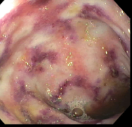
He underwent extensive testing and no monogenetic/immune dysregulation disorders were identified. After not improving with an elemental diet, his treatment ​consisted of prednisolone along with enteral antibiotics (vancomycin/gentamicin)​. His symptoms recurred with steroid taper and he was changed from antibiotic therapy to mesalamine. This was also ineffective. Subsequently, he started with vedolizumab therapy.

Due to ongoing symptoms, tacrolimus (along with PCP prophylaxis) was added two months later (this was 4 months after initial colonoscopy). The target level was between 8-10. He had a rapid and sustained response allowing him to stop corticosteroids which had been used intermittently for more than 6 months. His vedolizumab was stopped shortly thereafter.

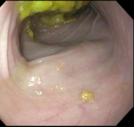
After a followup panendoscopy demonstrated resolution of his colitis (see image below), his diet was gradually advanced (no restrictions) while continuing tacrolimus.
His tacrolimus was discontinued after 2.5 years and he has continued to do well off therapy for two years at this point. He still requires periodic vitamin-micronutrient monitoring for his short bowel syndrome.

My take: Eosinophilic colitis is a rare disorder in pediatrics with sparse data to guide management. In this young pediatric patient, tacrolimus was effective. It is possible that vedolizumab would have been effective if given for a longer duration prior to administration of tacrolimus. At the time of presentation, dupilumab had not been approved for eosinophilic esophagitis. There have been case reports of its use off-label for eosinophilic colitis (T Sia et al. Clin Transl Gastroenterol. 2025 Aug 26;16(10):e00908. Dupilumab for Adult and Adolescent Patients With Primary Eosinophilic Colitis).

Related blog posts:

Disclaimer: This blog, gutsandgrowth, assumes no responsibility for any use or operation of any method, product, instruction, concept or idea contained in the material herein or for any injury or damage to persons or property (whether products liability, negligence or otherwise) resulting from such use or operation. These blog posts are for educational purposes only. Specific dosing of medications (along with potential adverse effects) should be confirmed by prescribing physician. Because of rapid advances in the medical sciences, the gutsandgrowth blog cautions that independent verification should be made of diagnosis and drug dosages. The reader is solely responsible for the conduct of any suggested test or procedure. This content is not a substitute for medical advice, diagnosis or treatment provided by a qualified healthcare provider. Always seek the advice of your physician or other qualified health provider with any questions you may have regarding a condition.

Immune Mediated Enterocolitis with CAR-T Cell Therapy

JA Damianos et al. Alimentary Pharmacology & Therapeutics, 2026; 0:1–12. Open Access! Review Article: Immune Effector Cell-Mediated Enterocolitis Following CAR-T Cell Therapy—Clinical Features, Pathophysiology and Management

Background: “Chimeric antigen receptor T-cell (CAR-T) therapy has revolutionized the treatment of hematologic malignancies, and its use is expanding rapidly into numerous other disease states including autoimmune diseases. However, CAR-T therapy is associated with a spectrum of immune-related toxicities…it has become apparent that rarely, CAR-T can cause gastrointestinal mucosal inflammation, termed immune effector cell-mediated enterocolitis (IEC-EC).”

Other treatments affecting the immune system can result in gastrointestinal inflammation. “Immune checkpoint inhibitors, including cytotoxic T lymphocyte associated anti-gen 4 (CTLA- 4) inhibitors, programmed death-1 (PD1) inhibitors, and PD-ligand 1 (PDL-1) inhibitors have been associated with colitis (with or without enteritis) in up to 54% of patients treated with these agents”
Key points:

  • “Occurring in up to approximately 6% of patients typically following B cell maturation antigen-targeted CAR-T therapy, IEC-EC presents with severe diarrhea and malabsorption, responds poorly to treatment, and portends a dire prognosis.”
  • “Step-up pharmacotherapy, often featuring biologics and small molecules drawn from the inflammatory bowel disease pharmacologic armamentarium.” Figure 6 presents an algorithm for evaluation and management.
  • “Patients typically present 1–3 months after CAR-T infusion with persistent watery, non-bloody diarrhea, often Grade 3 or higher [33]. Diarrhea is often severe and persistent, frequently accompanied by malnutrition and significant weight loss, and may necessitate prolonged parenteral nutritional support [25]. Diagnosis is often delayed due to the rarity and novelty of this condition, and the fact that patients receiving CAR-T have numerous other potential reasons to experience diarrhea.”

My take: There are a growing number of individuals needing CAR-T cell therapy. Recognizing this delayed complication quickly may help improve outcomes.

Related blog posts:

Puffins on the Edge of a Cliff Overlooking a Black Sand Beach

Federal Policies Changing Medical Standards

BA Barsky et al. N Engl J Med 2026;395:419-421. Medical Standards by Federal Fiat

An excerpt:

“Historically, the federal–state division of authority over medical practice has been strict. This division is evident in health care financing and medical product regulation, for example. States license physicians and set standards for medical care, often incorporating standards established by the medical profession while allowing broad discretion for physician judgment.1…

The Trump administration has attempted to alter the balance of federal–state authority in the area of gender-affirming care in two important ways. First, in June 2025, the U.S. Department of Justice (DOJ) issued administrative subpoenas to more than 20 health systems and clinics providing gender-affirming care, demanding unusually broad information related to off-label prescribing of puberty blockers and hormones…The DOJ’s subpoenas, however, demanded information at the heart of physician judgment and the patient–doctor relationship, including physicians’ case-specific clinical assessments justifying off-label prescriptions and information that could be used to identify patients, including children, by name…

The second federal disruption came in December 2025, when Secretary of Health and Human Services Robert F. Kennedy, Jr., issued a declaration stating that gender-affirming care for children and adolescents is unsafe and does not meet professionally recognized standards of care…

Much of the authority and discretion now held by medical professionals and the states would move upward to a federal government that would exert expanded control over the health care that patients receive. Such a shift would potentially force all states to accept a single federal standard of care in various areas of medicine while concentrating immense medical power in the hands of political appointees. Federal authority over health care standards is appropriate in some instances. But this authority should be established using a proper legal process — Congress acting within its enumerated constitutional powers, or the executive branch acting under properly delegated authority — not by fiat.”

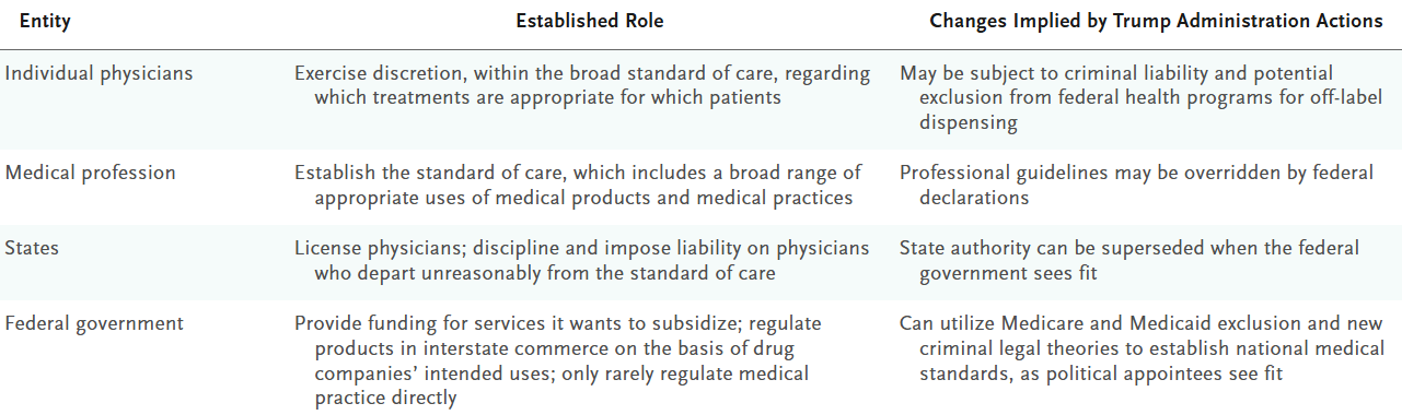
Established Roles for Setting the Medical Standard of Care
and Changes Implied by Trump Administration Actions.

My take: While this issue primarily affects a narrow area currently, it is not difficult to imagine that other administratioins could similarly impose policies affecting other sensitive decisions like abortion (expanding or limiting options), contraceptive availability, discussions about guns, and viability decisions.

Dr. Michael Wilsey: Endoscopy Pearls and Ergonomics (Part 2)

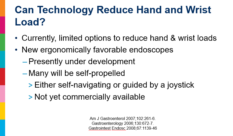
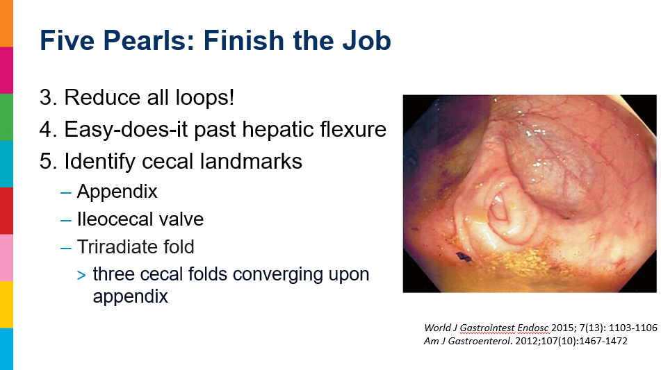
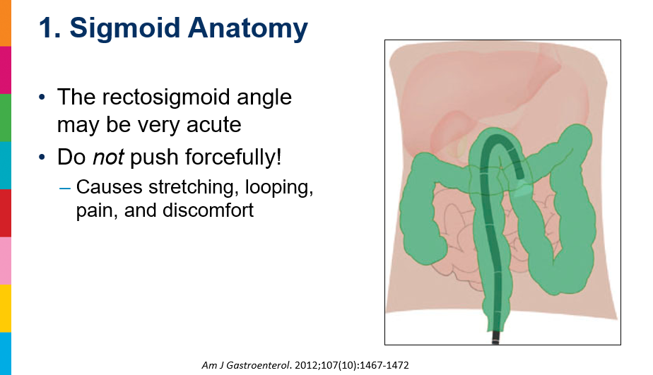
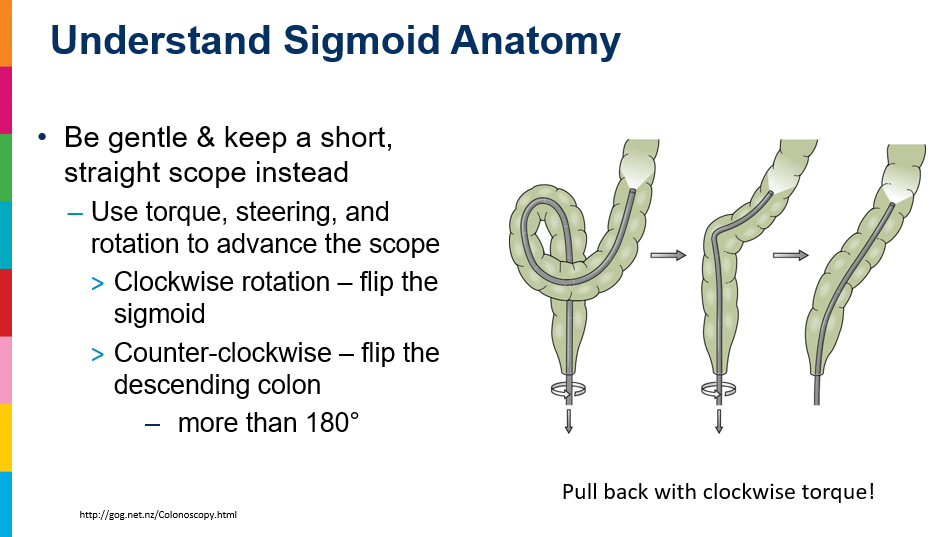
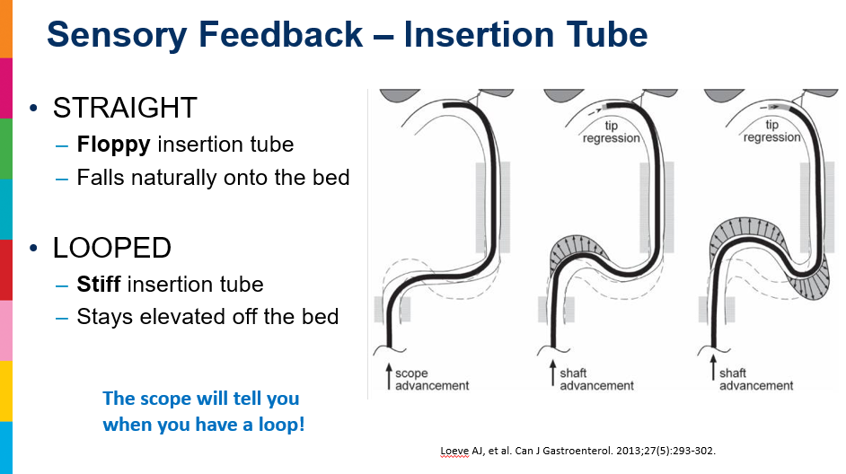
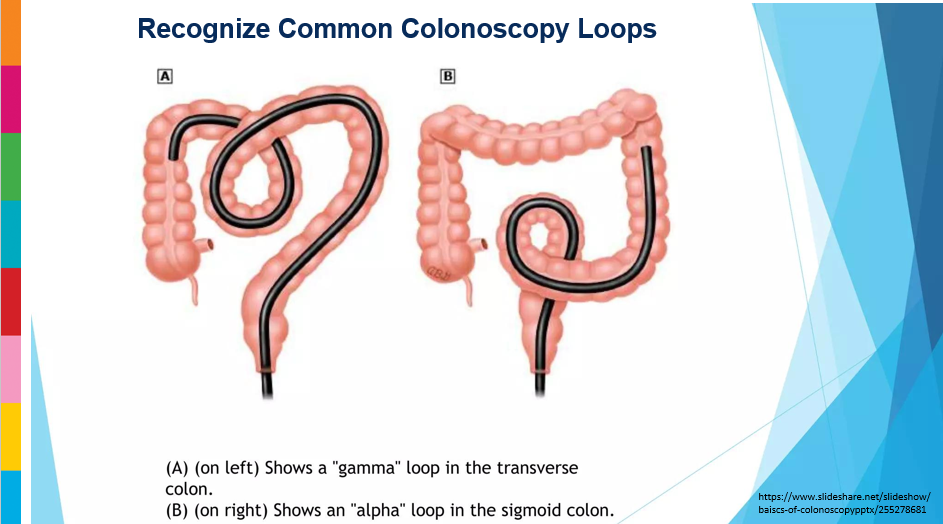
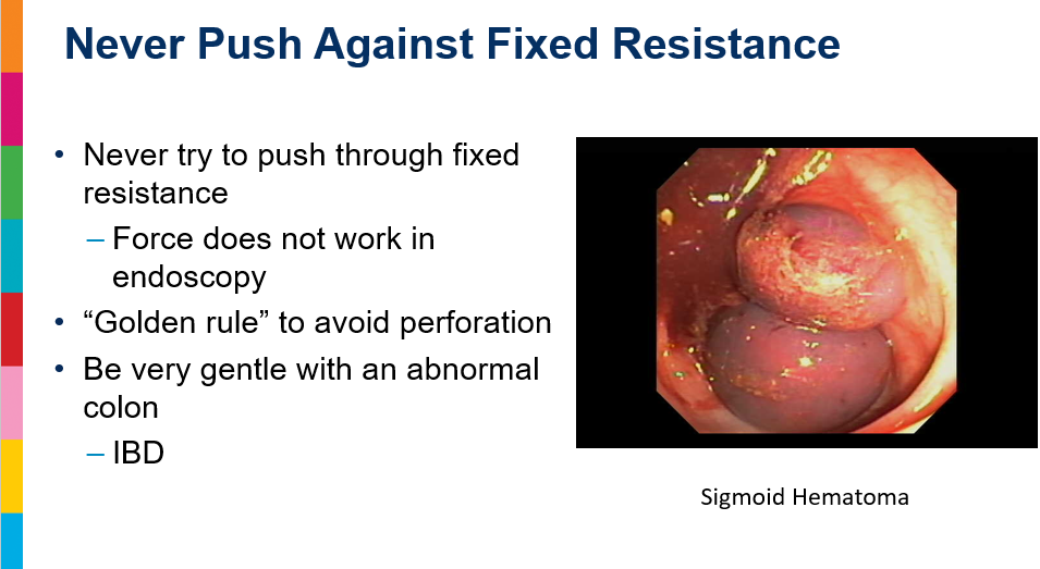
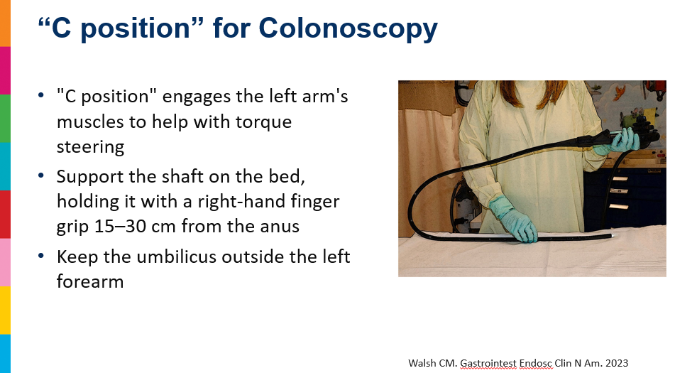
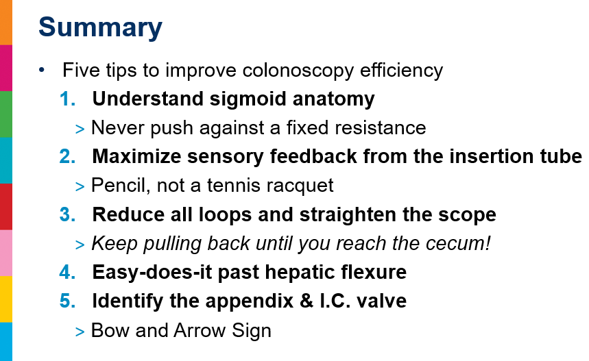
Dr. Michael Wilsey gave our group an excellent update on Endoscopy Pearls and Ergonomics. My notes below may contain errors in transcription and in omission. Along with my notes, I have included many of his slides.
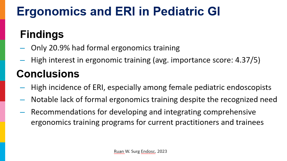
Yesterday’s post focused on endoscopy pearls. Today’s post focuses on ergonomics. Most of the attendees realized that we could use a lot of improvement in this area after listening to this presentation. This issue is likely of even greater importance in those with a high endoscopy case load.
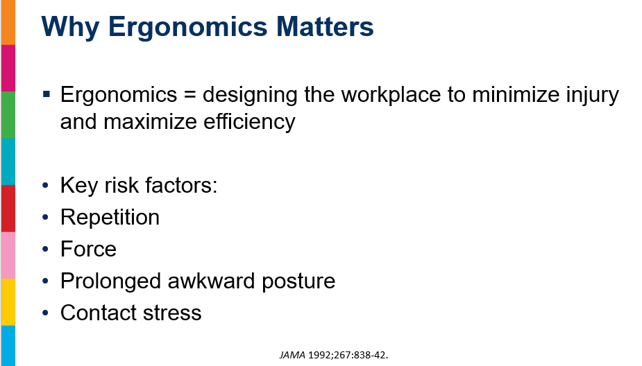
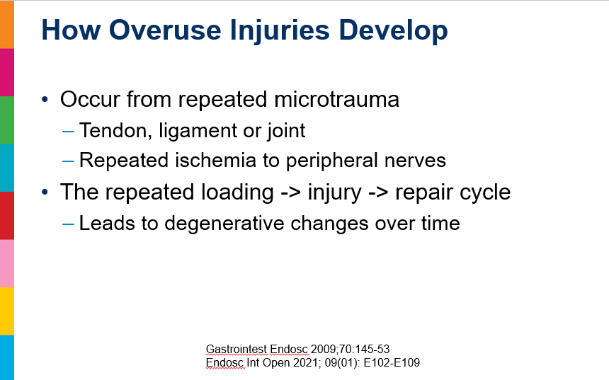
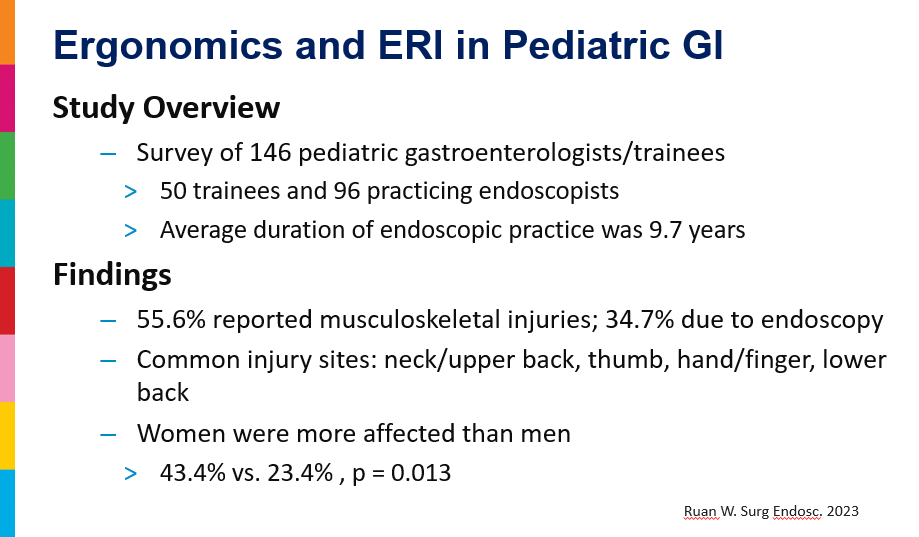
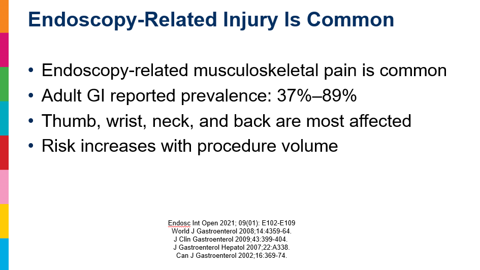
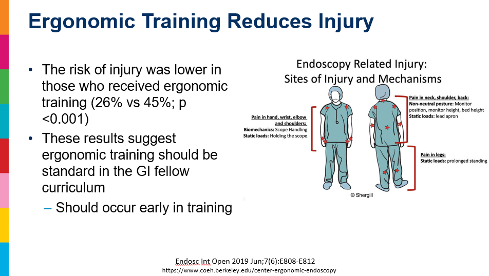
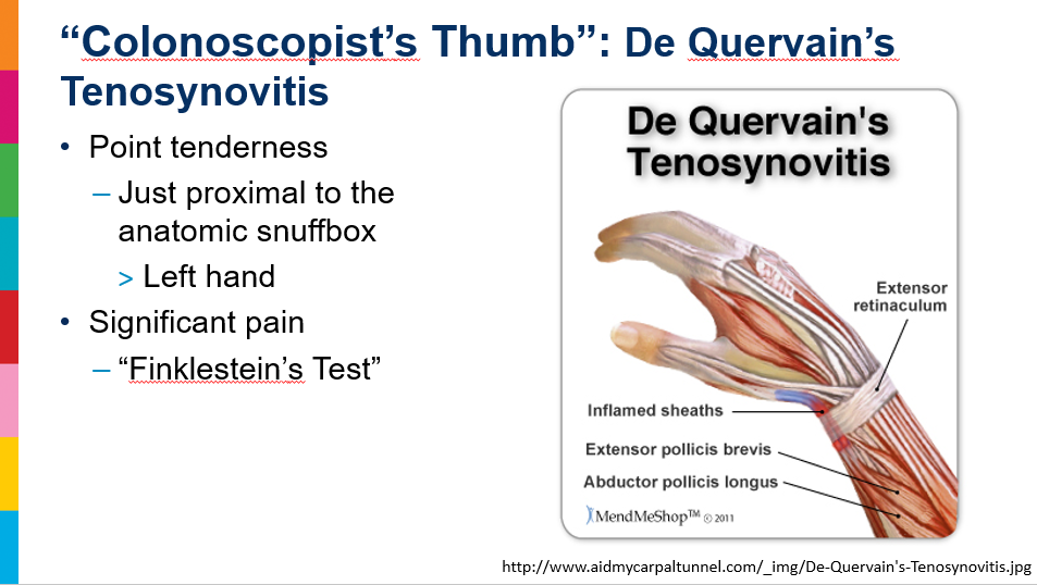
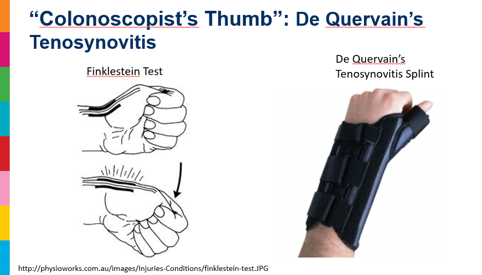
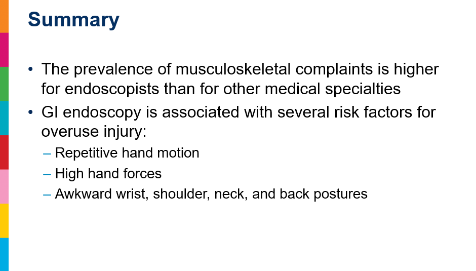
  • A high percentage of gastroenterologists/pediatric gastroenterologists develop injuries related to repetitive endoscopy procedures.  These can be mitigated using proper ergonomics.  Although this advice is pretty straight forward, a lot of endoscopists could benefit from careful attention to this.
Areas included neck/upper back (44.0%), thumb (42.0%), hand/finger (38.0%), lower back (36.0%).
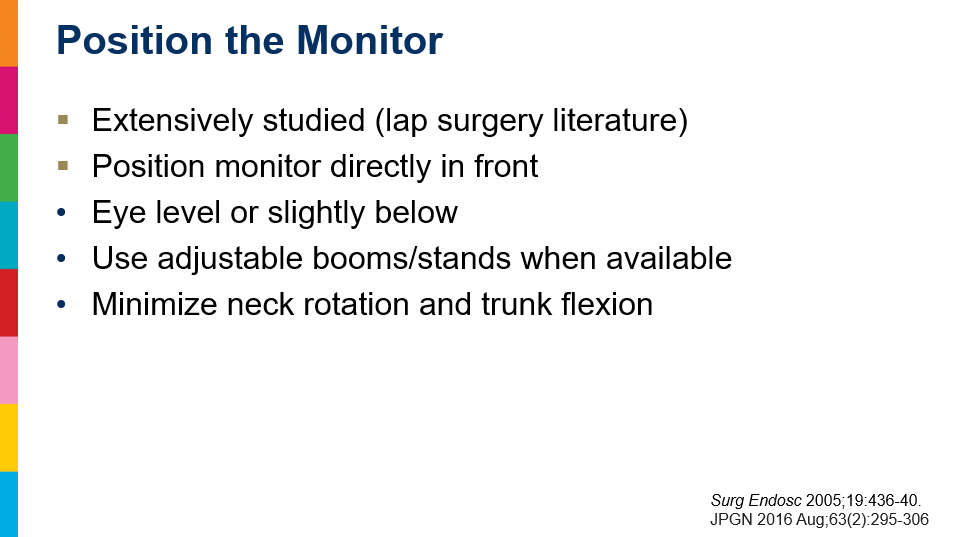
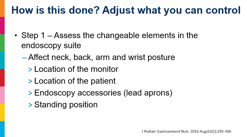
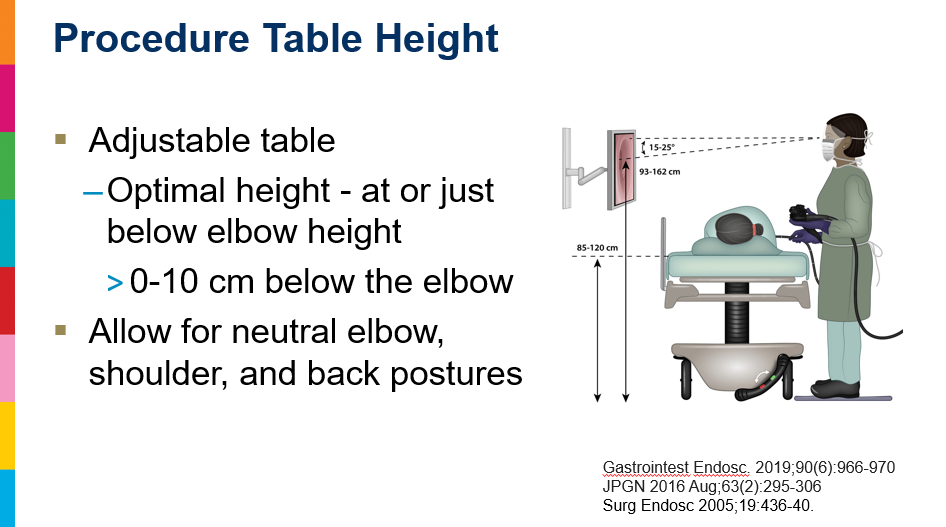
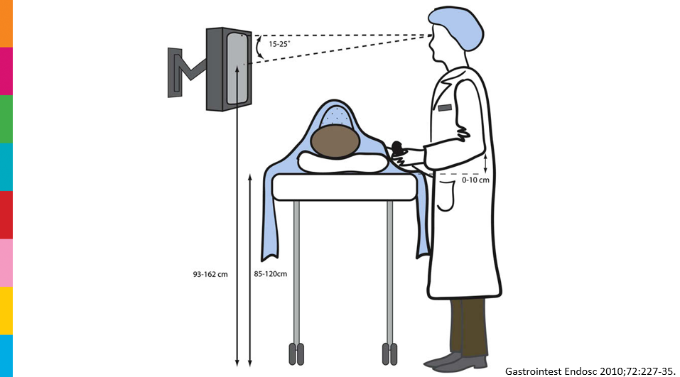
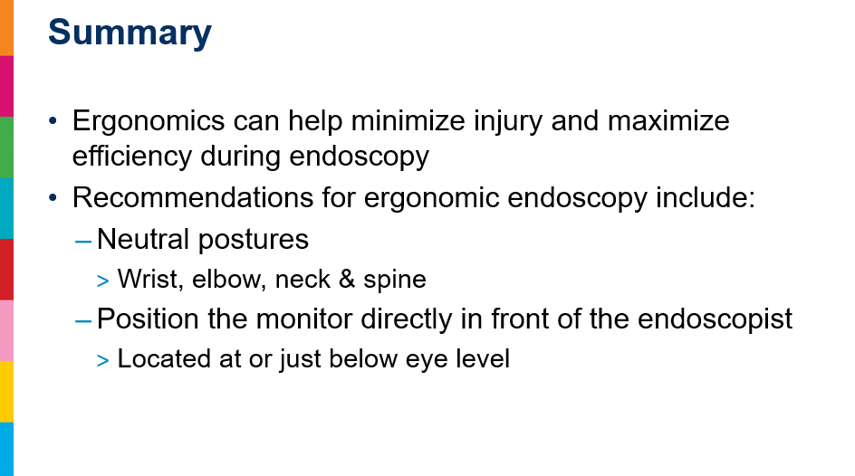
  • Keep Monitor near eye level and in front of you (don’t torque your neck)
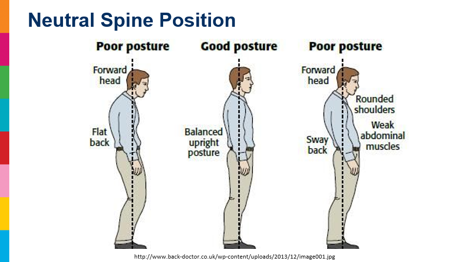
  • Maintain good posture. Can start by standing upright against a wall
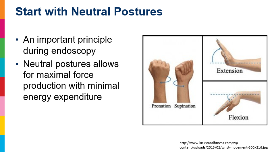
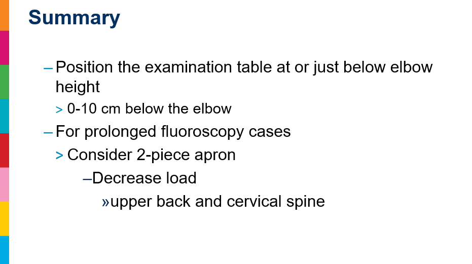
  • Position bed so elbows can be in a fairly neutral position with bed not more than 10 cm below elbows
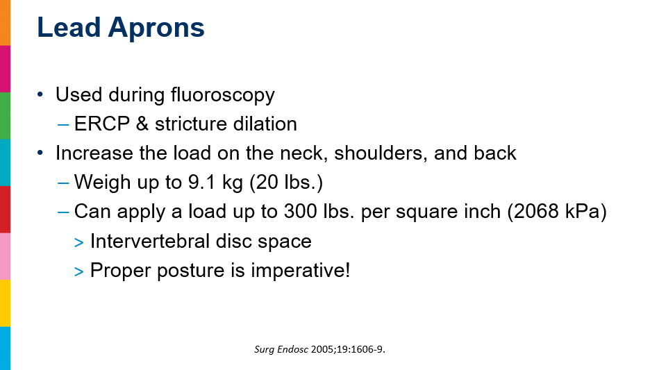
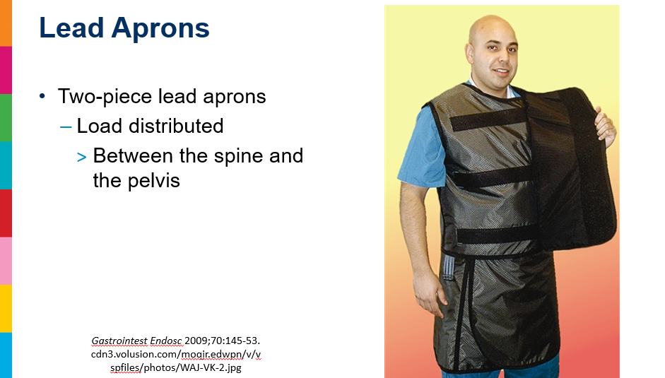
  • Two-piece lead aprons helpful to distribute weight better
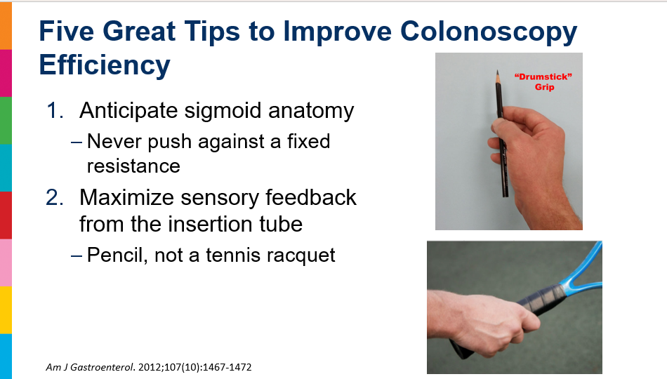
  • Handle endoscope more like holding a pencil than using a firm grip
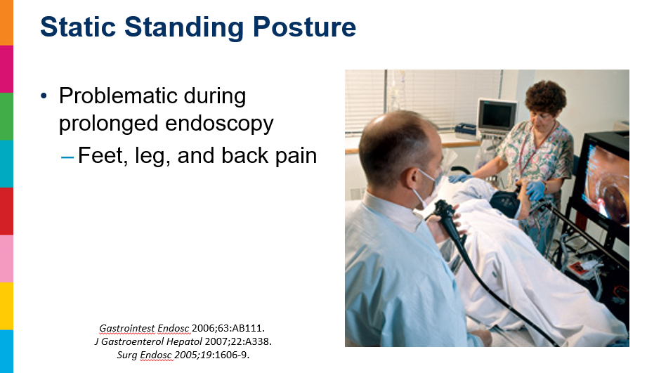
  • Cushioned mat and cushioned soles can help with standing posture/fatigue

Dr. Michael Wilsey: Endoscopy Pearls and Ergonomics (Part 1)

Dr. Michael Wilsey gave our group an excellent update on Endoscopy Pearls and Ergonomics. My notes below may contain errors in transcription and in omission. Along with my notes, I have included many of his slides.
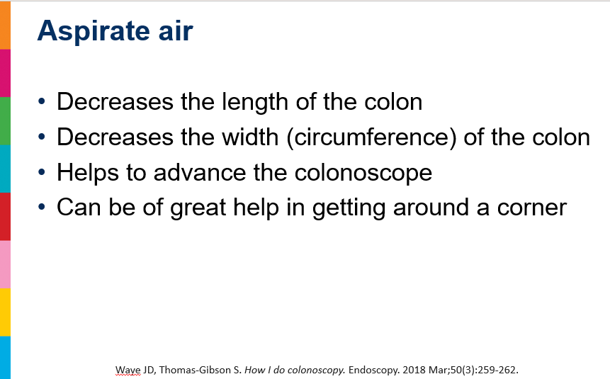
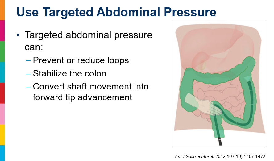
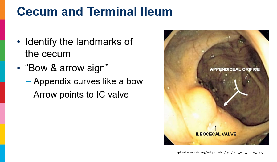
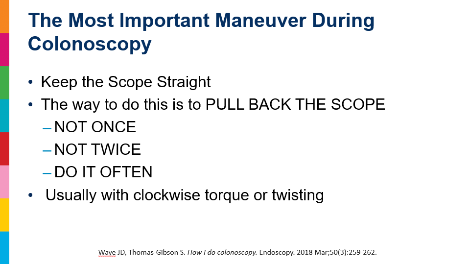
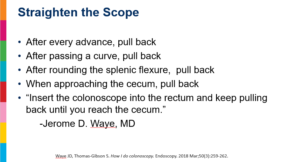
Endoscopy Pearls:

  • Video Link to show techniques:
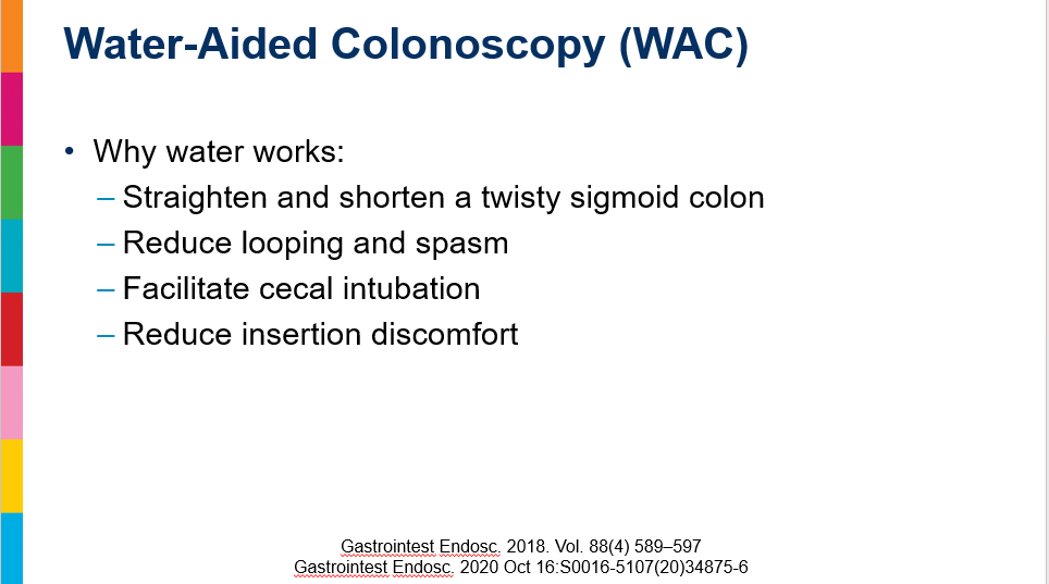
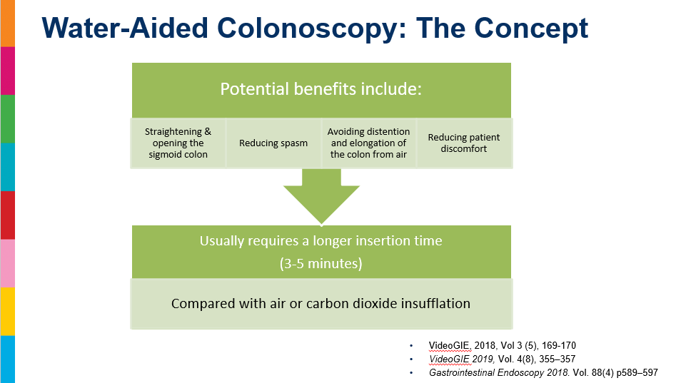
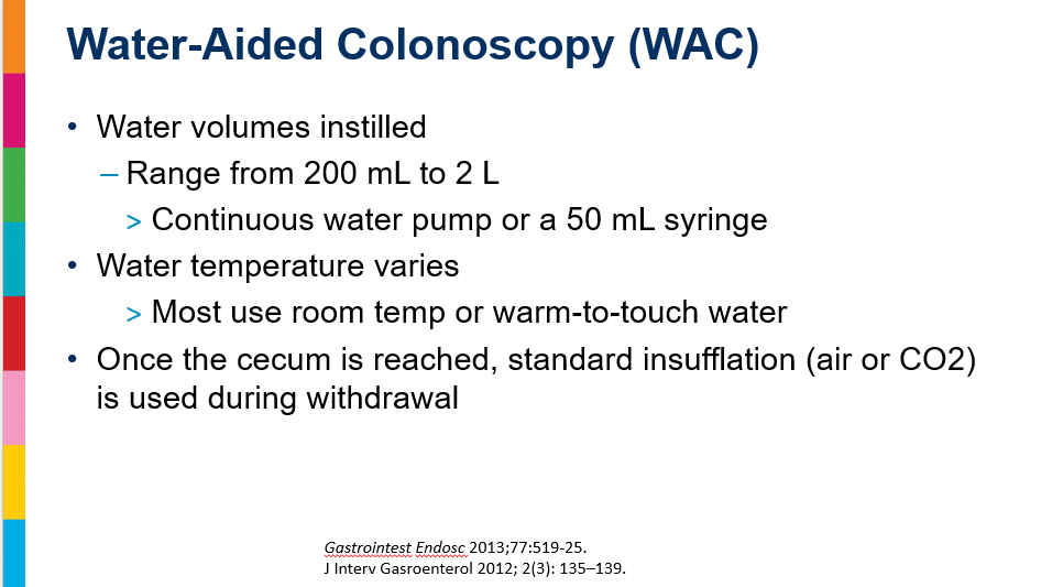
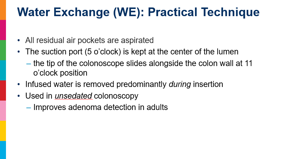
    • YouTube (~6 minute): How to Perform a Water-aided colonoscopy. Sergio Cadoni, Sauid Ishaq, VideoGIE, Volume 3, Issue 5, 2018; Pages 169-170.
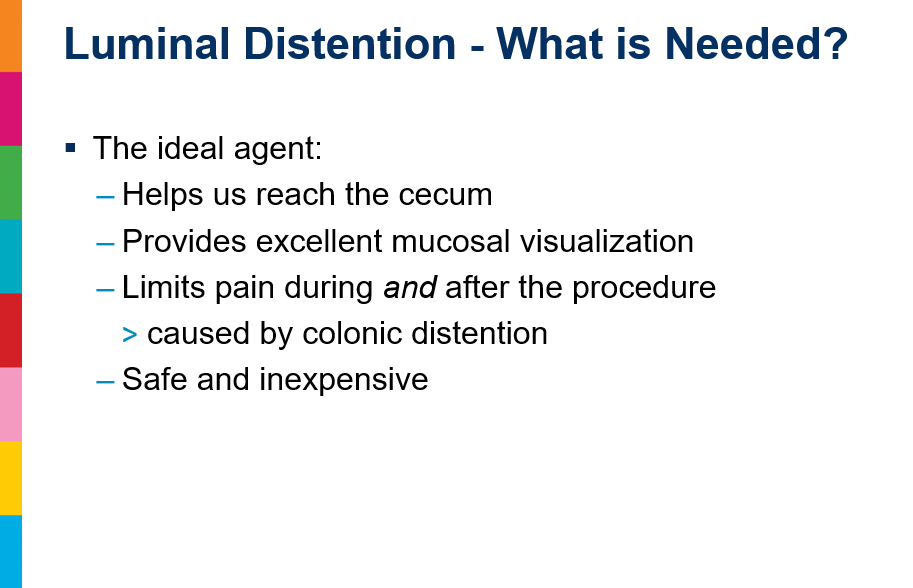
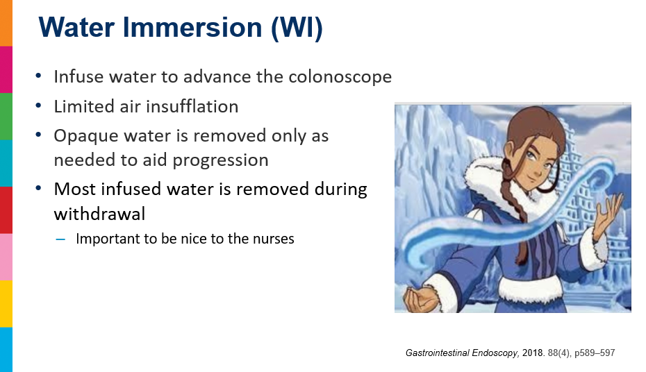
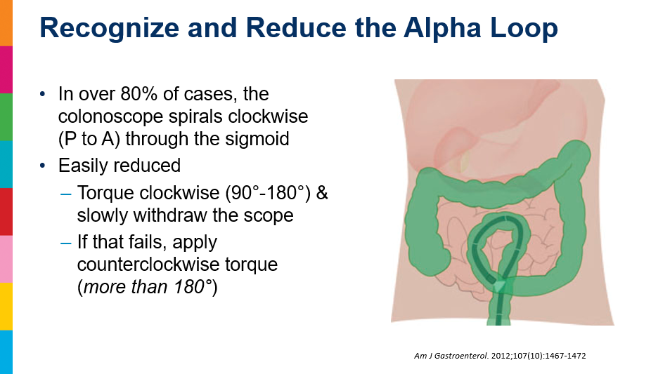
    • Residue can be suctioned while infusing more water, which helps to clean the colon, but if there is too much residue, it is very difficult to see clearly enough to insert the colonoscope properly.
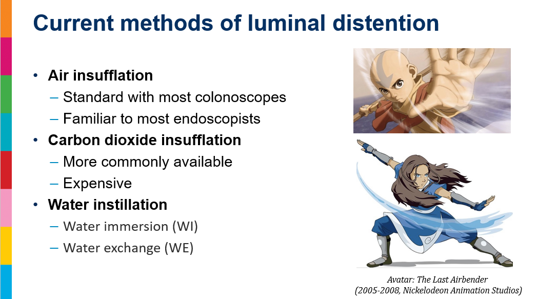
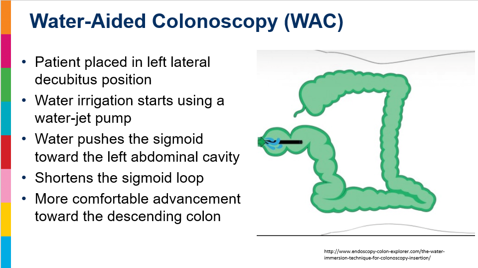
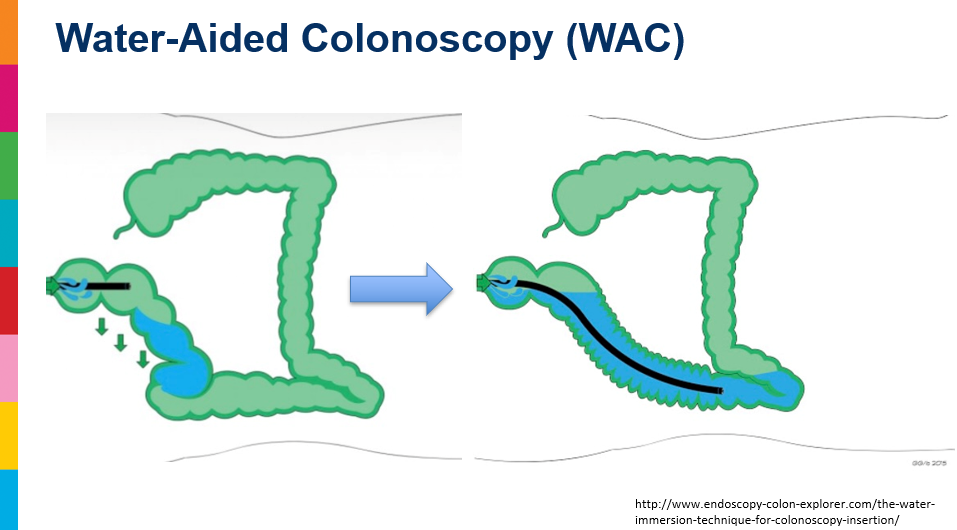
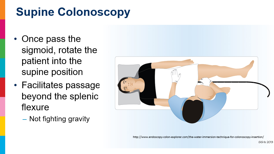
    • The water immersion technique starts with the patient in the left lateral position so that the progressive irrigation of water eases down the sigmoid by gravity on the left abdominal quadrant. Instead of syringes, the use of water flushing pumps allows one to better adjust the quantity of water needed according to the morphology of the sigmoid and does not delay the maneuvers of scope progression.
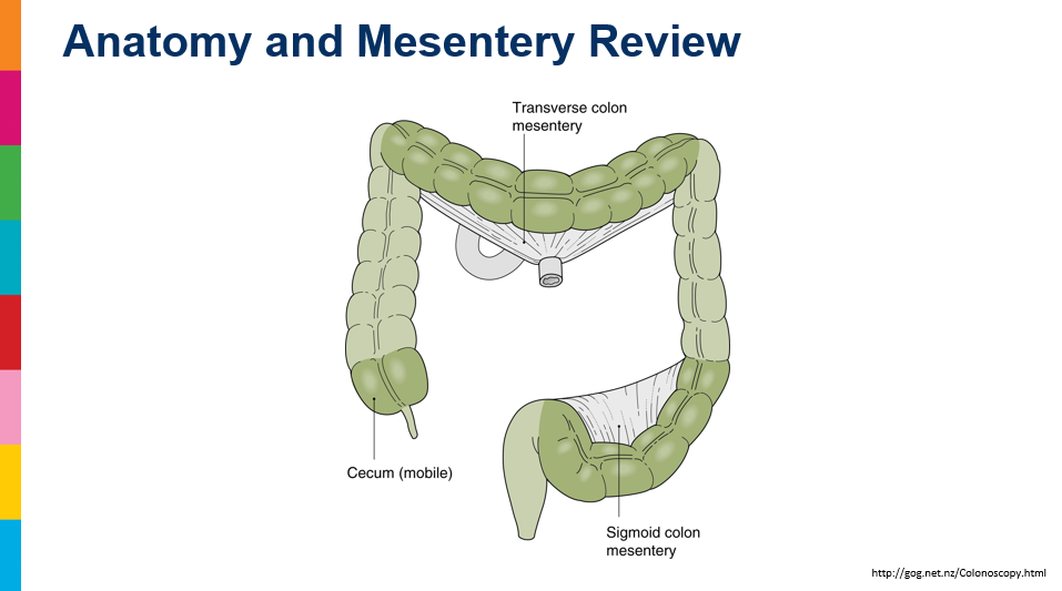
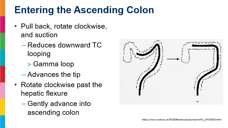
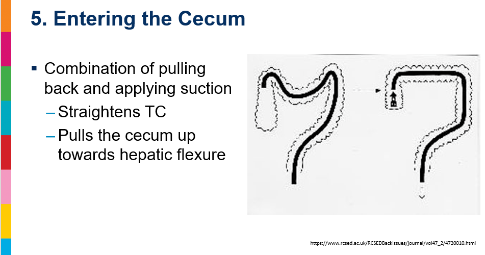
  • Anatomy: sigmoid and transverse colon are more mobile. They are suspended by mesenteries (the transverse mesocolon and sigmoid mesocolon), which allow them to swing or shift freely in the abdomen, unlike the other fixed parts of the colon.

Related blog posts:

Optimizing PPI Therapy for Eosinophilic Esophagitis with CYP2C19 Genotyping

P Bose et al. American Journal of Gastroenterology. DOI: 10.14309/ajg.0000000000004117 Dose May Matter: CYP2C19 Genotype and Proton-Pump Inhibitor Response in Pediatric Eosinophilic Esophagitis

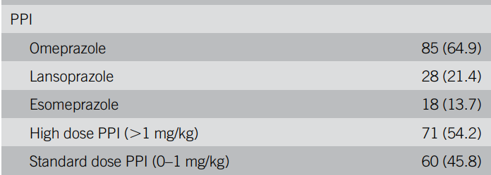
Methods: A cohort study of pooled data from 2 tertiary-care pediatric centers (Riley Children’s Health,Indianapolis, IN, and Children’s Hospital of Philadelphia, Philadelphia, PA) was conducted. N=131, mean age 8.5 yrs. Individuals with certain CYP2C19 polymorphisms were classified as normal (NMs), intermediate/slow(IMs), and rapid metabolizers (RMs) of PPIs based on the presence of normal function (*1), loss-of-function (*2), or gain-of-function (*17) alleles. PPI choice and dosing:

Key findings:

  • PPI response occurred in 22.1% of subjects (29/131)
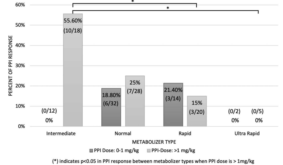
  • Overall, IMs had the highest proportion of PPI response at 33.3% (10/30), followed by NM of 21.7% (13/60) and RM of 14.6% (6/41) (P 5 0.205).
  • No ultra-rapid metabolizers had a response.

Discussion: “Atypical dosing for PPI use in EoE may be related to mechanisms of action apart from gastric acid suppression, such as inhibiting eosinophil migration or restoring esophageal mucosal barrier integrity, which have been previously proposed.”

There may have been a selection bias in this population. The methods section does not detail these cohorts precisely. It is unclear if all patients in these centers undergo CYP2C19 genotype testing. In most centers, CYP2C19 genotype testing is uncommon and may be more likely in those who have not responded to therapy.

My take:

  1. CYP2C19 genotype testing may help determine whether PPI therapy is likely to work for a patient with EoE and influence the dosage selected. This is not a new concept. It was noted at a NASPGHAN meeting in 2017.
  2. In those with unfavorable CYP2C19 genotype, either an alternative therapy or a PPI that is not metabolized with CYP2C19 (eg. rabeprazole) should be considered.

Related blog posts:

AI Diagnosis of Achalasia on Plain Chest X-ray

T Ochaiai et al. Clin Gastroenterol Hepatol 2026; 24: 2308-2310. Open Access! Artificial Intelligence-Based Detection of Achalasia on Plain Chest Radiography

Methods: This retrospective study collected posteroanterior plain chest radiographs of
patients with and without achalasia. The training and validation datasets comprised 447
chest radiographs taken between January 2017 and March 2023.

Key findings:

  • In the validation dataset, the area under the curve for identifying achalasia was 0.971, and using Youden’s index, the sensitivity, specificity, accuracy, and positive and negative predictive values were 0.950, 0.917, 0.932, 0.905, and 0.957, respectively
  • In the temporal test dataset, the area under the curve for detecting achalasia was 0.964, and using Youden’s index, the sensitivity, specificity, accuracy, and positive and negative predictive values were 0.941, 0.891 , 0.901, 0.696 , and 0.983, respectively.
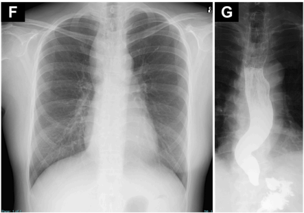
In Figure F, the CXR corresponds to the findings in the barium esophagogram in G

Discussion:

  • It may help detect patients with early achalasia with mild symptoms who are unlikely to undergo EGD. However, the rarity of the condition may result in many false positives.

My take: In this cohort, AI was developed and validated to detect achalasia on chest radiographs. This is yet another example of how AI can yield additional information from routine testing. Previously, AI has been shown to potentially identify diabetes from routine CXR (Emory News 2023: AI model enables earlier detection of diabetes through chest x-rays).

Related blog posts:


Complimentary, Alternative, and Integrative Medical Therapie Use in a Pediatric DGBI Cohort

A VonAxelson et al. J Pediatr Gastroenterol Nutr. 2026;83:282–284. Patterns in integrative medicine usage among pediatric patients in a disorders of gut–brain interaction clinic

In a retrospective cohort (n=331, mean age 15 years) from a DGBI clinic, the patterns of integrative medicine usage were evaluated. The most common comorbidities in the sample were anxiety (61%), sleep disturbance (36%), and depression (32%). The most common DGBI diagnoses in the study were FD (58%) and IBS (57.4%), with FAP-NOS being third most frequent 10.6%).

Key findings:

  • In total, 59% of patients were receiving some form of integrative treatment
  • Peppermint oil, caraway oil, and acupuncture were the most used in this practice. 

Limitations: “While this study does identify trends in IM strategies for management of DGBI, it is limited by retrospective design, so identifying use of many IM techniques may be underrepresented (ginger, L-glutamine, aloe vera, etc). These might have been used but were not reported or recorded in charts.”

My take: Whatever you want to call it, complementary, alternative or integrative, it is clear that many patients are using non-traditional therapies trying to improve their symptoms. This cohort may have higher use as they are from a specialty multidisciplinary DGBI clinic rather than a general pediatric gastroenterology or general pediatric cohort.

Related blog posts:

Discordinate Recommendations for Celiac Screening with Down Syndrome

VN Dargenio et al. J Pediatr Gastroenterol Nutr. 2026;83:304–313. A systematic review of celiac disease recommendations for children with Down syndrome

Background: “CD [celiac disease] prevalence in DS is higher than in the general population.10 The overlapping symptoms between CD and DS, such as growth failure, fatigue, GI disturbances, and, less frequently, neurological and behavioral problems, pose significant diagnostic challenges, as these are often attributed to underlying DS comorbidities, leading to delays in recognizing CD.4, 11 

Guidelines:

The authors note wide variation in the prevalence of CD in DS in various studies. “A meta-analysis of 31 studies including 4383 individuals found a pooled prevalence of biopsy-confirmed CD of 5.8% (95% CI 4.7%–7.2%), with slightly higher rates in children (6.6%) compared to mixed-age samples (5.1%)31…these findings align with the 5%–13% prevalence range documented in earlier European studies2“

The authors recommend the following strategy:

“Given the high prevalence of asymptomatic and atypical presentations, reliance on symptoms alone is insufficient. A pragmatic strategy should initiate with universal serological screening for all children with DS after gluten introduction (12–24 months of age), followed by periodic re-screening every 2–3 years, using tTG-IgA with total IgA assessment, supplemented by IgG-based assays in the context of the high rate of selective IgA deficiency in DS.”

My take: There are wide discrepancies in the recommendations for screening for celiac disease in asymptomatic individuals with Down syndrome.

Related blog posts: