Celiac Disease: Pro Tips (Part 3)

In June 2024 (special issue), Gastroenterology published an entire issue (193 pages) focused on celiac disease. There was a lot of useful information on almost every aspect of this disease. Below I have summarized some of the points.

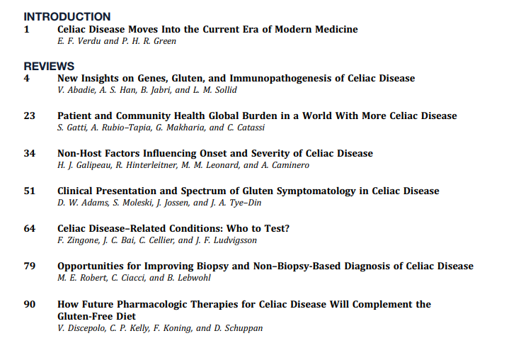
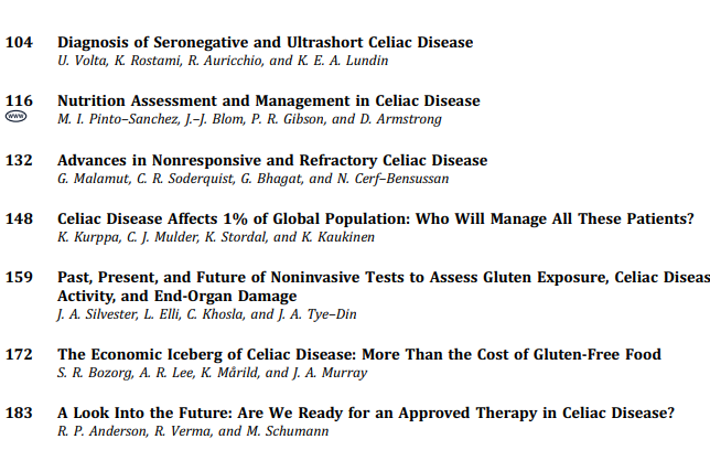
ME Robert et al. Open Access! Opportunities for Improving Biopsy and Non–Biopsy-Based
Diagnosis of Celiac Disease

This article has the most pragmatic advice in the entire issue.

Key points regarding the Non-Biopsy Approach should be applicable in adults:

  • “Retrospective studies in adults found that, in different clinical settings, TTG IgA raised to 10 or more times the upper normal value predicts the presence of mucosal atrophy from 95.2% in low-risk populations to 100% in high-risk populations. There is evidence that the magnitude of the TTG increase correlates with the risk of more severe damage at histology.”
  • “A subsequent prospective multicenter, international study added further evidence for a non-biopsy approach. This study confirmed that a 5-fold and a 10-fold increase of TTG in a high-risk population predicted mucosal atrophy in 97.4% and 97.5%, respectively, when the biopsy was interpreted locally. However, after a central expert pathologist re-evaluated local histology, the PPV of a 10-fold TTG elevation was 99.4%…the only patient [without initial diagnosis of CeD] was eventually diagnosed as having CeD.”
  • The authors discount the rationale for endoscopy in those with TTG IgA >10-fold ULN. 1. Missed CeD-related diagnosis (eg. EoE, lymphocytic gastritis): “The concern about missed endoscopic diagnoses in a biopsy-free approach appears to be a theoretical concern without empiric data.” 2. Concern for CeD complications: “Ulcerative jejunitis and enteropathy-associated T cell lymphoma are rare and, generally, not detected by the initial endoscopy” 3. Overdiagnosis (identification of Potential CeD):”PCeD is rarely found in cases of very high levels of TTG…Moreover, concern about the long-term implications of untreated PCeD may lead to a recommendation for a GFD, rendering moot the need for a biopsy.”
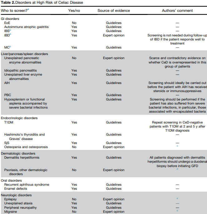
  • Certain populations like those with type 1 diabetes should undergo a biopsy as the serology has a “lower specificity” in this group.
  • There is concern that widespread adoption of a biopsy-free approach may lead to an over-reliance on serologies that fall short of the criteria that would lead to an accurate diagnosis.

Key points about Biopsy-Based Approach:

  • At present, there is broad consensus that among individuals with an elevated TTG IgA that falls short of a 10-fold elevation, a biopsy-based approach is necessary.
  • Obtain appropriate biopsies: guidelines recommend 2 biopsies from the first portion of duodenum and 4 biopsies from the distal duodenum (1 biopsy per pass recommended)

Related blog posts:

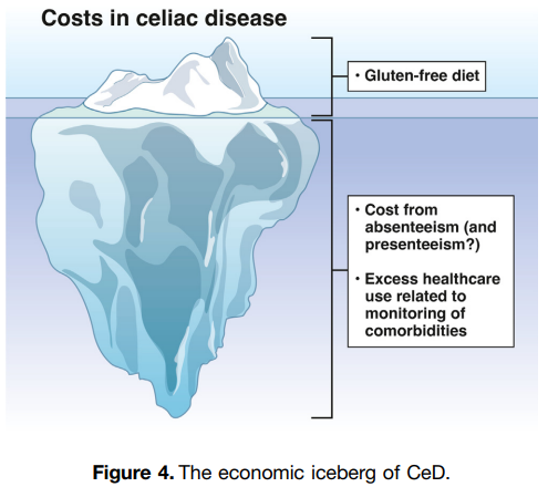
SR Bozorg et al. Open Access! The Economic Iceberg of Celiac Disease: More Than the Cost of Gluten-Free Food

Key points:

  • Long-term data from Sweden have revealed a persisting excess use of health care, with health care costs estimated to be 1.79 times higher than in reference individuals up to 5 years after diagnosis. Similar observations were made by Violato et al, who found UK primary health care costs in patients with incident CeD to be approximately 1.9 times higher than in reference individuals from 5 to 10 years after diagnosis.
  • Data collected from the Swedish national social insurance register showed that working-age patients with prevalent CeD had 1.49 times more work loss than matched reference individuals (42.5 days vs 28.6 days), equivalent to $2800 in lost productivity in 2015

Related blog post: Work Disability with Celiac Disease

Celiac Disease: Pro Tips (Part 2)

In June 2024 (special issue), Gastroenterology published an entire issue (193 pages) focused on celiac disease. There was a lot of useful information on almost every aspect of this disease. Below I have summarized some of the points.

V Disepolo et al. Open Access! How Future Pharmacologic Therapies for Celiac Disease Will Complement the Gluten-Free Diet

  • This article focuses on the emerging pharmacologic treatments
One of the treatment strategies has been to try to sequester gluten. This has included using enzymes to degrade dietary gluten. Other approaches include genetically modifying diet, tight junction modulation, immune modulation and tolerance induction.
Some therapies being developed in adults may have unacceptable risk profiles for children. This could include blocking cytokine signaling (anti–IL-15, anti–IL-15/IL-21) and blocking intestinal T-cell recruitment

Related blog posts:

G Malamut et al. Advances in Nonresponsive and Refractory Celiac Disease

Celiac Disease: Pro Tips (Part 1)

In June 2024 (special issue), Gastroenterology published an entire issue (193 pages) focused on celiac disease. There was a lot of useful information on almost every aspect of this disease. Below I have summarized some of the points.

F Zingone et al. Open Access: Celiac Disease–Related Conditions: Who to Test?

  • The authors detail disorders with increased risk for CeD and which merit screening (see Table 2 below). Some disorders that merit screening that are more obscure include idiopathic pancreatitis, autoimmune hepatitis, delayed menarche, and chronic fatigue.
  • They note that type 1 diabetes mellitus could require serial screening. “Because CeD can manifest at any time and with greater frequency during the initial 5 years, conducting additional screenings in CeD-negative T1DM patients 2 and 5 years after T1DM diagnosis and those who later develop gastrointestinal (GI) symptoms may be advisable.” In addition, it is important to recognize that CeD serology testing is less reliable in patients with T1DM.

S Gatti et al. Patient and Community Health Global Burden in a World With More Celiac Disease, describes the worldwide burden of celiac disease and how to improve detection.

  • They note that the worldwide prevalence is between 0.7% and 2.9%. In this issue, most authors estimate the prevalence to be about 1% with more than 50% undetected.
  • There are many places with higher rates. In U.S. “children in Colorado had a 2.5-fold higher risk compared to Washington State…similar regional differences were seen …in Sweden, Finland, and Germany.”
  • They note the burden before and after diagnosis. Before diagnosis/undetected, there can be persistent symptoms, complications (eg. osteoporosis, decreased fertility) and impaired quality of life. Afterwards, there are increased costs of a GFD and psycho-social burden of GFD.
  • In terms of generalized screening compared to case-finding, the authors note that given the number of at-risk groups, the case-finding approach could entail screening >50% of the population.

V Abadie et al. New Insights on Genes, Gluten, and Immunopathogenesis of Celiac Disease, reviews the intricate details of genetic, biochemical, and immunologic studies, which together have revealed mechanisms of gluten peptide modification and HLA binding, thereby enabling a maladapted anti-gluten immune response.

  • What I was most interested in was the mechanisms behind ‘potential’ celiac disease (PCeD) in which patients have autoimmunity (+serology) but normal histology. In potential CeD, the anti-CD4+ T-cell response is present but decoupled from tissue cytotoxicity. However, notably, IL-21, a cytokine produced by gluten-specific CD4+ T cells in active CeD, is not up-regulated in potential CeD…In addition, patients with potential CeD lack the presence of an epithelial stress response associated with IL-15, HSP70, and HSP27 upregulation in epithelial cells.” “The presence of epithelial stress is a crucial prerequisite for the development of tissue damage.”

How Closely Related Are Eosinophilic Gastrointestinal Disorders To Isolated Eosinophilic Esophagitis

H Sato et al. Clin Gastroenterol Hepatol 2024; 22: 1531-1534. (Research Letter) Eosinophil Involvement Outside the Esophagus in Eosinophilic Esophagitis

The authors retrospectively studied a cohort of children (n=782) with eosinophilic gastrointestinal disorders (EGID) including 592 with isolated eosinophilic esophagitis (EoE), 190 with EGID which included esophageal involvement (“EI”) and 35 EGD without esophageal involvement (“Non-EI”).

Key findings:

  • Abdominal pain was more frequent in EI than isolated EoE: 61% vs 45%, p=.002)
  • EI patients had more dense esophageal eosinophil infiltrate (peak) than isolated EoE: 115 vs 92; P=.036) and higher peripheral blood eosinophil level (0.44 vs 0.38; p=0.027)
  • 94-Gene expression profiles from esophageal biopsies from a 168 subgroup showed that EoE and EI were not significantly different. The heat map for upregulated and downregulated gene expression was different based on disease activity but not significantly different between EoE or EI in either state.

My take (borrowed from authors): The similar molecular transcriptome between EI and EoE are indicative of a shared pathogenesis.

Related blog posts:

Colorado River near Moab

Tirzepatide for Metabolic Dysfunction–Associated Steatohepatitis (MASH) & Uptick in GLP1 Use

R Loomba et al. NEJM 2024; 391; 299-310. Tirzepatide for Metabolic Dysfunction–Associated Steatohepatitis with Liver Fibrosis

F Kanwal. N Engl J Med 2024;391:371-372. Dual Agonists for Management of Metabolic Dysfunction–Associated Steatohepatitis (Associated editorial)

Background: The efficacy and safety of tirzepatide, an agonist of the glucose-dependent insulinotropic polypeptide and glucagon-like peptide-1 receptors, in patients with MASH and moderate or severe fibrosis is unclear.

Methods: This was a phase 2, dose-finding, multicenter, double-blind, randomized, placebo-controlled trial involving participants with biopsy-confirmed MASH and stage F2 or F3 (moderate or severe) fibrosis. Participants were randomly assigned to receive once-weekly subcutaneous tirzepatide (5 mg, 10 mg, or 15 mg) or placebo for 52 weeks. 

My take: Until very recently, there were no effective pharmacologic agents for steatotic liver disease (SLD). Now it appears that there will be multiple effective agents. When newer agents for hepatitis C became available (Harvoni was FDA approved 10 years ago), there were concerns about the high cost. With SLD, this is an even bigger concern give the indefinite treatment period.

From editorial: “Overall, these data are encouraging. Clinicians providing care for patients with MASH will probably have an increasing number of options in their armamentarium. With a growing menu of effective treatments, harms and unacceptable side effects will be important considerations in making treatment decisions.”

Related blog posts:

ACG Review (Zobair Younassi, MD): NAFLD and NASH

Related article: YH Yeo et al. Annals of Intern Med 2024; https://doi.org/10.7326/M24-00. Shifting Trends in the Indication of Glucagon-like Peptide-1 Receptor Agonist Prescriptions: A Nationwide Analysis

Risankizumab Outperforms Ustekinumab

L Peyrin-Biroulet et al. NEJM 2024; 391:213-223. Risankizumab versus Ustekinumab for Moderate-to-Severe Crohn’s Disease

Background: “Interleukin-23 is a heterodimeric proinflammatory cytokine comprising a p40 subunit shared with interleukin-12 and a unique p19 subunit that plays a key role in skin, joint, and gastrointestinal inflammation.16 Ustekinumab and risankizumab are humanized IgG1 monoclonal antibodies; ustekinumab selectively binds p40, and risankizumab selectively binds p19…In head-to-head trials directly comparing their efficacy in psoriasis, risankizumab was superior to ustekinumab, which suggests greater efficacy with p19 blockade than with p40 blockade.”

This “SEQUENCE” trial was a phase 3b, multicenter, open-label, randomized controlled trial with 527 patients with moderate-to-severe Crohn’s disease who either had an inadequate response or had intolerance to anti-TNF agents, received either risankizumab or ustekinumab.

Key Findings:

  • A higher percentage of patients in the risankizumab group than in the ustekinumab group completed all the assigned treatment (90.2% [230 of 255 patients] vs. 72.8% [193 of 265 patients]).  The primary reason for discontinuation of risankizumab was an adverse event (3.1% [8 of 255 patients]), and the primary reason for discontinuation of ustekinumab was lack of efficacy (13.2% [35 of 265 patients]
  • Clinical remission at 48 weeks was 60.8% with risankizumab and 40.8%% with ustekinumab (P<0.001); there were similar rates of glucocorticoid-free clinical remission, 60.8% vs 40.4% respectively. Endoscopic response at 48 weeks was 45.1% and 21.9% respectively.

My take: These head-to-head results showed the superiority of risankizumab over ustekinumab across numerous clinical and endoscopic end points, including glucocorticoid-free clinical remission and endoscopic remission. However, it is still concerning to me that endoscopic remission rates were only 32% at 1 year and that less than half had an endoscopic response.

Related blog posts:

Liver Briefs: Hereditary Angioedema/Liver Transplantation, Bulevirtide/PEG for HDV, and AASLD Cystic Fibrosis Guidance

NE Peters et al. NEJM 2024; 391; 56-59. Normalization of C1 Inhibitor in a Patient with Hereditary Angioedema

“An infant with genetically confirmed hereditary angioedema and low C1 inhibitor levels (but without previous episodes of angioedema) underwent liver transplantation for biliary atresia, an unrelated condition. Liver transplantation led to normalization of the C1 inhibitor level and function. To our knowledge, this represents the first patient to be potentially cured of hereditary angioedema.” This case report shows that liver-directed therapy can reverse hereditary angioedema.

Related blog posts:

T Asselah et al. NEJM 2024; 391:133-143. Bulevirtide Combined with Pegylated Interferon for Chronic Hepatitis D

Key finding: At 24 weeks after the end of treatment, HDV RNA was undetectable in 17% of the patients in the peginterferon alfa-2a group, in 32% of those in the 2-mg bulevirtide plus peginterferon alfa-2a group, in 46% of those in the 10-mg bulevirtide plus peginterferon alfa-2a group, and in 12% of those in the 10-mg bulevirtide group. 

My take: This is a long (48 weeks) and difficult treatment (2 injection meds and lots of peginterferon side effects). However, there is a fairly good response rate.

Related blog post: Image Only: World Hepatitis Day Infographic

ZM Sellers et al. Hepatology 2024; 79(5):p 1220-1238, May 2024. Open Access! Cystic fibrosis screening, evaluation, and management of hepatobiliary disease consensus recommendations

This link to the article also has links to related AASLD guidelines (eg. management of portal hypertension). Table 2 summarizes the ~34 recommendations which include yearly evaluation with labs, ultrasound at least every 2 years in pediatric patients (age 3 yrs and older) and against routine use of ursodeoxycholic acid.

Delivery Vehicle and Outcomes for Budesonide-Treated Eosinophilic Esophagitis

N Lendner, et al. J Pediatr Gastroenterol Nutr. 2024;79:92‐99. Comparison of budesonide vehicles in inducing histologic remission in pediatric eosinophilic esophagitis.

This retrospective study with 111 patients with EoE examined histologic remission with oral viscous budesonide (OVB) and various delivery vehicles (Splenda, honey, syrup or applesauce). Key findings:

  • Overall rate of histologic remission with OVB was 52.6% (“which is less than the reported response of approximately 66% for topical steroid therapy”).
  • There was no difference in rates of histologic remission or response in mid and distal esophagus, respectively) among the different vehicle types or treatment regimens. Similarly, there was no difference in endoscopic remission or response

My take: It does not seem to matter which delivery vehicle is used for OVB; thus, clinicians should aim for more palatable and cost‐effective vehicles.

Related blog posts:

Arches National Park

Updated Diagnostic Accuracy of Serum Matrix Metalloproteinase-7 (MMP-7) for Biliary Atresia

S Pandurangi et al. Hepatology 2024; 80: 152-162 Open Access!.Diagnostic accuracy of serum matrix metalloproteinase-7 as a biomarker of biliary atresia in a large North American cohort

Methods: MMP-7 was measured in serum samples of 399 infants (North America)18 with cholestasis in the Prospective Database of Infants with Cholestasis study of the Childhood Liver Disease Research Network, 201 infants with BA and 198 with non-BA cholestasis (age median: 64 and 59 days, p = 0.94). MMP-7 was assayed on antibody-bead fluorescence (single-plex) and time resolved fluorescence energy transfer assays.

Key findings:

  • On the single-plex assay, MMP-7 generated an AUROC of 0.90. At cutoff 52.8 ng/mL, it produced sensitivity = 94.03%, specificity = 77.78%, positive predictive value = 64.46%, and negative predictive value = 96.82% for BA.
  • MMP-7 outperformed other parameters. AUROC for gamma-glutamyl transferase = 0.81 (CI: 0.77–0.86), stool color = 0.68 (CI: 0.63–0.73), and pathology = 0.84 (CI: 0.76–0.91).  Obstructive features on pathology were the second-best predictor of BA.
  • GGT cutoff was 267.5 U/L (per personal communication with senior author) with sensitivity of 86.6%, and specificity of 77.4%
  • Similar results were found with TR-FRET assay with cut-off of 18.2 ng/mL.
  • 6% (False-negatives) of BA patients had MMP-7 levels below the cutoff
  • 22% (False-positives) of non-BA patients had MMP-7 levels above the cutoff. This included 7 of 8 choledochal cyst patients, 8 of 17 with A1AT, and 13 of 98 with indeterminate cholestasis

In the discussion, the authors note that MMP-7 has performed better in studies with Asian populations, MMP-7 could be useful for dried blood spots in newborns, and could be useful as a measure of successful HPE; continued elevation of MMP-7 has been associated with hepatic fibrosis.

My take: This study shows that MMP-7 is not a perfect assay but often quite helpful. The exact cutoff depends on the specific assay that is utilized. Also, this study shows that checking for A1AT and checking an ultrasound to exclude choledochal cyst need to continue to be done early in the evaluation process.

Related blog posts:

Disclaimer: This blog, gutsandgrowth, assumes no responsibility for any use or operation of any method, product, instruction, concept or idea contained in the material herein or for any injury or damage to persons or property (whether products liability, negligence or otherwise) resulting from such use or operation. These blog posts are for educational purposes only. Specific dosing of medications (along with potential adverse effects) should be confirmed by prescribing physician.  Because of rapid advances in the medical sciences, the gutsandgrowth blog cautions that independent verification should be made of diagnosis and drug dosages. The reader is solely responsible for the conduct of any suggested test or procedure.  This content is not a substitute for medical advice, diagnosis or treatment provided by a qualified healthcare provider. Always seek the advice of your physician or other qualified health provider with any questions you may have regarding a condition.

How to Save a Life

From Caitlin Rivers Newsletter (Force of Infection):

Each year, I share a series of first aid videos to help remind everyone of these life-saving skills. I specifically chose these videos because they are only 2-5 minutes long, so you can get through the entire set on your coffee break. There are plenty of high-quality, longer tutorials on YouTube if you want a deeper dive. Either way, I hope you’ll find some time to review these important lessons.

(Also note that CPR and choking procedures are different for infants, so if you have babies in your life, please look up specific instructions for them!)

These videos are best as a refresher. If first aid skills are new to you, I recommend taking an in-person course. Most community centers offer classes for free or at a low cost. Don’t skimp on these valuable skills—they could make all the difference.

This post is public so feel free to share it.


Her newsletter allowed links by clicking image. To access the videos from this post, clink on the link rather than the image.

Link: Recognizing drowning

Link: CPR for adults and children + using an AED

Link: CPR 1-12 yrs of age

Link: How to Use an AED

Link: How to Stop Severe Bleeding

Link: Choking rescue