How Quickly Does Upadacitinib Work for Crohn’s Disease Symptoms?

JF Colombel. et al. Clin Gastroenterol Hepatol 2024; 22: 1668-1677. Open Access! Upadacitinib Reduces Crohn’s Disease Symptoms Within the First Week of Induction Therapy

This study was a post hoc analysis included pooled data from 2 phase 3, multicenter, double-blind, 12-week induction trials (U-EXCEL and U-EXCEED) and 1 maintenance trial (U-ENDURE). The study included 1021 patients with Crohn’s disease (CD) (n = 674 UPA45; n = 347 PBO).

Key findings:

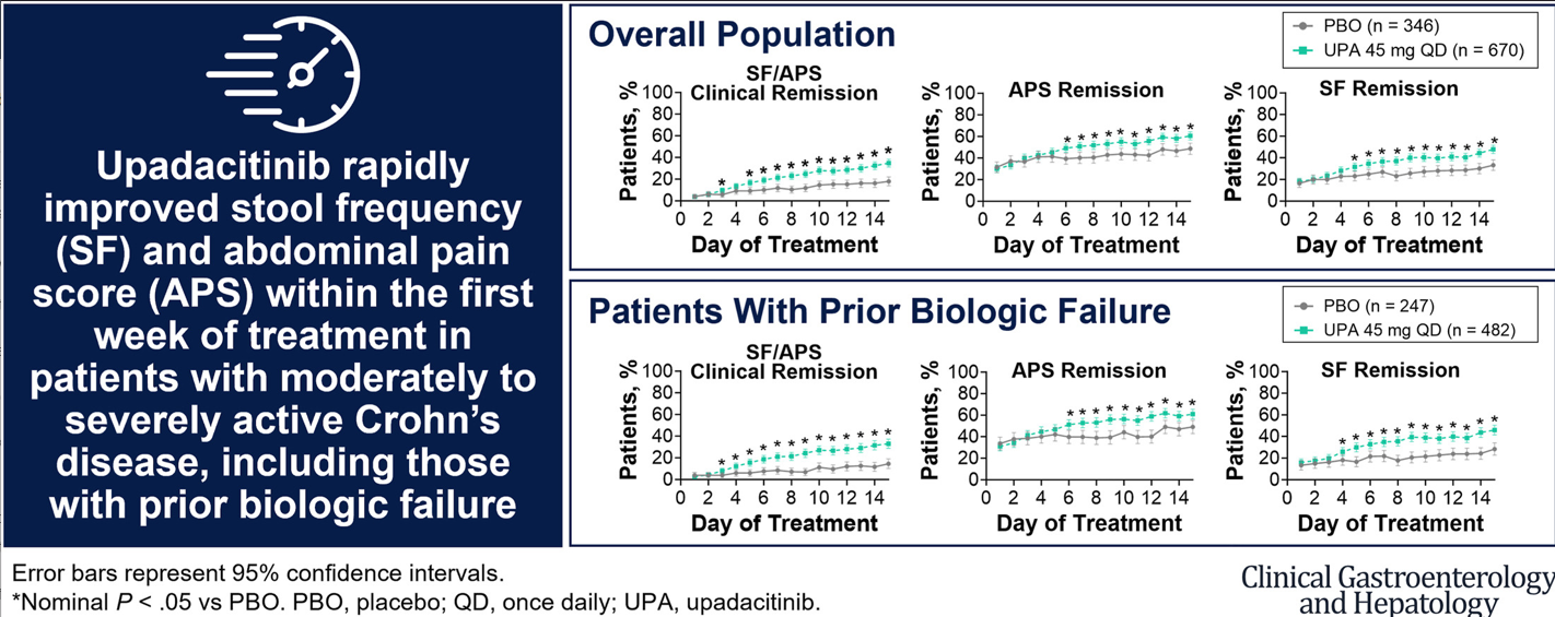
  • Upadacitinib 45 mg taken once daily resulted in rapid relief from CD symptoms within 5 to 6 days of treatment initiation and improved clinical outcomes starting at week 2.
  • The present analysis demonstrates symptomatic relief as early as day 5 to 6 for patients receiving UPA, with 16.7% of patients experiencing daily SF/APS clinical remission by day 5. 
  • The first achievement of daily stool frequency/abdominal pain score (SF/APS) clinical remission occurred earlier with UPA45 (median, 13 d) vs PBO (median, 32 d)
  • Patients treated with UPA45 showed improved rates of SF/APS clinical remission (21.1% UPA45 vs 8.9% PBO) and clinical response (58.8% UPA45 vs 37.9% PBO) starting at week 2 (both P ≤ .01).

In their discussion, the authors note that time to response to treatment with upadacitinib compares favorably to other advanced therapies:

“Vedolizumab resulted in symptomatic improvement within 2 to 4 weeks of treatment initiation16; ustekinumab led to clinical response and remission at week 3 or 6, depending on the dose.17 Similarly, of the time points analyzed, clinical response and/or clinical remission was observed as early as week 2 for risankizumab, 5 infliximab,18 and certolizumab pegol,19 and as early as week 1 for adalimumab.20,21

My take: The rapid response seen in many patients indicate that upadacitinib can be a steroid-sparing therapy in patients with Crohn’s disease.

Unrelated article: E Louis et al. JAMA 2024; doi:10.1001/jama.2024.12414. Risankizumab for Ulcerative Colitis: Two Randomized Clinical Trials

Key findings:  Among the 975 patients with moderate to severe ulcerative colitis, analyzed in the induction trial, 1200 mg of risankizumab significantly increased the rates of clinical remission at 12-week follow-up compared with placebo (20.3% vs 6.2%, respectively). Among 548 patients included in the primary efficacy analysis for the maintenance trial, 180 mg of risankizumab and 360 mg of risankizumab significantly increased the rates of clinical remission (40.2% and 37.6%, respectively) compared with placebo (25.1%).

Related blog posts:

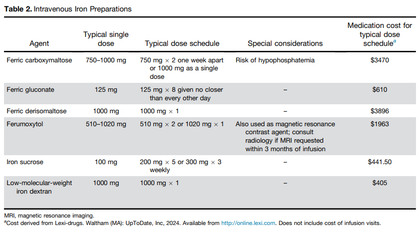
AGA Recommendations For Iron Deficiency Anemia

TG DeLoughery et al. Clin Gastroenterol Hepatol 2024; 22: 1575-1583. Open Access! AGA Clinical Practice Update on Management of Iron Deficiency Anemia: Expert Review

This guideline was developed with adults in mind; however, much of the practice advice is applicable in the pediatric population as well. Here are some of the recommendations:

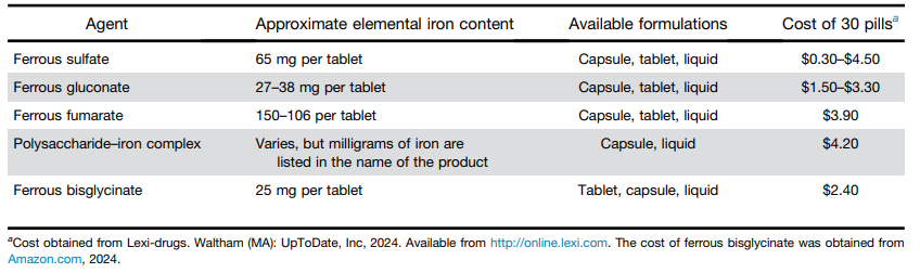
  • Best Practice Advice 1: No single formulation of oral iron has any advantages over any other. Ferrous sulfate is preferred as the least expensive iron formulation.
  • Best Practice Advice 2: Give oral iron once a day at most. Every-other-day iron dosing may be better tolerated for some patients with similar or equal rates of iron absorption as daily dosing.
  • Best Practice Advice 3: Add vitamin C to oral iron supplementation to improve absorption.
  • Best Practice Advice 4: Intravenous iron should be used if the patient does not tolerate oral iron, ferritin levels do not improve with a trial of oral iron, or the patient has a condition in which oral iron is not likely to be absorbed.
  • Best Practice Advice 5: Intravenous iron formulations that can replace iron deficits with 1 or 2 infusions are preferred over those that require more than 2 infusions.
  • Best Practice Advice 6: All intravenous iron formulations have similar risks; true anaphylaxis is very rare. The vast majority of reactions to intravenous iron are complement activation–related pseudo-allergy (infusion reactions) and should be treated as such.

With regard to iron infusion reactions, the authors note the following:

Being truly allergic to IV iron is very rare—almost all reactions are complement activation–related pseudo-allergy, which are idiosyncratic infusion reactions that can mimic allergic reactions.26 For mild reactions, simply stopping the infusions and restarting 15 minutes later at a slower rate will suffice. For more severe reactions, corticosteroids may be of benefit. Diphenhydramine should be avoided because its side effects of mouth dryness, tachycardia, diaphoresis, somnolence, and hypotension can be mistaken for worsening of the reaction.27 Studies have shown that rates of mild reactions are approximately 1:200 and rates of major reactions are approximately 1:200,000.28


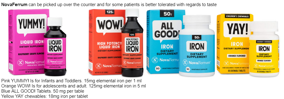
Related information: Our hematologists often recommend Novaferrum (polysaccharide-iron complex) products in children.

Food/diet items with plenty of iron:

  • beef, pork, poultry, and seafood
  • tofu
  • dried beans and peas
  • dried fruits
  • leafy dark green vegetables
  • iron-fortified breakfast cereals, breads, and pastas
  • Use of “lucky fish” (also available at Amazon) while cooking and cooking with cast iron pan can increase iron intake. The lucky fish can be used for 5 years.

Limiting milk consumption can help improve iron absorption.

My take: Iron deficiency anemia is a common issue in pediatric gastroenterology that usually merits evaluation. The AGA practice update provides helpful information with regard to management.

Related blog posts:

Disclaimer: This blog, gutsandgrowth, assumes no responsibility for any use or operation of any method, product, instruction, concept or idea contained in the material herein or for any injury or damage to persons or property (whether products liability, negligence or otherwise) resulting from such use or operation. These blog posts are for educational purposes only. Specific dosing of medications (along with potential adverse effects) should be confirmed by prescribing physician.  Because of rapid advances in the medical sciences, the gutsandgrowth blog cautions that independent verification should be made of diagnosis and drug dosages. The reader is solely responsible for the conduct of any suggested test or procedure.  This content is not a substitute for medical advice, diagnosis or treatment provided by a qualified healthcare provider. Always seek the advice of your physician or other qualified health provider with any questions you may have regarding a condition.

A Treatment for Severe Fatigue Associated with Inflammatory Bowel Disease?

The proverb “Necessity is the mother of invention” is often attributed to Plato. In the dialogue of The Republic, Plato wrote, “our need will be the real creator”. This quote came to mind as I was reading about the use of f for fatigue in inflammatory bowel disease (IBD).

CD Moulton et al. Clin Gastroenterol Hepatol 2024; 22: 1737-1740. Open Access! Modafinil for Severe Fatigue in Inflammatory Bowel Disease: A Prospective Case Series

Background: “Fatigue is highly prevalent in patients with IBD, affecting 72% of patients with active inflammatory bowel disease (IBD) and 47% in remission, and is associated with poor quality of life, absenteeism and presenteeism.1 However, understanding the mechanisms of IBD fatigue remains limited, as reflected in a lack of effective treatments.1

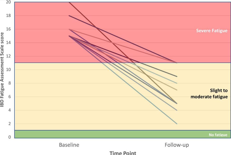
Methods: “Ten patients with IBD and severe fatigue were referred to a consultant psychiatrist. In all cases, mucosal inflammation and organic causes of fatigue (anemia, B12 deficiency, hypothyroidism) had been investigated and treated as much as possible. We measured fatigue severity using the IBD Fatigue Assessment Scale (IBD-FAS), designed specifically for IBD.5 Scores of 11 or higher out of 20 indicate severe fatigue and we only included patients scoring in this range.5

Dosing (for adults) of modafnil is described in the article.

Key findings:

  • “At baseline, the mean IBD-FAS score was 16.0 (SD, 1.7) of 20. After modafinil treatment [at 6 months], the mean IBD-FAS score was 6.7 (SD, 3.0), representing a mean improvement of 58.1% from baseline.”
  • “Although all 10 patients were severely fatigued at baseline, only 2 patients were still in the severe fatigue range after treatment.”
  • “Tolerability was good: 1 patient reported transient headache and 1 patient reported transient dizziness; another patient reported mild palpitations; but none of the patients reported gastrointestinal side effects.”

My take: Perhaps, modafinil will be helpful –pharmacologic therapy for severe fatigue is an unmet need. More studies are needed as this is a small study without a control group.

Related blog posts:

Getting Rid of H pylori Does Not Increase the Risk of Esophageal Adenocarcinoma

A-K Wiklund, et al. Gastroenterol 2024; 167: 485-492. Risk of Esophageal Adenocarcinoma After Helicobacter pylori Eradication Treatment in a Population-Based Multinational Cohort Study

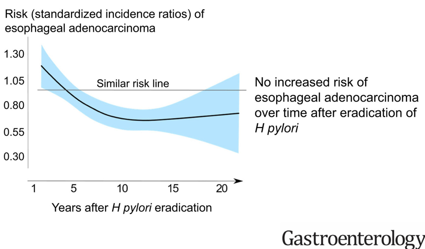
Background: Helicobacter pylori infection is associated with a decreased risk of esophageal adenocarcinoma, and the decreasing prevalence of such infection might contribute to the increasing incidence of this tumor. For this reason, the authors examined the hypothesis that eradication treatment of H pylori increases the risk of esophageal adenocarcinoma.

Methods: Using national registries with Nordic population adults (≥18 years, n=661,987) receiving H pylori eradication treatment from 1995–2018, the authors evaluated 5,495,552 person-years after eradication treatment.

Key findings:

  • The standardized incidence ratios (SIR) did not increase over time after eradication treatment, but rather decreased and was 0.73 at 11–24 years after treatment
  • The overall SIR of esophageal squamous cell carcinoma, calculated for comparison, showed no association (SIR = 0.99)

My take: Eradication of H pylori lowers the risk of gastric cancer. This study shows that treatment does NOT result in an unintended consequence of increasing esophageal cancer.

Related blog posts:

The Link Between Ultra-Processed Foods and Irritable Bowel

S Wu et al. Clin Gastroenterol Hepatol 2024; 22: 1497-1507. Ultra-Processed Food Consumption and Long-Term Risk of Irritable Bowel Syndrome: A Large-Scale Prospective Cohort Study

Methods: Participants (N = 178,711) who completed 24-hour dietary recalls during 2009 to 2012 from the UK Biobank, and free of IBS, celiac disease, inflammatory bowel disease, and any cancer at baseline, were included. During a median of 11.3 years of follow-up, 2690 incident IBS cases were identified.

Key findings:

  • The mean UPF consumption was 21.0% (SD, 11.0%) of the total diet
  • An 8% higher risk of IBS (hazard ratio, 1.08; 95% CI, 1.04–1.12) was associated with every 10% increment of UPF consumption
  • Compared with the lowest quartile of UPF consumption, the highest quartile was associated with a significantly increased risk of incident IBS (hazard ratio, 1.19)

Discussion:

  • “The worsening global epidemic of IBS has coincided with increased dietary consumption of UPF over the past few decades…A similar positive association with IBS (odds ratio of quartile 4 vs quartile 1 = 1.25; 95% CI, 1.12–1.39) was detected in…the French NutriNet-Santé study”
  • High UPF consumption has been associated with “a 42% greater risk of nonalcoholic fatty liver disease and a 22% greater risk of depression.”
  • The authors note that with an observational study, this limits the determination of causality between UPF and IBS

My take: Yet, another study showing that UPF are associated with negative health outcomes. This study reported that UPF consumption was 21% of participants’ diet. In U.S., the current estimate is 60-70% (Source: NPR May 2023: What we know about the health risks of ultra-processed foods). It would be helpful if these foods that are bad for our health did not taste so good!

Related blog posts:

Bryce Canyon

Celiac Disease: Pro Tips (Part 3)

In June 2024 (special issue), Gastroenterology published an entire issue (193 pages) focused on celiac disease. There was a lot of useful information on almost every aspect of this disease. Below I have summarized some of the points.

ME Robert et al. Open Access! Opportunities for Improving Biopsy and Non–Biopsy-Based
Diagnosis of Celiac Disease

This article has the most pragmatic advice in the entire issue.

Key points regarding the Non-Biopsy Approach should be applicable in adults:

  • “Retrospective studies in adults found that, in different clinical settings, TTG IgA raised to 10 or more times the upper normal value predicts the presence of mucosal atrophy from 95.2% in low-risk populations to 100% in high-risk populations. There is evidence that the magnitude of the TTG increase correlates with the risk of more severe damage at histology.”
  • “A subsequent prospective multicenter, international study added further evidence for a non-biopsy approach. This study confirmed that a 5-fold and a 10-fold increase of TTG in a high-risk population predicted mucosal atrophy in 97.4% and 97.5%, respectively, when the biopsy was interpreted locally. However, after a central expert pathologist re-evaluated local histology, the PPV of a 10-fold TTG elevation was 99.4%…the only patient [without initial diagnosis of CeD] was eventually diagnosed as having CeD.”
  • The authors discount the rationale for endoscopy in those with TTG IgA >10-fold ULN. 1. Missed CeD-related diagnosis (eg. EoE, lymphocytic gastritis): “The concern about missed endoscopic diagnoses in a biopsy-free approach appears to be a theoretical concern without empiric data.” 2. Concern for CeD complications: “Ulcerative jejunitis and enteropathy-associated T cell lymphoma are rare and, generally, not detected by the initial endoscopy” 3. Overdiagnosis (identification of Potential CeD):”PCeD is rarely found in cases of very high levels of TTG…Moreover, concern about the long-term implications of untreated PCeD may lead to a recommendation for a GFD, rendering moot the need for a biopsy.”
  • Certain populations like those with type 1 diabetes should undergo a biopsy as the serology has a “lower specificity” in this group.
  • There is concern that widespread adoption of a biopsy-free approach may lead to an over-reliance on serologies that fall short of the criteria that would lead to an accurate diagnosis.

Key points about Biopsy-Based Approach:

  • At present, there is broad consensus that among individuals with an elevated TTG IgA that falls short of a 10-fold elevation, a biopsy-based approach is necessary.
  • Obtain appropriate biopsies: guidelines recommend 2 biopsies from the first portion of duodenum and 4 biopsies from the distal duodenum (1 biopsy per pass recommended)

Related blog posts:

SR Bozorg et al. Open Access! The Economic Iceberg of Celiac Disease: More Than the Cost of Gluten-Free Food

Key points:

  • Long-term data from Sweden have revealed a persisting excess use of health care, with health care costs estimated to be 1.79 times higher than in reference individuals up to 5 years after diagnosis. Similar observations were made by Violato et al, who found UK primary health care costs in patients with incident CeD to be approximately 1.9 times higher than in reference individuals from 5 to 10 years after diagnosis.
  • Data collected from the Swedish national social insurance register showed that working-age patients with prevalent CeD had 1.49 times more work loss than matched reference individuals (42.5 days vs 28.6 days), equivalent to $2800 in lost productivity in 2015

Related blog post: Work Disability with Celiac Disease

Celiac Disease: Pro Tips (Part 2)

In June 2024 (special issue), Gastroenterology published an entire issue (193 pages) focused on celiac disease. There was a lot of useful information on almost every aspect of this disease. Below I have summarized some of the points.

V Disepolo et al. Open Access! How Future Pharmacologic Therapies for Celiac Disease Will Complement the Gluten-Free Diet

  • This article focuses on the emerging pharmacologic treatments
One of the treatment strategies has been to try to sequester gluten. This has included using enzymes to degrade dietary gluten. Other approaches include genetically modifying diet, tight junction modulation, immune modulation and tolerance induction.
Some therapies being developed in adults may have unacceptable risk profiles for children. This could include blocking cytokine signaling (anti–IL-15, anti–IL-15/IL-21) and blocking intestinal T-cell recruitment

Related blog posts:

G Malamut et al. Advances in Nonresponsive and Refractory Celiac Disease

Celiac Disease: Pro Tips (Part 1)

In June 2024 (special issue), Gastroenterology published an entire issue (193 pages) focused on celiac disease. There was a lot of useful information on almost every aspect of this disease. Below I have summarized some of the points.

F Zingone et al. Open Access: Celiac Disease–Related Conditions: Who to Test?

  • The authors detail disorders with increased risk for CeD and which merit screening (see Table 2 below). Some disorders that merit screening that are more obscure include idiopathic pancreatitis, autoimmune hepatitis, delayed menarche, and chronic fatigue.
  • They note that type 1 diabetes mellitus could require serial screening. “Because CeD can manifest at any time and with greater frequency during the initial 5 years, conducting additional screenings in CeD-negative T1DM patients 2 and 5 years after T1DM diagnosis and those who later develop gastrointestinal (GI) symptoms may be advisable.” In addition, it is important to recognize that CeD serology testing is less reliable in patients with T1DM.

S Gatti et al. Patient and Community Health Global Burden in a World With More Celiac Disease, describes the worldwide burden of celiac disease and how to improve detection.

  • They note that the worldwide prevalence is between 0.7% and 2.9%. In this issue, most authors estimate the prevalence to be about 1% with more than 50% undetected.
  • There are many places with higher rates. In U.S. “children in Colorado had a 2.5-fold higher risk compared to Washington State…similar regional differences were seen …in Sweden, Finland, and Germany.”
  • They note the burden before and after diagnosis. Before diagnosis/undetected, there can be persistent symptoms, complications (eg. osteoporosis, decreased fertility) and impaired quality of life. Afterwards, there are increased costs of a GFD and psycho-social burden of GFD.
  • In terms of generalized screening compared to case-finding, the authors note that given the number of at-risk groups, the case-finding approach could entail screening >50% of the population.

V Abadie et al. New Insights on Genes, Gluten, and Immunopathogenesis of Celiac Disease, reviews the intricate details of genetic, biochemical, and immunologic studies, which together have revealed mechanisms of gluten peptide modification and HLA binding, thereby enabling a maladapted anti-gluten immune response.

  • What I was most interested in was the mechanisms behind ‘potential’ celiac disease (PCeD) in which patients have autoimmunity (+serology) but normal histology. In potential CeD, the anti-CD4+ T-cell response is present but decoupled from tissue cytotoxicity. However, notably, IL-21, a cytokine produced by gluten-specific CD4+ T cells in active CeD, is not up-regulated in potential CeD…In addition, patients with potential CeD lack the presence of an epithelial stress response associated with IL-15, HSP70, and HSP27 upregulation in epithelial cells.” “The presence of epithelial stress is a crucial prerequisite for the development of tissue damage.”

Risankizumab Outperforms Ustekinumab

L Peyrin-Biroulet et al. NEJM 2024; 391:213-223. Risankizumab versus Ustekinumab for Moderate-to-Severe Crohn’s Disease

Background: “Interleukin-23 is a heterodimeric proinflammatory cytokine comprising a p40 subunit shared with interleukin-12 and a unique p19 subunit that plays a key role in skin, joint, and gastrointestinal inflammation.16 Ustekinumab and risankizumab are humanized IgG1 monoclonal antibodies; ustekinumab selectively binds p40, and risankizumab selectively binds p19…In head-to-head trials directly comparing their efficacy in psoriasis, risankizumab was superior to ustekinumab, which suggests greater efficacy with p19 blockade than with p40 blockade.”

This “SEQUENCE” trial was a phase 3b, multicenter, open-label, randomized controlled trial with 527 patients with moderate-to-severe Crohn’s disease who either had an inadequate response or had intolerance to anti-TNF agents, received either risankizumab or ustekinumab.

Key Findings:

  • A higher percentage of patients in the risankizumab group than in the ustekinumab group completed all the assigned treatment (90.2% [230 of 255 patients] vs. 72.8% [193 of 265 patients]).  The primary reason for discontinuation of risankizumab was an adverse event (3.1% [8 of 255 patients]), and the primary reason for discontinuation of ustekinumab was lack of efficacy (13.2% [35 of 265 patients]
  • Clinical remission at 48 weeks was 60.8% with risankizumab and 40.8%% with ustekinumab (P<0.001); there were similar rates of glucocorticoid-free clinical remission, 60.8% vs 40.4% respectively. Endoscopic response at 48 weeks was 45.1% and 21.9% respectively.

My take: These head-to-head results showed the superiority of risankizumab over ustekinumab across numerous clinical and endoscopic end points, including glucocorticoid-free clinical remission and endoscopic remission. However, it is still concerning to me that endoscopic remission rates were only 32% at 1 year and that less than half had an endoscopic response.

Related blog posts:

When To Take Fewer Biopsies With Eosinophilic Esophagitis

A Godat et al. Clin Gastroenterol Hepatol 2024: 22: 1528-1530. Eosinophil Distribution in Eosinophilic Esophagitis and its Impact on Disease Activity and Response to Treatment

In this post hoc analysis of the EOS-1 and EOS-2 trials with 263 adult patients, the authors analyzed eosinophil distribution and impact on treatment. Key findings;

  • Peak eosinophil count was highest in the distal esophagus (median 166 eos/mm2) followed by mid esophagus (142) and then proximal esophagus (113). 46% of patients had highest peak eosinophil count in the distal esophagus, 33% in the mid esophagus, and 21% of patients in the proximal esophagus
  • Diagnosis: a biopsy protocol using only distal esophagus would have missed EoE diagnosis in only 13 (4.9%) of patients
  • Remission rates stratified by histologic categories were not statistically different base on disease location: 73% distal esophagus, 76% mid esophagus, 64% proximal esophagus, and 64% diffuse esophageal disease
  • None of the following factors affected treatment outcome: histologic location category, histologic disease severity (peak eos count) and atopic status. For example, treatment failure occurred in 37% without atopy and 30% with atopy

My take: In this study population, separate evaluation of biopsies by location modestly increased the diagnostic yield at baseline. Thus, additional biopsies at disease onset is a good idea. However, the actual distribution of disease activity did not seem to help provide any insight into therapeutic response (to budesonide). Practical implications are that fewer biopsies on follow-up endoscopy may be reasonable to help determine a treatment response.

Related blog posts.