Fundamentals for Ostomy Management

K Mullin, RM Rentea, M Appleby, PT Reeves. Pediatrics in Review 2024; 45: 210-224. Gastrointestinal Ostomies in Children: A Primer for the Pediatrician

Like yesterday’s article on GTs, this is another terrific review with plenty of helpful images and advice regarding ostomy management.

  • Background: “There are upwards of 1 million people living with ostomies (ostomates) in the United States.” “Approximately 75% of all ostomies in the pediatric population are created in the neonatal or infant patient.”
  • Table 1 lists the purposes and types of ostomies including gastrostomy, jejunostomy, ileostomy, appendicostomy (Malone), cecostomy, colostomy and urinary diversions (eg. Mitrofanoff).
  • Surgical considerations are reviewed including optimizing nutrition preoperatively and minimizing corticosteroids. Biologics: “The most recent evidence does not support a delay in gastrointestinal surgery for children with IBD receiving biological therapy…[and] typically, biological therapy can be resumed 14 to 28 days after the operation.” For oral small molecules (with short half-lives), these may be restarted sooner if indicated.
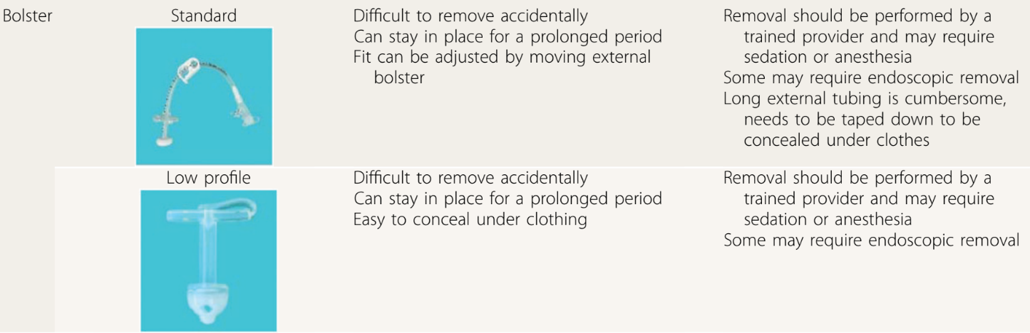
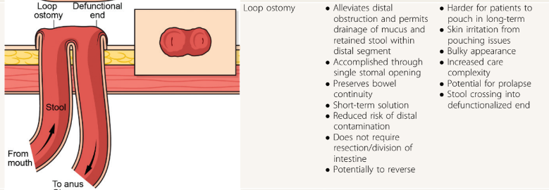
  • Table 2 provides pictures of the lower ostomies. For example:
  • Postoperative care is discussed including healing times, need for wound ostomy nurse input, and addressing self-image. Patients with motility disorders are “more likely to experience postoperative complications”
  • Table 4 details the products for pouch care including pouching systems, skin barriers, pouch liner, gas vent, pouch lubricant, pouch covers, and adhesive remover.
  • Table 5 summarized ostomy-related complications and treatments. Complications include stomal necrosis, stomal bleeding, stomal retraction, mucocutaneous separation, parastomal hernia, stoma prolapse (can apply cool compresses, apply osmotic agent (sugar) or manually reduce), stoma stenosis, and dermatitis.
  • Table 6 addresses medical management issues like odor, blockage, diarrhea, and constipation. This table also provides recipes for antegrade enemas (see below) and links including a very useful bowel management guide for families (28 pg from Boston Children’s) and enema ingredients and supplies (2 pg from Seattle Children’s); the latter has some overlapping information with the former.
  • At the conclusion of the article, there is further discussion of systemic and ostomy-related complications (much of which is summarized in Table 5). The article references the Ostomy Skin Tool as a metric to follow the clinical state of the ostomy. The United Ostomy Associations of America (ostomy.org) is listed as a good resource (which it is!).

My take: This is a very useful resource. Even a quick read will make clinicians appreciative of having the assistance wound ostomy nurses.

Related blog posts:

Disclaimer: This blog, gutsandgrowth, assumes no responsibility for any use or operation of any method, product, instruction, concept or idea contained in the material herein or for any injury or damage to persons or property (whether products liability, negligence or otherwise) resulting from such use or operation. These blog posts are for educational purposes only. Specific dosing of medications (along with potential adverse effects) should be confirmed by prescribing physician.  Because of rapid advances in the medical sciences, the gutsandgrowth blog cautions that independent verification should be made of diagnosis and drug dosages. The reader is solely responsible for the conduct of any suggested test or procedure.  This content is not a substitute for medical advice, diagnosis or treatment provided by a qualified healthcare provider. Always seek the advice of your physician or other qualified health provider with any questions you may have regarding a condition.

Fundamentals of Gastrostomy Tubes

I Novak, NK Velazco. Pediatrics in Review; 2024: 45: 175-187. Gastrostomy Tubes: Indications, Types, and Care

Thanks to Patrick Reeves for sharing this reference. While reading this article, I was reminded of Debbie Mason RN. She worked mainly for Colin Rudolph when I was a fellow at Cincinnati. She would teach the fellows the ‘ins and outs’ regarding gastrostomy tubes (GTs).

Some of the points from this article:

  • Background: In one large study, 25% of hospitalized pediatric patients in 63 U.S. hospitals required a temporary NG tube. GTs “preferred for longer-term access due to being less prone to accidental dislodgement, blockage, and interruption of feeds. They are also more durable, discrete, and avoid nasal trauma.” They are probably safer too for longer-term use. (An omission in the article: AMT bridles can help maintain NGs.)
  • Indications: Reviews the extensive list of reasons for GT placement, most related to inadequate nutritional intake (related to many chronic disease processes)
  • Contraindications are reviewed. “Absolute contraindications include active sepsis or peritonitis, massive ascites, uncorrectable coagulopathy, portal hypertension with significant varices, and history of total gastrectomy.” Transoral PEG tube is contraindicated, as well, if pharyngeal or esophageal obstruction, malrotation, or colonic interposition.
  • Preprocedural evaluation is described. Some have recommended UGI prior to placement in those with congenital anomalies. Others have stated that “even congenital anomalies should not mandate an upper GI series given how rare malrotation is in general.” Many children benefit from NG feeding trials prior to GT placement which can also improve nutritional status preoperatively.
  • The placement methods and types of GTs are reviewed. “As of now, there is no clearly identified optimal technique” (eg. laparoscopic GT vs PEG). The others note a meta-analysis of 22 studies (n >5000) found a higher rate of major complications with PEG placement. The authors recommend T-fasteners if GT balloon is used for initial placement.
  • Examples of bolster-type (non-balloon) GTs:
  • GT complications are discussed including infection, peritonitis, bleeding (rare to need a transfusion), injury to adjacent organs, pneumoperitoneum (usually benign and transient), hypergranulation, cellulitis (often treated with a first-generation cephalosporin or topical mupirocin), dislodgement, tube migration, and buried bumper syndrome. For early dislodgement (especially first 4-6 weeks after placement), blind reinsertion should be avoided.
  • GT care: The authors recommend starting feeds “not more than 3 to 6 hours” after placement to monitor for immediate postoperative complications, and cleaning site with warm water, saline or soap. Once the site has healed showering and bathing can resume; swimming can be permitted a few weeks after placement. Medications should be given via gastric port (if GJ) for better absorption of medications and lessen risk of tube clogging.
  • Troubleshooting: This is the most useful part of this article. Advice on peristomal leakage: “Placing larger tubes should be avoided because this will only enlarge the stoma tract…removal of the tube for a few hours can be considered because this permits the tract to start closing.” Leakage is often due to issues with balloon volume, poor fit, gastric pressure (eg dysmotility, gastroparesis) and poor wound healing. Clogging: “Carbonated beverages, juices, and meat tenderizer…studies have not shown these to be effective.” Lukewarm water, left to stand 20 minutes, is the first line agent for declogging. Other options include commercial enzymatic decloggers, and mechanical decloggers.
  • GT removal is discussed and I disagree with the authors that “persistent gastrocutaneous fistulas are rare after removal” (though the majority will close with conservative management). The authors do not recommend cutting GT bolsters due to risk of obstruction and note that endoscopic removal is often necessary.

My take: This article would be well-positioned as part of any GI fellows’ required curriculum and has a bunch of pointers for experienced clinicians as well. It could easily be used for material for ABP questions too. The article is much more detailed than the summary I have provided.

Also, another relevant resource (not discussed in article): Oley Foundation (oley.org). This foundation aims to help those living with home IV nutrition or tube feeding through advocacy, education, community and innovation.

Related blog posts:

Proactive Monitoring Associated with Higher Rates of Transmural Healing

SR Fernades et al. Inflammatory Bowel Diseases, izad272, https://doi.org/10.1093/ibd/izad272 Proactive Infliximab Monitoring Improves the Rates of Transmural Remission in Crohn’s Disease: A Propensity Score–Matched Analysis 

Methods: Retrospective cohort study (n=195) including consecutive CD patients starting treatment with IFX. Rates of transmural remission were compared between patients with and without therapeutic drug monitoring (target level: 5-7 µg/mL).

Key findings:

  • The rates of transmural remission were higher in patients under proactive therapeutic drug monitoring (37.2% vs 18.3%; P = .004) with similar results in the propensity score–matched analysis (34.2% vs 17.1%; P = .025). 
  • In multivariate analysis, proactive therapeutic drug monitoring was independently associated with transmural remission (odds ratio, 2.95)

My take: Proactive therapeutic monitoring is beneficial in improving outcomes in patients with Crohn’s disease. Higher drug levels are likely to be particularly important to achieve adequate tissue penetration in transmural Crohn’s disease.

Related blog posts:

Ram Head Trail, St John

Increased Risk of Irritable Bowel Before and After the Diagnosis of Celiac Disease

K Marild et al. Clin Gastroenterol Hepatol 2024; Open Access (PDF)! Association Between Celiac Disease and Irritable Bowel Syndrome: A Nationwide Cohort Study

Methods: Using Swedish histopathology and register-based data, we identified 27,262 patients with CD diagnosed in 2002–2017 and 132,922 age- and sex-matched general population comparators.

Key Findings:

  • During an average of 11.1 years of follow-up, 732 celiac patients (2.7%) were diagnosed with IBS vs 1131 matched general population comparators (0.9%).
  • Compared with siblings (n= 32,010), celiac patients (n = 19,211) had >/= 2-fold risk of later IBS (aHR, 2.42)
  • Compared with celiac patients with mucosal healing, those with persistent villus atrophy on follow-up biopsy were less likely to be diagnosed with IBS (aHR, 0.66)

Interpretation of findings:

“We found celiac patients with persistent villus atrophy on follow-up biopsy less likely to be
diagnosed with IBS than those with mucosal healing. Traditionally, physicians have hesitated to diagnose IBS in patients with an organic gastrointestinal disorder (eg, CD), possibly underestimating the observed IBS risk in CD. This reluctance to diagnose IBS may be particularly true for celiac patients who have not achieved mucosal healing, because persistent villus atrophy may indicate that ongoing symptoms are due to gluten exposure
instead of IBS.”

Surveillance bias is another challenge of studies associating IBS with CD. From the outset of diagnosing and managing these conditions, they are often mutually excluded (eg, CD-specific serology tests are often part of the workup of IBS-like symptoms). Consequently, the
strength of the association between these conditions may be overestimated.” This is why the authors focused on IBS events beyond the first year of CD diagnosis and there continue to be an increased risk of IBS 10 years of follow-up.

Another limitation of this study: “a large proportion of IBS patients are cared for in
primary care or never seek care at all, and hence our study may have had a low sensitivity for IBS, particularly mild IBS.”

My take: While recurrent symptoms in patients with CD could indicate ongoing gluten exposure, recurrent symptoms can also be due to IBS which can occur even with mucosal healing.

Related blog posts:

Improved Efficacy with Vonoprazan for Severe Esophagitis

Briefly noted:

Q Zhuang et al. The American Journal of Gastroenterology  :10.14309/ajg.0000000000002714, March 22, 2024. Comparative Efficacy of P-CAB vs Proton Pump Inhibitors for Grade C/D Esophagitis: A Systematic Review and Network Meta-analysis

In this meta-analysis, 24 studies met criteria. Key findings:

  • Vonoprazan (20 mg) had the lowest rates of treatment failure: 6% in the initial treatment phase, and 21% in the maintenance phase of healing of grade C/D esophagitis
  • Vonoprazan had similar risk of incurring adverse events, severe adverse events, and withdrawal to drug when compared with PPI.

Related blog posts:

Tofacitinib vs Ustekinumab -Which is Better for Ulcerative Colitis?

RS Dalal et al. Inflammatory Bowel Diseases, Volume 30, Issue 3, March 2024, Pages 395–401. 1-Year Comparative Effectiveness of Tofacitinib vs Ustekinumab for Patients With Ulcerative Colitis and Prior Antitumor Necrosis Factor Failure

In this real-world cohort of anti-TNF-exposed patients with ulcerative colitis, tofacitinib (n=69) and ustekinumab (n=97) demonstrated similar effectiveness in achieving steroid-free clinical remission (SCFR) at 12 and 52 weeks. The median follow-up was 88.0 and 62.0 week, respectively. 35 of 66 in the tofacitinib cohort had dose reduction from the starting dose of 10 mg twice daily. This reduction occurred at a mean of 144 days. 59 of 97 in the ustekinumab cohort received either Q4W dosing (n=43) or Q6W (n=16).

Key findings:

  • 53% of patients receiving tofacitinib and 32% of patients receiving ustekinumab achieved SFCR at 12 weeks. Tofacitinib-treated patients had higher baseline Mayo endoscopic subscores and CRPs.
  • At 52 weeks, approximately 50% of patients in both treatment groups achieved SFCR. There were also high proportions (>60%) of patients in both treatment groups who had endoscopic response within 52 weeks.
  • Both drugs were well-tolerated, as only 1 patient in each treatment group discontinued therapy due to an AE during >260 patient-years of follow-up.

My take: This shows similar response to either tofacitinib and ustekinumab in a cohort that had refractory disease as patients were anti-TNF failures and most had prior vedolizumab as well.

Related blog posts:

AGA Guidance: Nutritional Therapies for Inflammatory Bowel Disease

JG Hashash et al.Gastroenterology 166; 521-532. Open Access! AGA Clinical Practice Update on Diet and Nutritional Therapies in Patients With Inflammatory Bowel Disease: Expert Review

There are 12 “best practice” recommendations. Here are a few of them:

  • Best Practice Advice 1: Unless there is a contraindication, all patients with IBD should be advised to follow a Mediterranean diet rich in a variety of fresh fruits and vegetables, monounsaturated fats, complex carbohydrates, and lean proteins and low in ultraprocessed foods, added sugar, and salt for their overall health and general well-being. No diet has consistently been found to decrease the rate of flares in adults with IBD. A diet low in red and processed meat may reduce ulcerative colitis flares, but has not been found to reduce relapse in Crohn’s disease.
  • Best Practice Advice 3: Exclusive enteral nutrition using liquid nutrition formulations is an effective therapy for induction of clinical remission and endoscopic response in Crohn’s disease, with stronger evidence in children than adults. Exclusive enteral nutrition may be considered as a steroid-sparing bridge therapy for patients with Crohn’s disease.
  • Best Practice Advice 6: In patients with IBD who have an intra-abdominal abscess and/or phlegmonous inflammation that limits ability to achieve optimal nutrition via the digestive tract, short-term parenteral nutrition may be used to provide bowel rest in the preoperative phase to decrease infection and inflammation as a bridge to definitive surgical management and to optimize surgical outcomes.
  • Best Practice Advice 7: We suggest the use of parenteral nutrition for high-output gastrointestinal fistula, prolonged ileus, short bowel syndrome, and for patients with IBD with severe malnutrition when oral and enteral nutrition has been trialed and failed or when enteral access is not feasible or contraindicated.
  • Best Practice Advice 10: All patients with IBD should be monitored for vitamin D and iron deficiency. Patients with extensive ileal disease or prior ileal surgery (resection or ileal pouch) should be monitored for vitamin B12 deficiency.
  • Best Practice Advice 12: Breastfeeding is associated with a lower risk for diagnosis of IBD during childhood. A healthy, balanced, Mediterranean diet rich in a variety of fruits and vegetables and decreased intake of ultraprocessed foods have been associated with a lower risk of developing IBD.

AGA has a summary and video here: What you need to know about diet and nutritional therapies for IBD patients

Related blog posts:


IBD Updates: Newer biologics for post-op Crohn’s, Vedolizumab-exposed newborns, and Anti-TNF injections for Orofacial Granulomatosis

FH Mourad et al. Inflammatory Bowel Diseases, Volume 30, Issue 3, March 2024, Pages 459–469. Open Access! Are the New Biologics Effective in the Management of Postoperative Crohn’s Disease?

In this  a systematic review, the authors identified 32 relevant studies. The literature review revealed some encouraging, although conflicting, results on the role of the newer biologic agents, ustekinumab and vedolizumab, in the prevention and treatment of postoperative Crohn’s disease. My take: More high-quality studies are needed to determine the effectiveness of these newer biologics at preventing recurrence disease activity after resection.

This is their recommended management algorithm:

 ¥Other factors to consider are duration of Crohn’s disease,
age of patient at diagnosis, and length of stricture.

—————————————

JH Gorodensky et al. Inflammatory Bowel Diseases, Volume 30, Issue 3, March 2024, Pages 496–498. Serious Infections in Offspring Exposed in Utero to Vedolizumab

Background/Methods:  “Offspring exposed to vedolizumab in utero are born with lower blood drug (relative to maternal drug levels) compared with offspring exposed to TNFi or ustekinumab.4,5 In addition, offspring exposed to vedolizumab are known to clear the drug quickly after birth.6” In this IBM MarketScan database cohort of 8507 offspring to women with IBD, 43 offspring were exposed to vedolizumab. Key findings:

Key finding: The cumulative incidence of serious infection at 1 year was 2.3% in the vedolizumab group. This was lower than in those who had no drug exposure (3.0%) and similar to the rate in the TNFi (2.9%), traditional immunosuppressants (2.5%), and groups. Discussion: “This aligns with data from the PIANO study,5 a French retrospective cohort study,8 and the pan-European CONCEIVE study9

My take: This article title is textbook clickbait. A more accurate title would be “Lack of Serious Infections in Offspring Exposed in Utero to Vedolizumab”

——————————————

J Lee et al. Inflammatory Bowel Diseases, Volume 30, Issue 3, March 2024, Pages 499–500. Intralesional Injections of a TNF-α Inhibitor to Treat Orofacial Granulomatosis

This case report demonstrated successful injection of orofacial granulomatosis in a 24 yo with moderate-to-severe CD of the small and large intestines who presented with facial edema, painful lip sores with crusting, and tongue fissures. After numerous other failed treatments (topical steroids, adalimumab, hydroxychloroquine, prednisone, and methotrexate), the patient who had a mild treatment response to certolizumab had three injections split between thigh and lower lip (intralesional). After improvement, the patient continued to maintain response with injections into the thigh.

My take: This is an interesting case report; however, this is not an established therapy for orofacial granulomatosis.

Therapeutic Drug Monitoring with Ustekinumab

C McDonald et al. Inflammatory Bowel Diseases, Volume 30, March 2024, Pages 423–428. Open Access! Higher Ustekinumab Levels in Maintenance Therapy are Associated with Greater Mucosal Healing and Mucosal Response in Crohn’s Disease: An Experience of 2 IBD Centers

This retrospective study enrolled 47 patients receiving maintenance ustekinumab (UST) for Crohn’s disease. Over one-third of patients (n = 18, 38.3%) were on higher than standard dosing of 90 mg every 8 weeks. The study utilized drug-tolerant ELISA assay for UST trough levels and drug antibody titers.

Key findings:

  • In this observational cohort of patients with CD on maintenance UST, 63.8% of patients (n = 30 of 47) had achieved mucosal healing at time of level assay, and 85.1% (n = 40 of 47) had achieved at least mucosal response.
  • Patients with mucosal healing (n = 30) had significantly higher mean serum UST levels (5.7 µg/mL, SD 6.4) compared with those with no response (1.1 µg/mL, SD 0.52; n = 7, P < .0001).
  • Patients with mucosal healing (n = 30) had significantly higher mean serum UST levels (5.7 µg/mL, SD 6.4) compared with those with no response (1.1 µg/mL, SD 0.52; n = 7, P < .0001)
  • Similarly, for patients with mucosal response (n = 40), we observed a higher mean serum UST trough level (5.1 µg/mL, SD 6.1) compared with those with no response (1.1 µg/mL, SD 0.52; n = 7, P < .0001).
  • There were no antidrug antibodies detected in the cohort.

Discussion:

Unlike anti-TNF therapeutic drug monitoring, “there are few data supporting a correlation between serum ustekinumab levels and MH. The STARDUST randomized control trial34 is studying standard of care compared with treat-to-target ustekinumab therapy and has reported preliminary data at 1-year showing superiority of treat-to-target approaching achieving endoscopic response.”

My take: In this study, patients with UST levels above 2.3 μg/mL had a 10-fold level higher likelihood of mucosal healing and mucosal response. UST therapeutic drug monitoring can help “determine true nonresponse rather than insufficient dosing in patients who have not responded to UST.”

Other studies have suggested higher target levels. Mayo clinic lab site: “Concentrations > or =4.5 mcg/mL are associated with mucosal healing.” Ref: Papamichael K, Cheifetz AS, Melmed GY, et al. Appropriate therapeutic drug monitoring of biologic agents for patients with inflammatory bowel diseases. Clin Gastroenterol Hepatol. 2019;17(9):1655-1668.e3

 Mean through serum ustekinumab levels with standard deviation in patients who had achieved mucosal healing (n = 30), mucosal response (n = 40) or nonresponse (n = 7).

Related blog posts:

“Real-World” Dupilumab for Eosinophilic Esophagitis

CJ Lee, DS Dellon. Clin Gastroenterol Hepatol 2024; 22: 252-258. Open Access! Real-World Efficacy of Dupilumab in Severe, Treatment-Refractory, and Fibrostenotic Patients With Eosinophilic Esophagitis

Rationale for the retrospective study: ” Although it is the first Food and Drug Administration–approved treatment for EoE, eligibility criteria for the clinical trial program excluded several characteristics of the most severe EoE patients seen in clinical practice…Therefore, the purpose of this study was to determine the real-world efficacy of dupilumab in patients with severe, treatment-refractory, and fibrostenotic EoE.”

This cohort of 46 patients with severe disease including 39 (85%) who had prior esophageal dilatation (mean of 9). Patients had a mean age of 39 and had had symptoms for a mean of 13 years. Patients were considered treatment-refractory as all had received PPIs and topical steroids; in addition, most (87%) had tried elimination diets.

Key findings:

  • The peak eosinophil counts decreased markedly, and postdupilumab histologic response rates were 80% and 57% for fewer than 15 eosinophils per high-power field and 6 or fewer eosinophils per high-power field, respectively. Mean eosinophil count dropped from 70 to 9 following dupilumab treatment.
  • The Endoscopic Reference Score (EREFS) decreased from 4.62 to 1.89 with improvement in all categories: exudates, rings, edema, furrows and strictures.
  • Global symptom improvement was reported in 91% (P < .001).

My take: Many clinical studies are not representative of typical patients with various ailments, often excluding those with the most severe manifestations. This study indicates that dupilumab is an effective agent for patients with severe fibrostenotic eosinophilic esophagitis.

Related blog posts:

Where I Want to Be Right Now (Honeymoon Beach, St Johns)