Low Risk of Malignancy with Celiac Disease and Even Lower After Starting a Gluten Free Diet

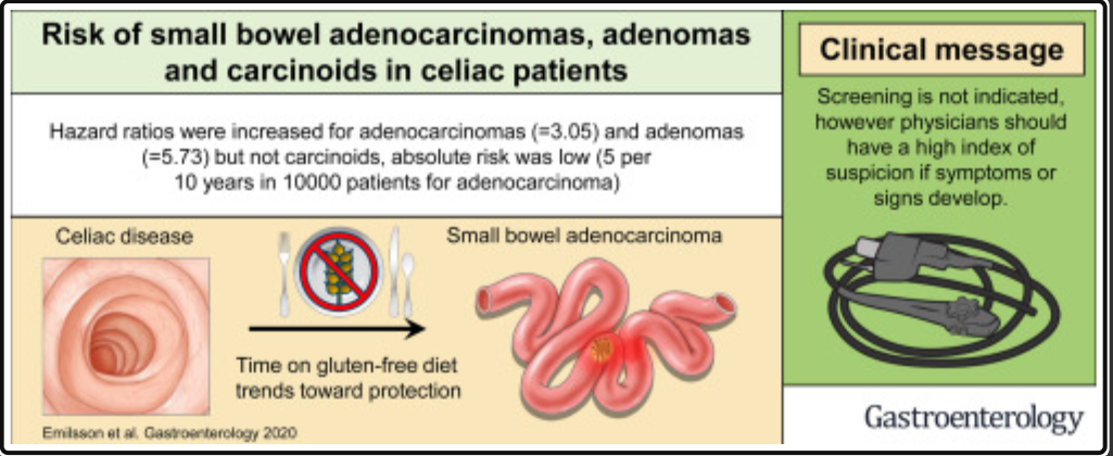
A recent study (L Emilsson et al. Gastroenterol 2020; 159: 1686-1694. Full/open access: Risk of Small Bowel Adenocarcinoma, Adenomas, and Carcinoids in a Nationwide Cohort of Individuals With Celiac Disease) quantifies the risk of small bowel adenocarcinoma in individuals with celiac. Using a Swedish nationwide cohort with a median follow-up of 11 years, the authors identified 48,119 individuals with CD (patients) and 239,249 reference individuals.

Key findings:

  • Beginning at 1 year after a diagnosis of CD, 29 patients (0.06%) received a diagnosis of small bowel adenocarcinoma vs 45 reference individuals (0.02%).
  • HRs were small bowel adenocarcinoma 3.05, carcinoids 0.59, and adenomas 5.73.).
  • Overall, there was 1 extra case of small bowel adenocarcinoma in every 2944 patients with CD followed for 10 years.
  • There was an inverse association between mucosal healing risk of future small bowel adenocarcinoma (HR, 0.18; 95% CI, 0.02–1.61), although the HR failed to attain statistical significance.

It is important to note that lymphoma is much more common malignancy than adenocarcinoma in celiac disease. The authors, in their discussion, state: “compared with lymphomas, small bowel adenocarcinomas were approximately 10 times less common in patients with CD.” At the same time, with the discovery of milder cases of Celiac disease, lymphoma risk is not nearly as high as previously suggested. A large cohort study (Clinical Gastroenterology and Hepatology 2012; 10: 30-36) of ~45,000 did not see an increased risk of GI cancers beyond the first year after diagnosis. In addition, another study (Gastroenterology 2010; 139: 763), found that mortality NOT worsened in undiagnosed celiac disease (identified by review of serology) in Olmstead County, though bone density decreased. n=129 of 16,847. (?milder cases undiagnosed). Related post: Good News For Celiac Disease

My take (mostly borrowed from authors): There is a tiny increase in risk of “small bowel adenomas and adenocarcinomas in patients with diagnosed CD, but only a very marginal increase in terms of absolute risk. Our results do not imply a need for surveillance but celiac individuals with signs or symptoms of malignancy should merit further investigation for small bowel adenocarcinoma. Mucosal healing was strongly associated with lower risk of small bowel adenocarcinoma, although the association failed to reach statistical significance.’ Lymphoma is a more common malignancy associated with CD but the absolute risk remains low.

Related blog posts:

Getting Lost in the Pathophysiology of Inflammatory Bowel Disease

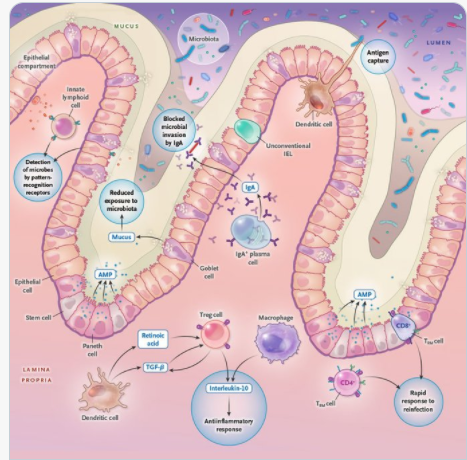
A recent review (JT Chang. NEJM 2020; 383: 2652-2664. Pathophysiology of Inflammatory Bowel Diseases) provides an in-depth description of the pathophysiology of inflammatory bowel disease (IBD). Digesting the article is akin to putting together a 1000 piece puzzle due to the complex interactions.

Some of the Key Points:

  • Based on genomewide association studies, there are “more than 240 risk variants that affect intracellular pathways recognizing microbial products (eg. NOD2); the autophagy pathway, which facilitates recycling intracellular organelles and removal of intracellular microorganisms (eg. ATG16L1); genes regulating epithelial barrier function (eg. ECM1); and pathways regulating innate and adaptive immunity (eg. IL23R and IL10).”
  • In this article, Figure 1 and 2 describe the intestinal mucosal immune system in health and disease. At baseline, this system promotes an antiinflammatory state “by virtue of active down-regulation of immune responses. For example, unlike macrophages in other parts of the body, intestinal macrophages do not produce inflammatory cytokines” after exposure to bacteria.

Other points:

  • Dysbiosis is present with IBD; however, studies have been “unable to infer causal relationships.”
  • Germ-free mice, when given fecal material from patients with IBD have increased susceptibility to colitis as compared to those who received fecal material from a healthy person.
    • Thus, this leads to potential for mitigating intestinal inflammation by modulation of the microbiome.
    • However, the authors note that humans are colonized by trillions of viral, fungal, bacterial, and eukaryotic microbes.
  • Other components of IBD pathophysiology: reduced mucus layer, increased microbial adherence, dysregulation of tight junctions/increased permeability, dysfunctional Paneth cells, TNF, IL23, IL12, IL6, IL 17A, IL17F, IL22, Interferon-gamma, integrins, JAK inhibitors, T-cells

My take: This article is a useful reference detailing the complexity of IBD pathophysiology and tries to summarize a whole textbook of information into 12 pages.

Related blog posts:

Expecting Change in Eosinophilic Esophagitis Treatment

A recent study (EJ Laserna-Mendieta et al. Clin Gastroenterol Hepatol 2020; 18: 2903-2911. Full text: Efficacy of Therapy for Eosinophilic Esophagitis in Real-World Practice) highlights the disconnect between clinical practice and outcomes.

  • Methods: This study relied on the multicenter EoE CONNECT database—with 589 patients.
    • Clinical remission was < 50% in Dysphagia Symptom Score; any improvement in symptoms = clinical response.
    • Histologic remission was eosinophil count below 5 eosinophils/hpf; 5-14/hpf = histologic response.

Key findings:

  • Topical steroids were most effective in inducing histologic remission: 54.8% compared to 36.1% for PPIs and 18.5% for empiric elimination diet; histologic remission and response was 67.7%, 49.7%, and 48.1% respectively.
  • Topical steroids were most effective in inducing clinical and histologic remission or response (in 67.7% of patients), followed by empiric elimination diets (in 52.0%), and PPIs (in 50.2%).
  • However, PPIs were the first-line treatment for 76.4% of patients, followed by topical steroids (for 10.5%) and elimination diets (for 7.8%).

My take: This data (and others) indicate that topical steroids are most effective pharmacologic therapy; at some point, I expect that they will become the most frequently used.

Related blog posts:

“Layering two less specialized masks on top of each other can provide comparable protection [to N95]. Dr. Marr recommended wearing face-hugging cloth masks over surgical masks, which tend to be made with more filter-friendly materials but fit more loosely. An alternative is to wear a cloth mask with a pocket that can be stuffed with filter material, like the kind found in vacuum bags.”

Unrelated from NY Times: One Mask Is Good. Would Two Be Better? (Yes)

Deluge of Liver Disease Due to COVID-19?

Two articles in a recent issue of Hepatology describe both direct and indirect effects of COVID-19 on the liver.

The first study with 2273 patients (MM Phipps et al Hepatology 2020; 72: 807-817. Full Text: Acute Liver Injury in COVID‐19: Prevalence and Association with Clinical Outcomes in a Large U.S. Cohort), with retrospective data, describes how most cases of COVID-19 are mild. Severe cases of liver disease are generally a marker for elevated inflammatory markers and severe systemic disease. Key findings:

  • 45% had mild (ALT <2 x ULN), 21% moderate (ALT 2-5 x ULN), and 6.4% severe liver injury (SLI) (ALT >5 x ULN).
  • Patients with SLI had a more severe clinical course, including higher rates of intensive care unit admission (69%), intubation (65%), renal replacement therapy (RRT; 33%), and mortality (42%).
  • In multivariable analysis, peak ALT was significantly associated with death or discharge to hospice (OR, 1.14; P = 0.044), controlling for age, body mass index, diabetes, hypertension, intubation, and RRT

Going into this new year, the more concerning effects of COVID-19 pandemic for the liver is likely to be the increase is severe chronic liver disease related to alcohol (and perhaps fatty liver disease too). The second article (BL Da et al. Hepatology 2020; 72: 1102-1108. Coronavirus Disease 2019 Hangover: A Rising Tide of Alcohol Use Disorder and Alcohol‐Associated Liver Disease) discusses the expectation of increased liver disease due to alcohol use disorder (AUD) and alcohol-associated liver disease (ALD). Key points:

  • In China, reports indicate a “>2-fold increase in harmful drinking after COVID-19, an effect likely repeated in the United States where an estimated 12.7% of the population has AUD and ALD is responsible for the highest hospitalization cost burden among all chronic liver diseases (CLDs).”
  • Increased alcohol use is likely to worsen other chronic liver diseases in addition to ALD
  • In addition, all of these effects are compounded by avoidance of health care facilities and delays in care

My take: COVID-19 infections have direct effects on the liver. However, the increased use of alcohol as well as weight gain are likely to be more important in terms of liver-related morbidity and mortality.

More Often Than Not Esophagitis in Children with Esophageal Atresia is NOT due to Reflux

A long time ago in a galaxy far far away, I was taught that children with esophageal atresia would have reflux for life due to dysmotility following repair. Thus, these children presumably should remain on acid blockers indefinitely. It turns out that this was fiction (just like Star Wars).

R Tambucci et al J Pediatrics 2021; 228: 155-165. Full text: Evaluation of Gastroesophageal Reflux Disease 1 Year after Esophageal Atresia Repair: Paradigms Lost from a Single Snapshot?

In this retrospective study with 48 children, the authors had the following key points:

  • Microscopic esophagitis was found in 33 (69%)
  • Pathological esophageal acid exposure on MII-pH was detected in 12 (25%)
  • The presence of long-gap esophageal atresia was associated with abnormal MII-pH.

The authors conclude that “histological esophagitis is highly prevalent at 1 year after esophageal atresia repair, but our results do not support a definitive causative role of acid-induced GERD. Instead, they support the hypothesis that chronic stasis in the dysmotile esophagus might lead to histological changes.”

My take: Along with endoscopy, pH probe testing can be helpful in selecting which children with esophageal atresia should continue with PPI therapy.

Related blog posts:

CAM Use in Functional Abdominal Pain

From Journal of Pediatrics Twitter Feed

SL Ciciora et al. J Pediatr 2020; 227: 53-59. Complementary and Alternative Medicine Use in Pediatric Functional Abdominal Pain Disorders at a Large Academic Center

Related blog posts:

Most Popular 2020 Posts

I want to thank all of you who take an interest in my blog, particularly those who give suggestions, references, and encouragement. The following posts were the most popular from the past year.

Related post: Favorite Posts of 2020

Sandy Springs at Sunrise

Favorite Posts of 2020

These are some of my favorite posts of the past year.

Humor:

GI:

Endoscopy:

Liver:

Nutrition

COVID-19:

Other:

From Picnic Island, Tampa Bay

Effects of Fecal Microbiome Transfer in Adolescents With Obesity

KSW Leong et al. JAMA Netw Open. 2020;3(12):e2030415. doi:10.1001/jamanetworkopen.2020.30415 (full text): Effects of Fecal Microbiome Transfer in Adolescents With Obesity Thanks to John Pohl’s twitter feed for this reference.

Methods: Single course of oral encapsulated fecal microbiome from 4 healthy lean donors or saline placebo.

Key findings:

  • In this randomized, double-masked, placebo-controlled trial of 87 adolescents with obesity, FMT alone did not lead to weight loss at 6 weeks.
  • There were no observed effects on insulin sensitivity, liver function, lipid profile, inflammatory markers, blood pressure, total body fat percentage, gut health, and health-related quality of life
  • In post-hoc exploratory analyses among participants with metabolic syndrome at baseline, FMT led to greater resolution of this condition (18 to 4) compared with placebo (13 to 10) by 26 weeks (adjusted odds ratio, 0.06; 95% CI, 0.01-0.45; P = .007)

Related blog posts::

Sandy Springs, GA

Converting to Monotherapy for Children with Inflammatory Bowel Disease

W El-Matary et al. JPGN 2020; 71: 740-743. Discontinuation of Immunosuppressive Medications in Children With Inflammatory Bowel Disease on Combination Therapy

This study looked at 105 patients receiving combination therapy; the a median duration of combination therapy was 2.1 years, with infliximab and either methotrexate  (53) or azathioprine (52). 89 patients had Crohn’s disease.

Key findings:

  • 11 (10.5%) patients experienced a clinical relapse over a median duration of follow-up of 12.0 months after stopping the immunomodulator.
  • In the patients who did not relapse, the median IFX trough level at IM discontinuation was 6.2; the IFX trough level was 3.8 μg/mL in those who relapsed.

In their discussion, the authors urge caution in discontinuation of immunomodulators in those with clinically-severe Crohn’s disease and those with low infliximab levels.

Related blog posts:

Tons of shells on Picnic Island, Tampa Bay

Disclaimer: This blog, gutsandgrowth, assumes no responsibility for any use or operation of any method, product, instruction, concept or idea contained in the material herein or for any injury or damage to persons or property (whether products liability, negligence or otherwise) resulting from such use or operation. These blog posts are for educational purposes only. Specific dosing of medications (along with potential adverse effects) should be confirmed by prescribing physician.  Because of rapid advances in the medical sciences, the gutsandgrowth blog cautions that independent verification should be made of diagnosis and drug dosages. The reader is solely responsible for the conduct of any suggested test or procedure.  This content is not a substitute for medical advice, diagnosis or treatment provided by a qualified healthcare provider. Always seek the advice of your physician or other qualified health provider with any questions you may have regarding a condition