Collinson S, Deans A, Padua-Zamora A, Gregorio GV, Li C, Dans LF, Allen SJ. Link to website with PDF availability: Probiotics for treating acute infectious diarrhoea. Cochrane Database of Systematic Reviews 2020, Issue 12. Art. No.: CD003048. DOI: 10.1002/14651858.CD003048.pub4. Thanks to Kipp Ellsworth for this reference.
This review identified “82 studies in 12,127 people (included 11,526 children) with acute diarrhea.” Key findings:
The number of children with diarrhea longer than 48 hours was not different between those taking a probiotic and those taking a placebo
“It was unclear whether taking probiotics shortened the time spent in hospital compared with taking a placebo or no additional treatment .”
My take: Probiotics probably make little or no difference in the setting of acute gastroenteritis/diarrhea. This analysis is based on large trials with low risk of bias.
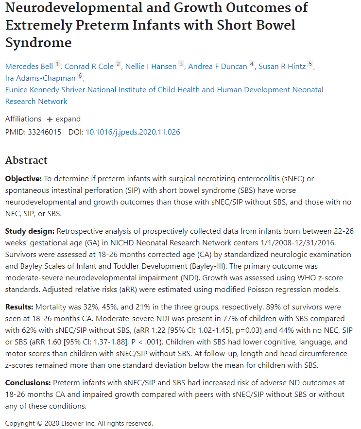
Moderate-severe neurodevelopmental impairment was present in 77% of children with extreme prematurity and with short bowel syndrome compared to 44% with extreme prematurity without necrotizing enterocolitis, spontaneous intestinal perforation or short bowel syndrome.
One of the authors, Ira Adams-Chapman, recently passed away (link to obituary: Ira Adams-Chapman, 1965-2020). She and I were residents together in Cincinnati. She was a terrific person.
“Randomized controlled trials published more than a decade ago demonstrated that exclusive enteral nutrition, wherein all table foods are eliminated from a diet and the patient relies on an elemental diet alone for nutrition, was effective in not just inducing clinical remission but also improving inflammatory biomarkers.”
“More recent rigorous studies have demonstrated that the effects of exclusive enteral nutrition can be mimicked either by a selected, less-restrictive diet (such as CD-TREAT4), which is more sustainable, or by combining partial enteral nutrition with an elimination diet that is quite diverse (such as CDED5).”
“Exclusive enteral nutrition (EEN) has been studied the most rigorously of all diets in IBD and has demonstrated the greatest benefit, compared with other diet studies in IBD. EEN requires the intake of elemental, semi-elemental, or polymeric formulas to meet all nutritional requirements without additional intake of food for 6-8 weeks. Studies have been performed mostly in pediatric populations and have shown effectiveness in induction of remission with reduction in inflammatory markers, including C-reactive protein, erythrocyte sedimentation rate, and fecal calprotectin, and even mucosal healing. EEN has not worked out as well for adult populations, because of the poor tolerability of exclusive intake of enteral formulas.”
“Beyond EEN, there are many diets that have been considered … only the SCD and Crohn’s disease exclusion diets have shown improvement in clinical remission and reduction in inflammatory markers.”
“Most dietary studies are underpowered, lack a control arm, and do not include endoscopic endpoints. The current body of evidence remains insufficient to support the use of diet alone for the treatment of IBD.”
My take: Except for exclusive enteral nutrition (EEN) which is quite challenging, dietary therapies have not been proven as effective long-term stand-alone treatments. In patients who choose dietary therapy, careful monitoring is particularly important.
In this systematic review, a total of 995 adult patients were included from 18 observational studies (4 prospective and 14 retrospective), 1 nonrandomized controlled trial, and 1 subgroup analysis of a randomized controlled trial.
Key findings:
Biologic dose de-escalation was associated with relapse rates as high as 50% at 1 year. Overall, clinical relapse occurred in 0%–54% of patients who dose de-escalated biologic therapy (17 studies).
Lower rates of relapse (10%–25%) were reported in studies involving patients with endoscopic and/or histologic remission
These results are in agreement with a previous meta-analysis, which found a 1-year risk of relapse after discontinuation of anti-TNF therapy of 36% in CD and 28% in UC ( Gisbert JP, et al.. Am J Gastroenterol 2016;111:632–47).
My take: This study shows that dose de-escalation of biologic therapy in IBD seems to be associated with high rates of clinical relapse
In this national multicenter retrospective cohort study in 207 adult patients with either active or inactive perianal Crohn’s disease (pCD) who received ustekinumab (2017-2018). The majority had received multiple biologics (~85% had at least 2 anti-TNF agents, 28% had received vedolizumab) and prior anal surgeries (mean 2.8).
Methods: Success of ustekinumab was defined by (i) clinical success at 6 months of treatment assessed by the physicians’ judgment, with (ii) no need for dedicated medical treatment for perianal lesions (antibiotics and/or topics) nor (iii) unscheduled surgical treatment. For perianal disease evaluation, clinical success was defined in the study protocol, by the absence of draining pus for fistulas, and no anal ulcers
Key findings:
In patients with active pCD, success was reached in 57/148 (38.5%) patients.
Among patients with setons at initiation, 29/88 (33%) had a successful removal.
In patients with inactive pCD at initiation, the probability of recurrence-free survival was 86.2% and 75.1% at weeks 26 and 52, respectively.
The absence of ustekinumab optimization was associated with upper odds of success (OR 2.74). “We can suppose in our present study that optimization of treatment was needed in severe refractory patients with no or insufficient response to ustekinumab. Thus, in these nonresponders, success was not achieved despite optimization.”
My take (partly borrowed from authors): “This large multicenter dedicated study adds substantial evidence to the growing literature on ustekinumab effectiveness in refractory CD.” For pCD, optimization of ustekinumab has a low likelihood of improving response.
This was a small study with 15 patients, with 9 patients having very elevated fecal calprotectin (FC) (>250) and the median level was 713 mcg/g for the entire cohort at baseline. After treatment with lumacaftor/Ivacaftor, median FC level was 102 mcg/g.
This pilot study is intriguing and makes the point that intestinal inflammation is commonly seen in children with cystic fibrosis. However, this study has some striking differences from prior studies.
My take: This study suggests that Lumacaftor/Ivacaftor is associated with a significant decrease in intestinal inflammation. When one looks at prior studies, the FC values presented in this study at baseline are much greater which could indicate significant selection bias.
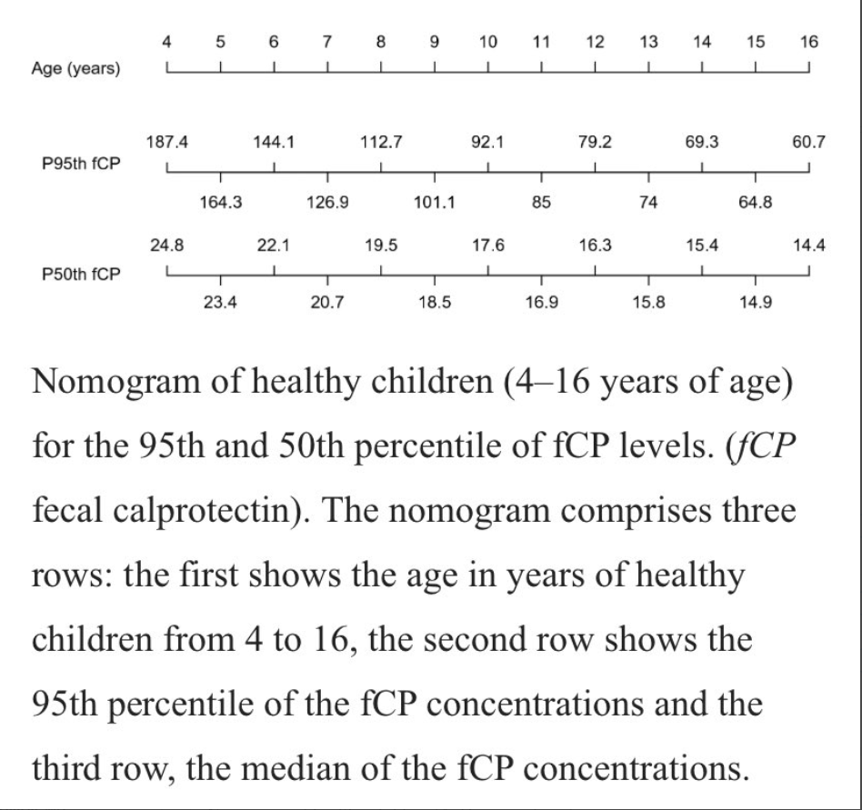
This study collected prospective data from 212 children. Key finding: The median and 95th percentile for fCP were 18.8 mg/kg and 104.5 mg/kg, respectively. “We found a statistically significant association between the 95th percentile of fCP concentrations and age (p < 0.001).”
My take: This is another study showing that calprotectin cut off values need to be higher in younger children.
The study and associated editorial highlight the effectiveness of GLP-2 in a prospective cohort of 17 patients with short bowel syndrome. It is noted that Dr. Hill has received funding from the pharmaceutical manufacturer of the product.
Key findings:
A total of 12 of 17 patients achieved parenteral independence: 3 patients after 3 months of treatment, 4 patients at 6 months, and 5 after 12 months.
The percentage able to wean off parenteral nutrition was 17%, 44%, and 60% at 3, 6, and 12 months respectively. Only 1 patient did not exhibit improvement
Plasma citrulline levels, a marker for enteral autonomy, increased from a baseline average of 20 micromol/l to 37.5, 46.75, and37.9at 3, 6, and 12 months respectively.
Adverse reactions included abdominal pain 30%, nauseas 18%, injection-site reactions 22%, and headache 16%.
Both the editorial and the study comment briefly on the cost of the therapy. The editorial also notes the current recommendation for surveillance endoscopy in view of a hypothetical risk of malignancy.
My take: Is GLP2 Worth the Cost? It probably depends on who is paying and long-term safety data. Perhaps, we will develop tools to improve prediction of which patients will achieve enteral autonomy with GLP2 who would otherwise require ongoing parenteral nutrition.
When the 10th Circuit Court, with judges Gorsuch, Ebel, and Bacharah, rolled back high-powered magnet regulations in 2016, it was expected that this would result in more suffering in children. The referenced article by Reeves et al documents the effects of this decision.
Background: In 2016, the Zen Magnets decision resulted in magnets returning to the market with warning labels “but not performance standards favored by NASPGHAN (ie, making magnets either too large to swallow or too weak to cause harm).” In this study, the authors used data from the National Electronic Injury Surveillance System (NEISS), a database of consumer product injuries.
Key findings:
When stratified by time period, suspected magnet ingestion (SMI)per year was 1598 during off-market period (when product was banned) compared with 2826 during on-market period.
An estimated 23,756 children (59% males, 42% < 5 years old) presented with a SMI from 2009 to 2019.
There was an average annual case increase of 6.1% (P = 0.01).
After 2017, there was a 5-fold increase in the escalation of care for multiple magnet ingestions (estimated n = 1094; CI 505–1686). “Escalation of care” refers to cases designated as ‘treated and transferred,’ ‘treated and admitted/hospitalized,’ or ‘held for observation.’
My take: Regulatory action is needed to prevent harm in children from these high-powered magnets.There are two companion bills in Congress which are in committee, one entitled “Magnet Injury Prevention Act.” These are clearly needed given previous judicial branch ruling.
Background: “The FMT National Registry was designed to assess FMT methods and both safety and effectiveness outcomes from North American FMT providers.” n=259 with 222 who completed short-term follow-up.
Key findings:
All FMTs were done for CDI and 249 (96%) used an unknown donor (eg, stool bank).
90% (n=200) were considered cured at one month. Of these, 197 (98%) received only 1 FMT.
Among 112 patients with initial cure who were followed to 6 months, 4 (4%) had CDI recurrence.
Safety: Severe symptoms reported within 1-month of FMT included diarrhea (n = 5 [2%]) and abdominal pain (n = 4 [2%]); 3 patients (1%) had hospitalizations possibly related to FMT. At 6 months, new diagnoses of irritable bowel syndrome were made in 2 patients (1%) and inflammatory bowel disease in 2 patients (1%). Milder adverse events were noted in 45% with symptoms including diarrhea, abdominal pain, bloating or constipation.
My take: Overall, the findings from this prospective registry confirm that FMT works fairly well for CDI. Long-term follow-up will provide more answers on the safety of FMT.
Key finding: Lysophosphatidic acid (LPA)partially restored the brush border height and the localization of SGLT1 and NHE3 in small intestine of MYO5B-knockout mice and enteroids. There are a number of high quality figures that illustrate these effects:
From Figure 1. Changes in jejunal morphologies by MYO5B deletion and administration of LPA
Small bowel biopsies in MVID include enterocytes that have either sparse, blunted microvilli or lack microvilli completely. Large cytoplasmic inclusions with prominent luminal microvilli are, however, present along with increased numbers of subapical lysosomes and other small vesicles
Biochemically, brush border expression of the Na+-glucose cotransporter SGLT1 (Slc5a1), the N+-H+ exchanger NHE3 (Slc9a3), and aquaporin 7 are markedly decreased in patients with MVID
Myo5b knockout in mice induces histopathologic and clinical features of MVID, including villous blunting, growth failure, and increased stool water, that is, diarrhea
Previous studies have shown that NHE3 trafficking from the apical storage pool to the brush border could be triggered by lysophosphatidic acid (LPA)
Key points from editorial
Kaji et al treated adult Myo5bf/f x vil-CreERT2 mice with …oral or systemic LPA administration. Villous blunting, microvillous loss, and apical lysosomal expansion were substantially reversed after 4 days of systemic LPA treatment
Although the morphologic and physiologic responses induced by LPA are striking, the weight loss that began within 2 days of Myo5b knockout was not attenuated. Thus, despite being remarkably beneficial when assessed using laboratory assays, LPA has not yet been shown to be an effective therapeutic agent.
My take (borrowed from editorial): This study shows “there are multiple trafficking pathways to the brush border and that one of these can be exploited to overcome defects in another.”