Which FODMAPs are Most Difficult to Reintroduce in Patients with Irritable Bowel Syndrome

K Van de Houte et al. Gastroenterol 2024; 167: 333-342. Open Access! Efficacy and Findings of a Blinded Randomized Reintroduction Phase for the Low FODMAP Diet in Irritable Bowel Syndrome

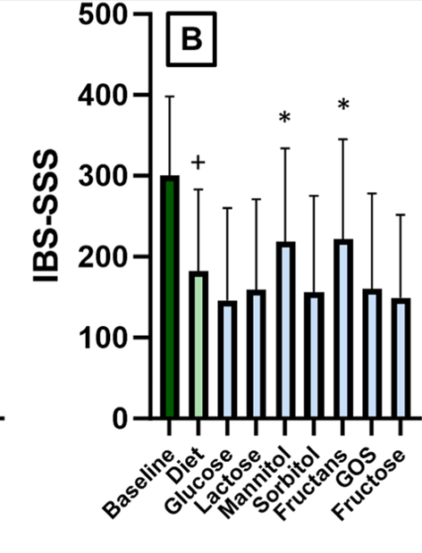
Methods: Responders (n=94 of 117) to a 6-week low FODMAP diet, defined by a drop in IBS symptom severity score (IBS-SSS) compared with baseline, entered a 9-week blinded randomized reintroduction phase with 6 FODMAP powders (fructans, fructose, galacto-oligosaccharides, lactose, mannitol, sorbitol) or control (glucose). A rise in IBS-SSS (≥50 points) defined a FODMAP trigger. Patients were challenged with 6 FODMAPs or glucose as a control (3/day x 7 days) while continuing with the low FODMAP diet.  At the end of the seventh day, patients entered 2 days of washout before starting with the next blinded FODMAP or control powder.

Key findings:

  • IBS-SSS improved significantly after the elimination period compared with baseline (150 vs. 301, P < .0001, 80% responders)
  • Symptom recurrence was triggered in 85% of the FODMAP powders, by an average of 2.5 FODMAPs/patient
  • The most prevalent triggers were fructans (56%) and mannitol (54%), followed by galacto-oligosaccharides, lactose, fructose, sorbitol, and glucose (respectively 35%, 28%, 27%, 23%, and 26%) with a significant increase in abdominal pain at day 1 for sorbitol/mannitol, day 2 for fructans/galacto-oligosaccharides, and day 3 for lactose.

One limitation of the study was selecting the dose for the challenge/reintroduction. “In comparison to clinical practice, our selected dose was higher, intended to maximize the potential of inducing symptoms. On the other hand, if that information was available, we aimed to stay below a dose for an individual FODMAP that was shown to elicit symptoms in healthy controls.”

My take:

  1. Fructans and Mannitol had the highest prevalence rate as trigger foods upon reintroduction. However, the other groups all had at least a 23% chance of being a food trigger as well.
  2. Having available powders of the FODMAP grouping could potential expedite and standardize reintroduction in clinical practice. If a patient did well with the specific FODMAP powder, there is a good likelihood that the related foods would be tolerated as well.

Related blog posts:

Disclaimer: This blog, gutsandgrowth, assumes no responsibility for any use or operation of any method, product, instruction, concept or idea contained in the material herein or for any injury or damage to persons or property (whether products liability, negligence or otherwise) resulting from such use or operation. These blog posts are for educational purposes only. Specific dosing of medications (along with potential adverse effects) should be confirmed by prescribing physician.  Because of rapid advances in the medical sciences, the gutsandgrowth blog cautions that independent verification should be made of diagnosis and drug dosages. The reader is solely responsible for the conduct of any suggested test or procedure.  This content is not a substitute for medical advice, diagnosis or treatment provided by a qualified healthcare provider. Always seek the advice of your physician or other qualified health provider with any questions you may have regarding a condition.

Firearms -Still the Leading Cause of Childhood Death in U.S.

Related blog posts:

Impact of Gene Mutation on Juvenile Polyposis Syndrome

S Cohen et al. J Pediatr Gastroenterol Nutr.2024;79:161‐167. Open Access! Juvenile polyposis syndrome in children: The impact of SMAD4 and BMPR1A mutations on clinical phenotype and polyp burden

One of the authors on this paper is Dr. Erdman (see yesterday’s post: Dr. Steve Erdman: Perplexing Polyposis Patients: a Case-Based Discussion).

Background/Methods: A constitutional disease‐causing variant (DCV) in the SMAD4 or BMPR1A genes is present in 40%–60% of patients with juvenile polyposis syndrome (JPS). A total of 124 children with JPS were included: 69 (56%) DCV‐negative and 55 (44%) DCV‐positive (53% SMAD4 and 47% BMPR1A) with a median (interquartile range) follow‐up of 4 (2.8–6.4) years

Key findings:

  • DCV‐positive children were diagnosed at an older age compared to DCV‐negative children [median 12 years vs. 5 years, respectively, p < 0.001], had a higher frequency of family history of polyposis syndromes (50.9% vs. 1.4%, p < 0.001), experienced a greater frequency of extraintestinal manifestations (27.3% vs. 5.8%, p < 0.001), and underwent more gastrointestinal surgeries (16.4% vs. 1.4%, p = 0.002). All operations in the DCV‐positive group were performed in patients with SMAD4 mutations
  • DCV-positive children had more frequent new polyps: average of 12.2 versus 2 new polyps for every year of follow‐up
  • DCV-positive children had a lower frequency of rectal bleeding (56% compared to 93%) which could be a factor in later presentation
  • There was no difference in the burden of polyps between patients with SMAD4 and BMPR1A mutations. A higher frequency of gastric polyps was observed in the SMAD4 group (55.3%vs. 9.1% for the patients in the BMPR1A group ,p = 0.004). . HHT was observed only in the SMAD4 mutations group (20.7% vs. 0 in the BMPR1Agroup. p = 0.024)

My take: Children with DCV-positive JPS likely require more frequent surveillance than DCV-negative JPS.

Related blog posts:

Arches National Park -Fiery Furnace Hike
Just outside Arches -Moab bridge that crosses Colorado River

Dr. Steve Erdman: Perplexing Polyposis Patients: a Case-Based Discussion

Recently, Dr. Steve Erdman gave our group a great update on polyposis disorders.  My notes below may contain errors in transcription and in omission. Along with my notes, I have included many of his slides.

Key points:

  • There has been breath-taking progress in understanding of polyposis disorders. It is important to have genetic counselors participate to optimize testing and evaluation
  • In patients with suspected polyposis syndromes, a genetic diagnosis is very important and can help guide management
  • Family history is very important. If several family members have had GI or other cancers at a young age, more aggressive interventions are usually indicated. However, individual family members can have a wide variation in presentation
  • In patients with many polyps, it is worthwhile to alert family to the fact that some polyps can be missed on colonoscopy and to contact medical team if there are recurrent symptoms like rectal bleeding
  • Some disorders, like juvenile polyposis syndrome (JPS), the connection between polyp presence and cancer risk is not clear. The increased risk for colon cancer my remain after polypectomy.
  • For most individuals with adenomatous polyps, removal of the polyps prevents cancer development (in the GI tract) as there is a well-described adenoma-to-cancer sequence that typically takes 7-10 years  to progress from adenoma to colon cancer
  • With FAP, the severity is related in part to the specific mutation. Mutations in the mutation cluster region are associated with an aggressive phenotype and mutations causing attenuated FAP are less aggressive
  • For FAP, timing of potential colectomy involves factors including severity as well as social factors. In teenagers in which there is a concern about being lost to follow-up, this is a factor that could influence earlier intervention
  • Many times a 2nd opinion in pathology can be helpful, especially if colon cancer is reported. However, histologic dysplasia can be tricky as well
  • Isolated CHRPE usually does not require evaluation. Dr. Erdman noted that sometimes genetic testing is offered to a family for reassurance. He discouraged colonoscopy in this setting unless a genetic diagnosis has been established or symptoms like rectal bleeding are present.  The penetrance of APC mutations (development of polyps) can be  quite variable (especially with attenuated form)
  • For JPS, after all polyps have been removed, consider surveillance every 3-5 years or for active symptoms (related post: ESPGHAN Juvenile Polyposis Syndrome in Children –Position Paper)

Case #1 presented a 14 yo with 50+ multilobulated pedunculated polyps which histologically were tubulovillus adenomas. Initial diagnosis was elusive despite extensive testing

Case#2 presented 8 yo twins. Aggressive management was indicated as 5 family members developed colorectal cancer prior to age 20 years.

Case#2 Improvements in testing allowed identification of a point mutation in the 1B promoter region of the APC gene

Case#3 presented a 16 yo with anemia and pain who was found to have a colonic mass related to mismatch repair mutation. Dr. Erdman indicated that obtaining adequate tissue for a diagnosis (“dig a hole”) is important. (As an aside, other colleagues have had the experience of tumors which were highly vascular and it is important to keep this possibility in mind)

Amsterdam II Criteria for Lynch Syndrome

Case#4 presented a 12 yo with neurofibromatosis (NF-1) who developed CRC and ultimately diagnosed with CMMR-D. This is a highly aggressive cancer susceptibility disorder with a very poor prognosis (see post: Are you familiar with CMMR-D?)

  • Case#5 presented two siblings (13 yo, 17 yo) who had half-sibling who died from CRC at age 25 yrs. This case illustrated “genetic anticipation” as each generation in this family with Lynch syndrome tended to develop CRC earlier in life. Amsterdam criteria can be helpful in identifying Lynch syndrome

Related blog posts:

Disclaimer: This blog, gutsandgrowth, assumes no responsibility for any use or operation of any method, product, instruction, concept or idea contained in the material herein or for any injury or damage to persons or property (whether products liability, negligence or otherwise) resulting from such use or operation. These blog posts are for educational purposes only. Specific dosing of medications (along with potential adverse effects) should be confirmed by prescribing physician.  Because of rapid advances in the medical sciences, the gutsandgrowth blog cautions that independent verification should be made of diagnosis and drug dosages. The reader is solely responsible for the conduct of any suggested test or procedure.  This content is not a substitute for medical advice, diagnosis or treatment provided by a qualified healthcare provider. Always seek the advice of your physician or other qualified health provider with any questions you may have regarding a condition.

Give the Right Dose (for H pylori) -It Works Better!

C Andrews et al. J Pediatr Gastroenterol Nutr. 2024;79:35–41. Impact of medication dosage on Helicobacter pylori eradication rates among pediatric patients

As I write this post on July 1st, I recollect having to learn the weight-based dosing of acetaminophen during my first day of pediatric internship. Even at this early stage, it was quite clear of the importance of getting the right dose. This article makes clear that there is a lot of room for improvement in dosing with regards to H pylori.

This retrospective study examined 144 children. Correct dosing was based on ESPGHAN-NASPGHAN guidelines:

Key findings:

  • The overall eradication rate was 73.6% (“well below the greater than 90% optimal eradication cut-off for H. pylori therapies”)
  • There was a high rate of improper weight‐based dosing: proton pump inhibitor(PPI) 31.2% (45/144), amoxicillin 31.7% (39/123), metronidazole (MET) 19.4% (12/62), clarithromycin (CLA) 23.9% (22/70), tetracycline 50% (6/12), bismuth 26.1% (6/23).
  • When PPIs were properly weight‐dosed, there was a 78.8% eradication rate that dropped to 62.2% with suboptimal dosing (p = 0.036, odds ratio [OR]: 2.26). The dose of PPI was incorrect in 45 patients.
  • When amoxicillin was properly weight‐dosed, successful eradication was achieved in 81% versus only 53.8% when improperly dosed (p = 0.002; OR: 3.64). The dose of amoxicillin was incorrect in 39 patients. There was no statistically significant impact on eradication rates with improper weight‐based dosing of MET, CLA, tetracycline, or bismuth.

My take: It is worthwhile to double-check on dosing for H pylori treatments -it results in better eradication rates. Also, in this study, as expected, quadruple therapy regimens had higher success rates (85% eradication); however, it was used in only 20 patients. In patients (virtually all of my patients) without known antimicrobial sensitivity, it has been my practice to use quadruple therapy (related post: Quadruple Therapy for Helicobacter Pylori Favored in Toronto Guidelines).

Related blog posts:

Many of the trees at Arches have this bark that feels like exposed firewood.
It doesn’t seem to have a typical outer shell
Delicate Arch (~90 ft tall)

More Data on Fazisiran for Alpha-One Antitrypsin

About two years ago, the NEJM published a phase 2 study of Fazisiran for Alpha-One Antitrypsin Deficiency (see: RNA Interference (Fazirsiran) for Liver Disease Associated with Alpha-1-Antitrypsin Deficiency). Now a larger placebo-controlled study which randomized 40 patients to subcutaneous placebo or fazirsiran 25/100/200 mg has been published:

VG Clark et al. Gastroenterol 2024; (in press). DOI:https://doi.org/10.1053/j.gastro.2024.06.028 Fazirsiran for Adults with Alpha-1 Antitrypsin Deficiency Liver Disease: A Phase 2 Placebo Controlled Trial (SEQUOIA)

Key findings:

  • At Week 16, least-squares mean percent declines in serum Z-AAT concentration were −61%, −83% and −94% with fazirsiran 25/100/200 mg, respectively, versus placebo (all P< .0001)
  • Efficacy was sustained through Week 52. At post-dose liver biopsy, fazirsiran reduced median liver Z-AAT concentration by 93% compared with an increase of 26% with placebo
  • All fazirsiran-treated patients had histological reduction from baseline in hepatic globule burden
  • Portal inflammation improved in 5/12 and 0/8 patients with baseline score >0 in the fazirsiran and placebo groups, respectively
  • Histological METAVIR score improved by >1 point in 7/14 and 3/8 patients with fibrosis >F0 at baseline in the fazirsiran and placebo groups, respectively

My take: This is an exciting development for patients with A1AT-associated liver disease. Longer duration data is needed to confirm whether fazirsiran will be a useful therapeutic agent for A1AT deficiency. If effective, selecting patients who benefit from treatment will need to be determined.

Huge Numbers of Long COVID Cases -Vaccination Helps

Y Xie, et al. NEJM 2024; DOI: 10.1056/NEJMoa2403211. Postacute Sequelae of SARS-CoV-2 Infection in the Pre-Delta, Delta, and Omicron Eras

Background: Postacute sequelae of severe acute respiratory syndrome coronavirus 2 (SARS-CoV-2) infection (PASC), also called “long Covid,” can affect many organ systems.1,2 The risk of PASC appears to increase with greater severity of infection and with the presence of preexisting medical conditions.

Methods: The authors used the health records of the Department of Veterans Affairs to build a study population of 441,583 veterans with SARS-CoV-2 infection between March 1, 2020, and January 31, 2022, and 4,748,504 noninfected contemporaneous controls.

Key findings:

NY Times, Pam Belluck 7/17/24: Vaccines Significantly Reduce the Risk of Long Covid, Study Finds

An excerpt:

The lowest rate of long Covid in the study, 3.5 percent, was among vaccinated people who were infected during the latest period in the study, between mid-December 2021 and January 2022. That compares with a rate of 7.8 percent for unvaccinated patients in the study who were infected during the same period…

To rule out other possible causes, the researchers factored in comparisons between uninfected people who developed similar symptoms…

Researchers found that among unvaccinated people infected between June 19 and Dec. 18, 2021, when Delta was the dominant variant, the rate of long Covid a year later decreased slightly to 9.5 percent from 10.4 percent among those infected in the first 15 months of the pandemic…

Among vaccinated people who had been infected, the rates of long Covid were markedly lower…About 5.3 percent of those infected during the Delta period had long Covid a year later, and 3.5 percent of those infected during the Omicron period did.

My take: A huge number of people in U.S. (and worldwide) have long COVID. This risk is markedly reduced with vaccination.

Related blog posts:

“Why It’s So Hard to Find a Pediatrician These Days”

Aaron Carroll, NY Times (July 1, 2024): Why It’s So Hard to Find a Pediatrician These Days

The author notes that increasingly there are long waits to get appointments to see pediatricians. In addition, pediatricians rely on a referral network (e.g. pediatric subspecialists, psychologists, psychiatrists) and there are shortages and delays in getting seen in these fields as well.

An excerpt:

There aren’t enough pediatricians right now, and because of that, some kids are unable to get the care they need…

Approximately 30 percent of pediatric training programs failed to fill their available residency slots, leaving 252 positions vacant — a notable increase from just 88 vacant spots last year. This isn’t a minor hiccup; it’s a warning for the future of pediatric care in the United States…Last year, a National Academies of Sciences, Engineering and Medicine committee published a report on the future of the pediatric work force and the issue of shortages, especially in rural areas. It underscored the fragmentation in care coordination between pediatric primary care and specialty care exacerbated by geographic barriers and inadequate financial support.

The elephant in the exam room, though, is that pediatricians earn less than specialists in almost every other medical field in the United States. A key reason is that so many children live in poverty and therefore qualify for Medicaid, which pays far less for care than private insurance and even less than Medicare.

Pediatricians attend the same medical schools as those who enter other specialties, and education is expensive. Almost half of those who graduated with over $150,000 in debt 20 years ago have still not paid it off completely. In 2020, the average debt of those completing pediatrics residencies was $264,000...

We need immediate action to address this crisis and find ways to attract more graduates to pediatrics. Our children are the future, but we sure don’t act like it when it comes to health care.

My take: Most pediatric physicians chose pediatrics to work with families and children. At this time, growing debt and pay inequity are factors causing many to choose other areas in medicine and needs to be addressed.

Related blog posts:

Getty Villa

Magnesium: The “Poorly Understood” Electrolyte

RM Touyz et al. NEJM 2024; 390: 1998-2009. Magnesium Disorders

Magnesium “is present in all cells in all organisms from plants to higher mammals and is indispensable for health and life because it is an essential cofactor for ATP, the cellular source of energy…All ATPase reactions require Mg2+–ATP, including those involved in RNA and DNA functions…Magnesium is involved in the control of neuromuscular function, regulation of cardiac rhythm.”

Key points:

  • “Hypomagnesemia is present in 3 to 10% of the general population, but its prevalence is increased among persons with type 2 diabetes and hospitalized patients.”
  • “Hypomagnesemia is usually associated with other electrolyte derangements, including hypocalcemia, hypokalemia, and metabolic alkalosis, and refractory hypokalemia” (& refractory hypocalcemia) is often responsive to treatment only after the magnesium concentration has been normalized.
  • “Patients with hypomagnesemia often present with nonspecific symptoms, such as lethargy, muscle cramps, or muscle weakness, and thus the diagnosis of magnesium deficiency may be overlooked.”
  • “Many drug classes, such as antibiotics, diuretics, biologic agents, immunosuppressants (including tacrolimus and rapamycin), proton-pump inhibitors, and chemotherapies, cause renal magnesium loss and hypomagnesemia.”
  • “Long-term use of PPIs causes magnesium deficiency in approximately 20% of patients receiving them, and these effects are dose-dependent.  PPIs reduce intestinal magnesium uptake.”
  • “Dietary sources rich in magnesium include cereals, beans, nuts, and green vegetables (magnesium is the central core of chlorophyll). Of the total dietary magnesium consumed, 30 to 40% is absorbed in the intestine.”
  • “Magnesium is a key component of bone — 60% of the total magnesium in the body is stored in this compartment.” Adequate magnesium intake can reduce fractures and osteopenia.
  • The intestines-bone-kidney axis regulate magnesium. “Hypomagnesemia may result from inadequate dietary intake, increased gastrointestinal loss, reduced renal reabsorption, or redistribution of magnesium from the extracellular to the intracellular space.” Rarely, hypomagnesemia is related to genetic causes.
  • Magnesium replacement is the basis for managing hypomagnesemia. “The most effectively absorbed forms are organic salts (magnesium citrate, aspartate, glycinate, gluconate, and lactate) rather than inorganic salts (magnesium chloride, carbonate, and oxide). However, a common side effect of oral magnesium supplementation is diarrhea, which poses a challenge for oral replacement.”
  • Intravenous magnesium (magnesium sulfate) is given for more severe deficiency and often therapeutically for torsades de pointes, acute asthma exacerbations, and preeclampsia or eclampsia.

This article is geared to adult medicine. Children less frequently have hypomagnesemia which is likely related to less frequent comorbidities.

My take: In pediatric patients taking PPIs as long-term therapy, checking magnesium levels is important particularly if there are multiple medications which could affect levels and if there are other comorbidities (e.g. renal disease).

Related study: A Stanford, R Rahhal. Gastro Open J. 2015; 1(4): 107-110. doi: 10.17140/GOJ-1-118 Effect of Chronic Proton Pump Inhibitor Use on Serum Magnesium Levels in Pediatric Patients

Key findings: This small retrospective pediatric study included 31 patients with a mean age of 7.8 years with 74 serum Mg levels. Only 2 patients, of adult age, had Mg levels below the normal reference limit of 1.6 mg/dL

Switzer Falls Trail (near Los Angeles)

Related blog posts:

Evidence-Based Algorithm for Surveillance in Esophageal Atresia Patients

JL Yasuda et al. J Am Coll Surg 2024; 238: 831-843. Surveillance Endoscopy in Pediatric Esophageal Atresia: Toward an Evidence-Based Algorithm

This retrospective study examined 546 children with esophageal atresia (EA) who underwent 1,473 surveillance endoscopies (2004-2023). The authors defined a hiatal hernia as at least 1 cm of gastric folds present above the level of the diaphragmatic pinch. “Actionable findings” were any finding that prompted a change in management.

Key findings:

  •  A total of 770 endoscopies (52.2%) in 394 unique patients (72.2%) had actionable pathology
  • The most common actionable finding was esophagitis which lead to escalation of therapy (484 endoscopies in 32.9% of patients). However, de-escalation of therapy was common as well; this occurred in 233 patients (after 310 endoscopies)
  • Barrett’s esophagus (intestinal metaplasia) was identified in 7 unique patients (1.3%) at a median age of 11.3 years. 6 of 7 patients with Barrett’s were receiving acid suppression at time of diagnosis
  •  Actionable findings leading to surgical intervention were found in 55 children (30 refractory reflux and 25 tracheoesophageal fistulas).
  • Significant predictors of actionable pathology included increasing age, long gap atresia, and hiatal hernia.
  • Symptoms were not predictive of actionable findings, except dysphagia (OR 5.80), which was associated with stricture.
  • Acid suppression was associated with a reduced odds of actionable findings (OR 0.78); however, there was still a high rate of actionable findings in this group. 51% of endoscopies while a patient had been receiving acid suppression had actionable findings.
  • Fundoplication was NOT associated with protection against actionable findings (OR 1.42)
  • Table 2 notes that 52 (3.5%) patients had eosinophilic esophagitis therapy escalated. Infrequently, non-esophageal diseases were identified: Celiac in 3 patients (0.2%), Crohn’s disease in 2 patients (0.1%), eosinophilic gastritis and/or duodenitis in 2 (0.1%), and H pylori in 4 patients (0.3%).

Proposed Management Algorithms:

Discussion Points (from authors):

Take multiple levels of esophageal biopsies: “Any surveillance esophagogastroduodenoscopy (EGD) should at minimum include 2 to 3 levels of esophageal biopsies”

Acid suppression recommendations: “Barrett’s esophagus is proposed to be an absolute contraindication to acid suppression wean; relative contraindications to weaning acid suppression include earlier erosive esophagitis, earlier failed acid suppression discontinuation attempt(s) with rebound esophagitis, or hiatal hernia; if acid suppression is weaned in these high-risk situations, we believe close follow-up endoscopy is warranted to assess for rebound pathology [which happened in nearly 20%].” And, “our practice is to empirically de-escalate acid suppression in children without contraindications to doing so when there is no gross esophagitis and biopsies show no or minimal inflammation (generally less than 5 eosinophils per high powered field).”

Consider doing endoscopy off acid suppression for approximately three months: “To maximize the information yield from the initial endoscopy, a patient may first wean off acid suppression approximately 3 months before endoscopy to permit evaluation for both anatomy and esophagitis off acid suppression. Subsequent surveillance is performed
according to our algorithm and within at most 6 to 12 months of any acid suppression changes.”

The authors do not generally recommend pH-Impedance. “There are no consensus “normal” values for pH-impedance reflux parameters in either healthy or EA children, and pH-impedance has failed to show any predictive correlation with actual esophagitis in multiple pediatric studies. In particular, children with EA with “normal” numbers of reflux events cannot adequately clear their refluxate in the context of poor esophageal motility and can still develop reflux injury. pH-impedance tracings in EA require manual review by an experienced reader, with automated analysis being highly unreliable in the setting of low baseline impedance values.”

Limitations: 1. Some of the actionable findings may have been expected based on prior endoscopies (e.g. prior stricture) and could influence value of symptoms like dysphagia. 2. This population was skewed to patients with more severe EA as it is a subspecialty center. One indicator was that their group had 24% with long-gap EA compared to an overall expected rate of 10% in the general EA population 3. Retrospective study 4. Many of the patients were not truly “surveillance” endoscopies but were done due to reported symptoms

My take: It’s unfortunate that this article did not get published in a more mainstream pediatric journal (e.g. JPGN, J Pediatr, Pediatrics) or a more mainstream gastroenterology journal (eg. Gastroenterol, Clin Gastroenterol Hepatol, AJG) as this article provides a lot of great data and useful advice. More than 70% of subjects had actionable findings during the course of their follow-up, including more than 40% of 1 year-olds.

The algorithms above suggest that at minimum, EA patients should have endoscopy every 5 years (likely starting between 12-18 months). More frequent endoscopy (every 2-3 years) may be worthwhile in those with risk factors (e.g. long gap EA, hiatal hernia, and prior esophagitis) and follow-up endoscopy is needed sooner if change in therapy (stricture dilation, esophagitis treatment or treatment de-escalation).

Related blog posts:

Disclaimer: This blog, gutsandgrowth, assumes no responsibility for any use or operation of any method, product, instruction, concept or idea contained in the material herein or for any injury or damage to persons or property (whether products liability, negligence or otherwise) resulting from such use or operation. These blog posts are for educational purposes only. Specific dosing of medications (along with potential adverse effects) should be confirmed by prescribing physician.  Because of rapid advances in the medical sciences, the gutsandgrowth blog cautions that independent verification should be made of diagnosis and drug dosages. The reader is solely responsible for the conduct of any suggested test or procedure.  This content is not a substitute for medical advice, diagnosis or treatment provided by a qualified healthcare provider. Always seek the advice of your physician or other qualified health provider with any questions you may have regarding a condition.