Disease extent and need for higher infliximab dosing

A recent study (JM Shapiro et al. JPGN 2016; 62: 867-72) reviewed 98 pediatric patients treated with infliximab (2012-2014) with inflammatory bowel disease (IBD).

The authors divided their patients into three groups, mainly based on extent of colonic involvement.  In those with limited colonic involvement, they were labelled “limited” disease (n=53).  In those with patchy inflammation involving the entire colon, they were considered to have “moderate” disease (n=27).  In contrast, those with continuous pancolitis were ascribed to have “extensive” disease (n=18).  Overall, Crohn’s disease accounted for 85 patients (87%),  ulcerative colitis for 11 patients (11%), IBDU for 2 patients (2%).  Interestingly, the majority (9 of 11) of those patients without Crohn’s disease were considered to have extensive disease.

Key findings:

  • “Patients with moderate and extensive disease, started taking 5 mg/kg per dose, showed statistically significant shorter times to escalation that those with limited disease.”
  • 70% of those with extensive disease required dose escalation, compared with 58.3% of those with moderate disease and 26.4% of those with limited disease.
  • The authors note that patients (n=8) with extensive disease who started with 10 mg/kg dosing  “exhibited longer treatment durability;” all 8 patients who started on 10 mg/kg dosing remained on this dose at the study’s conclusion.  Among the 10 patients that started on 5 mg/kg dosing, only 7 were on IFX therapy at the study conclusion–3 remained on 5 mg/kg, 4 were escalated to 10 mg/kg; there were 3 patients who stopped IFX therapy (1 infusion reaction, 2 with nonresponse).

My take: This is a small study. Yet, the implication is that early optimal dosing of IFX is likely  helpful, especially in the setting of extensive disease.

Related blog posts:

Chattahoochee River

Chattahoochee River


Identifying IBD Years Before Symptoms

A while back there was a movie called “Minority Report.”  The movie’s premise was that crimes could be predicted and stopped before they occurred.  A recent study (P Lochhead et al. Clin Gastroenterol Hepatol 2016; 14: 818-24) presents intriguing data suggesting a similar scenario for inflammatory bowel disease (IBD).

The authors used a prospective, nested case control study of participants in the Nurses’ Health Study I and II. Median age of patients with Crohn’s disease (CD) (n=83) and ulcerative colitis (UC) (n=90) was 52.7 years and 50.4 years respectively. Key findings:

  • Median prediagnostic hsCRP levels (mg/L) were 2.3 in CD, 2.2 in UC and 1.5 in controls (n=344).
  • Median prediagnostic IL6 levels (pg/mL) were 1.7 in CD, 1.2 in UC, and 1.0 in controls.
  • Median time interval between blood collection and diagnosis was 6.6 years for CD and 6.8 years for UC.
  • There was increased odds for developing disease even after adjustment for potentially confounding variables like smoking.  This analysis held up even when excluding disease that developed within 2 years of sampling.

Overall, this study suggests that there is a significant population of patients with subclinical IBD which precedes the diagnosis by several years.  This report adds to a number of other studies showing potential “preclinical phase” of many diseases including rheumatoid arthritis and type 1 diabetes.

My take: It is fascinating that bloodwork can be abnormal years before clinical symptoms. However, as in “Minority Report” the problem will be with identifying a crime/disease that might never occur.

Unrelated –Chart Depicting Car Temps:

car temp

IBD School Videos for Patients and Families

While these “IBD School” YouTube videos have been around for several years, I only became aware of them in the past few months.  I think they are good patient education resources.

Here are some links to a few of them:

There are a lot of these videos including the following:

My take: these videos are generally ~4 minutes and a good way to get a lot of information on IBD pretty quickly.

Screen Shot 2016-05-31 at 4.48.09 PM

Extent of Disease: Microscopic or Endoscopic Classification?

Several recent articles highlight the divergence between microscopic and endoscopic classification of disease.

  • Verstraete et al. JPGN 2016; 62: 242-5.
  • Asthon et al. JPGN 2016; 62: 246-51.
  • Pashankar et al. JPGN 2016; 62: 314-16.

The first two references describe histology in comparison to endoscopic extent of disease in pediatric Crohn’s and the 3rd reference provides information on the finding of “duodenitis.”

Verstraete et al selected 60 patients  randomly from their cohort for retrospective review.  Two physicians independently reviewed the patients.  In describing extent of disease, the extent of disease (Paris Classification) was discordant in 34 (56.6%) when comparing  macroscopic disease (imaging and endoscopy) to macroscopic/microscopic combined.  In addition, there was high interobserver variability of the physicians when the physicians reviewed just macroscopic findings (κ= 0.53).

Asthton et al examined data from 172 pediatric patients with inflammatory bowel disease.  They found that histologic disease was more extensive than endoscopic findings.  For example, among those with ileal biopsies, 49% had endoscopic findings compared with 71.3% having histologic disease.

Currently, the Paris classification relies on endoscopic findings; however, together these two studies suggest that the microscopic findings need to be considered as well. How often areas with microscopic disease will eventually develop endoscopic lesions is not clear.

Pashankar et al, reviewed pathology reports over a 5-year periods with 2772 children (mean age 10.6 years).  They identified duodenitis in 352 with a prevalence rate of 12.7%. Gastritis was seen in 64% of children with duodenitis. Interestingly, 63% of the cases with histologic duodenitis had normal endoscopic appearance. Reported reasons for duodenitis:

  • Celiac disease 32%
  • Crohn’s disease 13%
  • Ulcerative colitis 3%
  • Helicobacter pylori infection 6%
  • Functional dyspepsia 7%

The remaining children (36%) were considered to have nonspecific duodenitis.  The authors state: “this finding is similar to the high percentage of nonspecific duodenitis (60%) in adults.”

My take: It is difficult to know how important microscopic findings are in many cases.  With inflammatory bowel disease, whether/how to incorporate microscopic findings in classification is unclear.  With regard to the finding of microscopic duodenitis, when a specific etiology has not been identified, this leads to lots of questions:

  • How important is this finding?
  • How should this be treated?
  • How much additional workup and followup is needed?
  • How helpful is your pathologist –is the threshold for abnormality too low histologically?

Related blog posts:

marriage colonoscopy

More on Anti-TNF Drug Levels

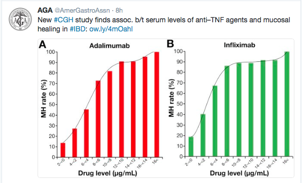
B Ungar et al (Clin Gastroenterol Hepatol 2016; 14: 550-7) report median serum levels of infliximab (n=78) or adalimumab (n=67) in correlation with mucosal healing.

In this retrospective cross-sectional study of adult patients with IBD (median age ~35 years), the authors found a correlation with higher drug troughs and mucosal healing.

“Levels of infliximab above 5 mcg/mL…and levels of adalimumab above 7.1 mcg/mL identified patients with mucosal healing with 85% specificity. Increasing levels of infliximab above 8 mcg/mL produced only minimal increases in the rate of mucosal healing, whereas the association between higher level of adalimumab and increased rate of mucosal healing reached a plateau at 12 mcg/mL”

The authors propose a “therapeutic window” of 6-10 for infliximab and 8-12 for adalimumab.

Clin Gastro Trough Levels

Remarks from DDW

Remarks from DDW

What happens when anti-TNF therapy is stopped

Another study (NA Kennedy et al. Aliment Pharmacol Ther 2016; 43: 910-23) has examined the issue of outcomes after anti-TNF therapy withdrawal among patients with inflammatory bowel disease.

This study included 166 UK patient cohort (117 with Crohn’s disease [median 31 yrs], 19 with ulcerative colitis [median 40 years]) as part of a retrospective observational study and a meta-analysis incorporating 11 further cohorts totalling 746 patients (624 with Crohn’s dissease, 122 with ulcerative colitis).

Key findings:

  • In the UK cohort, relapse rates were 36% at year and 56% at 2 years for Crohn’s disease
  • In the UK cohort, relapse rates were 42% at year and 47% at 2 years for ulcerative colitis
  • Increased relapse rates were noted for those with a diagnosis prior to age 22 years (hazard ratio (HR) 2.78), calprotectin >50 mcg/g (HR 2.95).
  • In meta-analysis, 1-year relapse rates were 39% for CD and 35% for UC/IBDU patients
  • Retreatment with anti-TNF was successful in 88% for CD and 76% of UC/IBDU patients

To understand this study, it is important to note some of the study criteria.  In the UK cohort, inclusion criteria required the patient to have had at least 12 months of ant-TNF therapy and be in corticosteroid-remission for at least 6 months.  In addition, the relapse rate is likely to be underestimated due to using a definition of relapse that required either commencement of steroids, immunomodulator or anti-TNF therapy.  The meta-anlaysis cohort studies also used clinical relapse rather than endoscopic or other objective markers.

My take: Relapse of clinical symptoms occur in about 40% after withdrawal in highly-selected groups who were doing well prior.  Significantly higher rates of endoscopic relapse are likely.  This study provides strong reasons for not interrupting therapy when it is working.

Related blog posts:

Cures Tshirt


Here’s Why Biologic Therapy for Crohn’s Helps Adolescents Grow

It is well-recognized that Crohn’s disease is associated with delays in the onset and progression of puberty with the potential for stunted growth, impaired bone accrual, and diminished quality of life.

Now, a study (MD DeBoer et al. J Pediatr 2016; 171: 146-52) shows that initiation of anti-tumor necrosis factor α (anti-TNFα) treatment results in a rapid increase in sex hormone and gonadotropin levels.

In 72 adolescents, this observational study followed levels of sex hormones, gonadotropin levels, dual-energy x-ray absorptiometry, along with cytokine/inflammatory markers at initiation of anti-TNFα therapy, at 10 weeks and at 12 months.

Key findings:

  • By week 10 , testosterone z scores in males increased from a median of -0.36 to 0.40 (P<0.05)
  • By week 10 , estradiol z scores in females increased from a median of -0.35 to -0.02 (P<0.01)

My take (from the authors): This study suggests that “systemic inflammation suppresses gonadotropin-stimulated production of sex hormones” and that treatment of this inflammation with anti-TNFα agents allows rapid resumption normal production.

Related blog posts:

Law Quad, Univeristy of Michigan

Law Quad, Univeristy of Michigan

Anti-TNF therapy and Pregnancy -More Data

G Broms et al (Clin Gastroenterol Hepatol 2016; 14: 234-41) provide more data on the ‘low risk of birth defects for infants whose mothers are treated with anti-tumor necrosis factor agents during pregnancy.”

From a Danish/Swedish cohort of 1,272,424 live births (2004-2012), the authors found the following (in comparison to healthy infants):

  • Birth defects were increased in chronic inflammatory bowel disease: 4.8% vs. 4.2%
  • 43 (6.3%) of the infants born to women with IBD who received anti-TNF therapy (683) had birth defects.  The OR for any defect was 1.32 (CI 0.93-1.82).  The types of defects were generally similar, including VSD, ASD, hypospadias, and hydronephrosis

Limitations:

  • In infants of mothers with chronic diseases, it is possible that more careful screening identified some less apparent defects.
  • Study did not examine rates of stillborn or abortions

My take: Overall there is a slightly but not significantly increased risk in birth defects based on the use of anti-TNF therapy.  Stopping anti-TNF therapy is likely to be more detrimental.

Briefly noted: P Wils et al. Clin Gastroenterol Hepatol 2016; 14: 242-50.  This retrospective study of 122 patients showed that 65% had a clinical benefit within 3 months of receiving ustekinumab for Crohn’s disease refractory to anti-TNF therapy.  Concomitant immunosuppressant therapy was associated with an increased likelihood of benefit (OR 5.43)

Related blog posts:

More on Hidradenitis Suppurativa and Inflammatory Bowel Disease

In a population-based inception cohort study (S Yada et al. Clin Gastroenterol Hepatol 2016; 14: 65-70) of 679 patients with inflammatory bowel disease (IBD) followed for a median of more than 19.8 years, it was determined that patients with IBD were ~9 times more likely to develop hidradenitis suppurativa (HS) compared with general population. 8 of 679 patients developed HS; only one had HS prior to IBD.

Other findings:

  • Most Crohn’s disease patients with HS had perianal disease.  Most ulcerative colitis patients developed HS after colectomy.
  • Female sex and obesity were risk factors for HS.

In a second retrospective study (N Kamal et al. Clin Gastroenterol Hepatol 2016; 14: 71-9), the authors identified 15 patients with CD and HS.  10 patients had perianal disease.  In this population, “both diseases were characterized by their severity, requirement of systemic medical therapies including anti-TNF and high operative rate.” this article contained some very helpful pictures.

Unrelated article: F Wang, JL Kaplan, BD Gold et al. Cell Reports; 2016: 14: 945-55.  This highly technical study used two independent cohorts of patients with Crohn’s disease and non-IBD controls.  One cohort, RISK, had over 700 patients and ~30,000 mean number of reads per sample; the other cohort, PIBD-CC, and 87 patients and ~3000 mean number of reads per sample.  Overall, the study showed associations between Crohn’s disease and bacteria in the lumen and the study helps provide an information-based method to depict dysbiosis.

Related blog post: Add it to the list

San Juan

San Juan

“Good Job Making the Diagnosis”

Briefly noted: JA Leon et al. Gastroenterol 2015; 149: 1697-99.  Case report of Job syndrome (Autosomal-Dominant Hyper-IgE syndrome) mimicking Crohn’s disease in a 37 yo with perianal fistula, and weight loss. Clues to the diagnosis: “Recurrent skin abscesses and respiratory infections, eczema, marked elevation of serum IgE, eosinophilia, and mucocutaneous candidiasis are the hallmark of the infectious and immunologic features…Boils and furuncles almost always occur and characteristically lack of the usual inflammatory findings…”cold” abscesses…associated with markedly increase IgE or eosinophil levels, should always raise a suspicion for AD-HIES.”

Related blog post: Add it to the list | gutsandgrowth