Elevated Ferritin Evaluation (in adults)

This is a useful article in evaluation of elevated ferritin levels in adults.  This approach is NOT applicable in young children but may have some use in adolescents.  In young children, other considerations include HLH and macrophage activation. In newborns, elevation of ferritin (along with liver dysfunction) may be indicative of GALD (gestational alloimmune liver disease).

W. Palmer et al. AJG 2020; 115: 1353-55. How I Approach Patients With Elevated Serum Ferritin

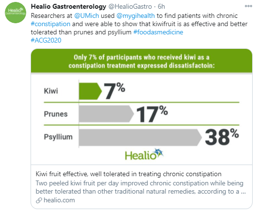
Image from ACG Twitter Link


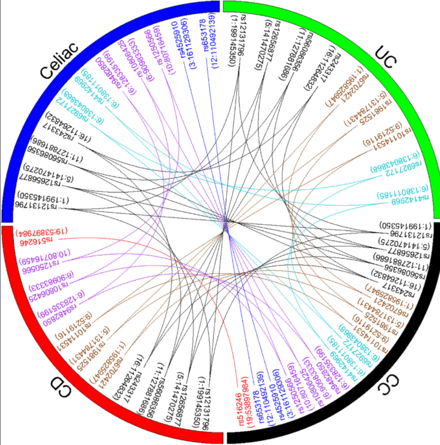
Shared Genetic Risk of Celiac Disease, Crohn’s Disease, Ulcerative Colitis, and Collagenous Colitis

E Stahl et al. Gastroenterol 2020; DOI:https://doi.org/10.1053/j.gastro.2020.04.063 Link: Collagenous Colitis Is Associated With HLA Signature and Shares Genetic Risks With Other Immune-Mediated Diseases

“In this largest genetic study of CC to date with histologically confirmed diagnosis, we strongly implicated the HLA locus and proposed potential non-HLA mechanisms in disease pathogenesis. We also detected a shared genetic risk between CC, celiac disease, CD, and UC.”


What GI Doctors Should Know About Anti-Reflux Surgery

This is a useful review -with helpful diagrams: Full text Ten Things Every Gastroenterologist Should Know About Antireflux Surgery (S Park et al. Clin Gastroenterol Hepatol 2020; 18: 1923-1929)

A couple excerpts:

Selecting Patients for Surgery: Current guidelines fall short in determining appropriate patients who would benefit most from surgery. For instance, the recommendation that a desire to discontinue PPI therapy is a suitable indication for antireflux surgery fails to recognize that 62% of patients end up back on PPIs within 9 years. Furthermore, indicating that those patients who failed medical management would benefit from surgery neglects the fact that the patients who respond best to antireflux surgery are those who have responded well to PPI therapy in the first place

Complications: Late postoperative complaints are more common and often are referred back to the referring gastroenterologist for diagnosis and management. These include late-onset dysphagia (3%–24%), recurrent heartburn (up to 62%), gas-bloat syndrome (up to 85%), and diarrhea (18%–33%).  Anatomic failure of the fundoplication (Figure Below) can present a unique challenge to the clinician because the symptoms and patient presentation (postoperative dysphagia, regurgitation, and heartburn) can be clinically indistinct from the issues seen commonly after this surgery even in the best of circumstances.  Therefore, the gastroenterologist should assess symptoms carefully in a stepwise approach with upper endoscopy, barium swallow, esophageal manometry, and/or ambulatory pH monitoring when appropriate and plan any interventions based on objective findings from focused testing.

Other points:
  • Antireflux Surgery Has No Significant Impact on the Progression of Barrett’s Esophagus to Esophageal Adenocarcinoma: Endoscopic Ablation of Dysplastic Barrett’s Esophagus Still Is Recommended
  • Medical Therapy Is More Cost Effective Than Surgical Treatment if the Cost of the Drug Is Low
  • Several New, Less-Invasive Surgical and Endoscopic Antireflux Procedures Are Now Food and Drug Administration Approved, Available, and Appear Promising

Related blog posts:

Nonceliac Gluten and Wheat Sensitivity: Review

A Khan et al. Clin Gastroenterol Hepatol 2020; 18: 1913-1922. Nonceliac Gluten and Wheat Sensitivity

This useful review notes that ” there is a great deal of skepticism within the scientific community questioning the existence of NCGS as a distinct clinical disorder.”

Key points:

  • The pathogenesis of NCGS is unclear and there is no known biomarker or diagnostic histologic lesion for this condition.
  • In these suspected patients, it is important to first exclude celiac disease and wheat allergy (especially if a rash with eating). If celiac disease is identified, this allows for appropriate longitudinal followup, strict dietary instructions, and potential screening of at-risk family members.
  • Recent studies have shown that GI symptoms in those labelled with NCGS are frequently due to dietary FODMAPs.
  • In a large meta-analysis study with 1312 adults, only 16% of participants experience gluten-specific symptoms using a double-blind placebo-controlled rechallenge.  In addition, 40% of participants experienced a nocebo response (ie. a greater negative effect than usual due to negative expectation from a dietary treatment)
  • In clinical practice, a single blind placebo-controlled rechallenge trial has been recommended for diagnosis

My take: GFD is often unnecessary and ineffective, even in those who have previously identified gluten as a potential food trigger.  Fructans are more likely to induce gastrointestinal symptoms.

Related blog posts:

Misty morning -Chattahoochee River, Island Ford

Alterations in Microbes and Impaired Psychological Function in Patients with Inflammatory Bowel Disease

Briefly noted: F Humbel et al. Clin Gastroenterol Hepatol 2020; 18: 2019-2029. Association of Alterations in Intestinal Microbiota With Impaired Psychological Function in Patients With Inflammatory Bowel Diseases in Remission

In a prospective study with 171 adults with IBD in remission, the authors combined

  1. measures of psychological comorbidities and quality of life (QoL)
  2. microbial analysis with 16S rRNA high-throughput sequencing

Key findings:

  • Microbiomes of patients with higher perceived stress had significantly lower alpha diversity
  • Anxiety and depressive symptoms were significantly associated with beta diversity

My take: This study adds another dimension to the idea of bidirectionality between psychological well-being and course of inflammatory bowel disease.  The microbiome may directly influence both psychological well-being and IBD activity.

Related blog posts:

Hemospray Efficacy and Rebleeding

A Ofusu et al. J Clin Gastroenterol 2020. doi:10.1097/MCG.0000000000001379. The Efficacy and Safety of Hemospray for the Management of Gastrointestinal Bleeding

This systematic review and meta-analysis included 19 studies and 814 patients.

  • 212 patients were treated with Hemospray as monotherapy
  • 602 patients were treated with Hemospray with conventional hemostatic techniques.

Key findings:

  • Overall pooled clinical success after the application of Hemospray was 92%
  • Overall pooled early rebleeding rates (<7 days) after application of Hemospray was 20%
  • Overall pooled delayed rebleeding rates after the application of Hemospray was 23% (<30 days)
  • There was no statistical difference in clinical success (RR, 1.02; 95% CI, 0.96-1.08; P=0.34) and early rebleeding (RR, 0.89; 95% CI, 0.75-1.07; P=0.214) in studies that compared the use of Hemospray as monotherapy versus combination therapy with conventional therapy.

Related study: D Chahal et al. Dig Liver Dis 2020. DOI: https://doi.org/10.1016/j.dld.2020.01.009 Full text: High rate of re-bleeding after application of Hemospray for upper and lower gastrointestinal bleeds Findings (n=86): Immediate hemostasis rate was 88.4%, but there was a high rate of re-bleeding (33.7%). Most re-bleeds occurred within 7 days (86.2%)

My take: Hemospray is effective in achieving immediate hemostasis but there are high rates of rebleeding. It may be eliminated by GI tract in as few as 24 hours after use.  Thus, for lesions at high risk for bleeding, hemospray is likely more of a last resort endoscopic option.

Related blog posts:

How Many Biopsies Are Sufficient for Diagnosis of Microscopic Colitis?

Briefly noted: Microscopic colitis is much more frequent in adults than in children; nevertheless, it is often important to exclude. A recent study (B Virine et al. Clin Gastroenterol Hepatol 2020; 18: 2003-2009. Full text: Biopsies From Ascending and Descending Colon Are Sufficient for Diagnosis of Microscopic Colitis) indicates that biopsies can be limited to the ascending and descending colon.

Methods: This was a retrospective study using biopsies from 101 consecutive patients with MC (52 cases of collagenous colitis, 42 cases of lymphocytic colitis, 7 combined cases),

Key finding:

  • In this study, microscopic colitis was detected with 100% sensitivity by analyzing biopsy specimens from the ascending and descending colon

My take: The authors note that previous guidelines have suggested taking 8 biopsy specimens.  Their findings support taking much fewer biopsies.

Related blog post/related article:


Intestinal Barrier Function and Risk of Crohn’s Disease

Several recent studies have examined biomarkers to predict Crohn’s disease.  A recent prospective study (W Turpin et al. Gastroenterol 2020; DOI: https://doi.org/10.1053/j.gastro.2020.08.005Increased Intestinal Permeability is Associated with Later Development of Crohn’s Disease) sought to determine whether increased intestinal permeability, as measured by urinary fractional excretion of lactulose to mannitol ratio (LMR), is associated with future development of CD.

Methods: 1420 asymptomatic first-degree relatives (6–35 years old) of patients with CD (collected from 2008 through 2015) had LMR measured and were then followed for a diagnosis of CD from 2008 to 2017, with a median follow up time of 7.8 years. We analyzed data from 50 participants who developed CD after a median of 2.7 years during the study period, along with 1370 individuals who remained asymptomatic until October 2017

Key findings:

  • An abnormal LMR (> 0.03) was associated with diagnosis of CD during the follow-up period (hazard ratio, 3.03; 95% CI, 1.64–5.63; P=3.97×10 -4).
  • This association remained significant even when the test was performed more than 3 years before the diagnosis of CD (hazard ratio, 1.62, 95% CI, 1.051–2.50; P=.029).

My take:  It remains unclear whether abnormal barrier function primarily precedes or follows CD development.  The authors state that these findings support a model in which altered intestinal barrier function contributes to pathogenesis.

Yellow or Blue for Cautery of Non-pedunculated Polyps

Almost all polyps that pediatric gastroenterologist manage are pedunculated polyps.  Nevertheless, a recent study (H Pohl et al. Gastroenterol 2020; 159: 119-28. Full text: Effects of Blended (Yellow) vs Forced Coagulation (Blue) Currents on Adverse Events, Complete Resection, or Polyp Recurrence After Polypectomy in a Large Randomized Trial) on cautery for non-pedunculated polyps was intriguing.

Methods: This multicenter, randomized, controlled, single-blinded study enrolled patients with a large colorectal polyp across 18 medical centers between April 2013 and October 2017. N=928.  ERBE device.

Key finding:

  • Equivalent results were noted with both blended current (Yellow) or forced coagulation (Blue)
    • “Serious adverse events occurred in 7.2% of patients in the Endocut (blended) group and 7.9% of patients in the forced coagulation group, with no significant differences in the occurrence of types of events.”
    • Proportions of polyps that were completely removed: 96% in the Endocut group vs 95% in the forced coagulation group
    • Proportion of polyps found to have recurred at surveillance colonoscopy: 17% for both groups
    • “Endocut more frequently caused intraprocedural bleeding that required treatment than forced coagulation (17% vs 11%). In contrast, small residual tissue islands were more frequently described in the forced coagulation group than in the Endocut group.”

Discussion: 

  • “We also did not include pedunculated polyps. Because these polyps have a greater risk of immediate bleeding, we may infer from our study that it may be safer to apply a coagulation current with a lower risk of immediate bleeding to these polyps.”

My take: Both of these settings yielded similar results.  For now, with pedunculated polyps, probably best to rely on the coagulation setting (Blue).

Related blog posts:

Disclaimer: This blog, gutsandgrowth, assumes no responsibility for any use or operation of any method, product, instruction, concept or idea contained in the material herein or for any injury or damage to persons or property (whether products liability, negligence or otherwise) resulting from such use or operation. These blog posts are for educational purposes only. Specific dosing of medications (along with potential adverse effects) should be confirmed by prescribing physician.  Because of rapid advances in the medical sciences, the gutsandgrowth blog cautions that independent verification should be made of diagnosis and drug dosages. The reader is solely responsible for the conduct of any suggested test or procedure.  This content is not a substitute for medical advice, diagnosis or treatment provided by a qualified healthcare provider. Always seek the advice of your physician or other qualified health provider with any questions you may have regarding a condition

Does a Low Vitamin D Level Increase the Risk of Crohn’s Disease? And Other Biomarkers

A recent study (BN Limketkai et al. Clin Gastroenterol Hepatol 2020; 18: 1769-76Levels of Vitamin D Are Low After Crohn’s Disease Is Established But Not Before) takes advantage of stored serum from U.S. military personnel.

Key finding: By examining 240 with Crohn’s disease (CD) along with 240 control patients, the authors show that vitamin D levels prior to CD diagnosis are not associated with the development of CD up to 8 years preceding the diagnosis.

Two other articles on predictive biomarkers for CD and an associated editorial:

  • N Nair et al. Gastroenterol 2020; 159: 383-5. Association Between Early-life Exposures and Inflammatory Bowel Diseases, Based on Analyses of Deciduous Teeth
  • J Torres, F Petralia et al. Gastroenterol 2020; 159: 96-104Serum Biomarkers Identify Patients Who Will Develop Inflammatory Bowel Diseases Up to 5 Years Before Diagnosis
  • New Biomarkers for Crohn’s Disease (editorial) C Bernstein. Gastroenterol 2020; 159: 30-32. Key points from editorial:
    • “In the article by Nair et al, the authors relate the presence of heavy metals in baby teeth to the later development of Crohn’s disease…The finding of metals that can be tracked to the in utero state suggests that the offspring who will ultimately present with IBD and have high values of these metals are likely acquiring these metals from their mothers.”
    • “In the study by Torres et al, a serum bank of Department of Defense recruits was accessed to study for microbial antibodies and immune-inflammatory markers for ≤5 years antedating diagnoses of either Crohn’s disease or ulcerative colitis. Anti-Flagellin X and ASCA-IgA were predictive of Crohn’s disease…The authors have convincingly showed that these microbial antibodies and immune-inflammatory mediators are present years before the first clinical manifestation of Crohn’s disease. These phenomena very likely are early biological manifestations of Crohn’s disease. They may not be risk factors that Crohn’s disease is coming, but rather that it is already present.”

My take: Stored tissue/blood eventually may help predict who will develop CD.  Given a lack of current treatment options in those at risk, the importance of these predictive markers is unclear.