IBD Update -September 2020

Briefly noted:

Safety of Thiopurine Use in Paediatric Gastrointestinal Disease. E Miele et al. JPGN 2020; 71: 156-62. Useful review of thiopurines for IBD and for autoimmune hepatitis

The Effect of Adalimumab Treatment on Linear Growth in Children With Crohn Disease: A Post-hoc Analysis of the PAILOT Randomized Control Trial. M Matar et al. JPGN 2020; 71: 237-42. This study showed that 66 (of 78) who completed 72 weeks of treatment had improved (but not normalized) linear growth (height z-score at baseline improved from -0.62 to -0.33 (P=0.005) and normalization of weight and BMI. The presence of perianal disease was associated with diminished growth velocity.  Overall, this study adds to the literature that anti-TNF agents can reverse growth failure associated with Crohn’s disease.

Full text: Deep Remission at 1 Year Prevents Progression of Early Crohn’s Disease  RC Ungaro et al. Gastroenterol 2020; DOI: https://doi.org/10.1053/j.gastro.2020.03.039 Key finding: When we adjusted for potential confounders, deep remission (adjusted hazard ratio, 0.19; 95% confidence interval, 0.07–0.31) was significantly associated with a lower risk of major adverse outcome.  This study is reinforced by recent data published at DDW 2020 -Abstract 401: N Plevris et al. “Early Mucosal Healing Key to Long-Term Success.”  This was highlighted by Miguel Regueiro in Gastroendonews.com.  Among 375 patients, those who achieved a fecal calprotectin (FC) <250 mcg/g within one year of diagnosis, the disease progression was 65% slower than those with FC values that did not normalize within a year.  Initiation of a biologic within 3 months of diagnosis, more than quadrupled the likelihood of FC normalization within one year.


Weak Support For Probiotics in Acute Gastroenteritis

Two recent well-controlled studies (D Schnadower et al.N Engl J Med 2018; 379:2002-2014, SB Freedman et al. N Engl J Med 2018; 379:2015-2026) showed that probiotic-treated children with acute gastroenteritis (AGE) did not have better outcomes than placebo-treated children. In addition, a recent AGA practice guideline recommended against the use of probiotics for most GI conditions, including in AGE.

However, a new report (H Szajewska et al. JPGN 2020; 71; 261-69) from an ESPGHAN  working group recommends that probiotics should have a role for AGE.  Several points about this report:

  1. Their recommendations are very qualified: “weak recommendation” with “low to very low certainty of evidence” for the following in descending order: S boulardiii, L rhamnosis GG, L reuteri DSM 17938, and L rhamnosus 19070 & L reuteri DSM 12246
  2. It is noted that this report has a disclaimer from ESPGHAN: “it does not represent ESPGHAN policy and is not endorsed by ESPGHAN”
  3. The authors have extensive disclosures
  4. The report notes that “despite large number of identified trials, we could not identify 2 randomized controlled trials of high quality for any strain that provided benefit when used for treating acute gastroenteritis”

Of note, the associated editorial (pg 146-47) also favors probiotics in the setting of AGE.  “These recommendations…have clarified that there is a role for probiotics in treating” AGE.

Related article (just published): F Mourney et al. The Pediatric Infectious Disease Journal: August 7, 2020 – Volume Online First – Issue –doi: 10.1097/INF.0000000000002849 A Multicenter, Randomized, Double-Blind, Placebo-Controlled Trial of Saccharomyces boulardii in Infants and Children With Acute Diarrhea (n=100) Key findings: The time of recovery from diarrhea was significantly shorter in the probiotic group compared with the placebo group (65.8 ± 12 hours vs. 95.3 ± 17.6 hours, P = 0.0001).

My take: Overall, probiotic effectiveness is overstated; though, some strains may be helpful for AGE.  Still, there are concerns about variation in production and quality standards even in these strains.

Related blog posts:

Briefly Noted: How to Approach Crohn’s Disease Complicated by an Intra-abdominal Abscess

A recent review (T Qazi, M Regueiro. Practical Gastroenterology 2020: June Issue, 10-18. Full PDF Link: Crohn’s Disease Complicated by an Intra-abdominal Abscess: Poke, Prod, or Cut?)

The article is a good review & the algorithm below provides some good guidance -if difficult to visualize, then it may be worthwhile to look at source article.

The authors propose initial management with antibiotics, minimization of steroids, nutritional support and drainage.

Medical treatment is favored after initial management:

  • Newly diagnosed Crohn’s disease
  • Extensive disease
  • No fibrostenoting disease
  • Active perianal disease

Surgical treatment is favored after initial management:

  • Long-standing disease
  • Stricture with dilatation
  • Abscess >6 cm in size
  • Prior surgical intervention

The authors note that “recent studies have suggested that roughly 30% of patients
treated with PD are able to avoid future surgical resection.”

Related blog posts:


Trichobezoar: Don’t Do What They Did

A recent review and case report (AF Nita et al JPGN 2020; 71: 163-70) describe an anguishing outcome after an endoscopy.  This publication is a useful, cautionary tale. In my view, the associated editorial (“MacGyver and Rapunzel in the Pediatric Endoscopy Suite” by JR Lightdale, pg 147-8) tiptoes on the issue of safety concerns of the endoscopic approach described in the review.

Case report: The authors describe a 9 year old with trichobezoar/Rapunzel syndrome who underwent a 3 hour endoscopic procedure with APC to remove the trichobezoar which was found to be composed of both human hair and doll’s hair (the latter may have contributed to complications).  In addition, the child had undiagnosed celiac disease.  Subsequent to the procedure, the child required a laparotomy; she had 18 small intestinal perforations and a gastric perforation.  The child went on to need an extensive small bowel resection (107 cm) and a 3 month hospitalization.

The authors state that a previous review had indicated only a 5% success rate for trichobezoar endoscopic removal (RR Gorter et al. Pediatr Surg Int 2010; 26: 457-63). However, they claim increased success more recently by identifying 16 of 52 (30.7%) removal rate from recent case reports.  Interestingly, one of the successful endoscopic removals cited by the authors had a respiratory arrest during the procedure (Esmali et al).

Bad advice from this article:

  • #1 The authors repeatedly suggest now that there is a >30% endoscopic success rate for endoscopic removal and thus they suggest that “it remains reasonable to attempt endoscopic retrieval” as long as a gastric trichobezoar occupies less than two-thirds of the stomach and has limited to no extension into the small bowel.
  • #2 The authors believe that “the skill mix of the endoscopist” is an important issue.

My take on their ‘learning points’:

  1. This 30% success rate should not be taken seriously due to publication bias (many unsuccessful cases are not reported) and due to treatment bias.  Many clinicians would never attempt to remove a very large trichobezoar.  Thus, the 30% success rate likely includes bezoars that may be more amenable to removal and by centers with more advanced endoscopists.
  2. The second claim about endoscopist skill is also bad advice.  First of all, some of the authors of this study have extensive endoscopic experience and yet this did not preclude a bad outcome for this child.  Secondly, in my experience, ~85% of individuals (including GI doctors) consider themselves above average; thus, it may be difficult to know if the ‘skill mix’ of the endoscopist is suitable.  Large trichobezoars are rare and no individuals will have enough experience to be considered experts.
  3. My advice: Don’t try to be MacGyver in the endoscopy suite.  Most trichobezoar cases are more suitable for surgical removal.  The most important skill of a good endoscopist is good judgement and the ability to identify cases in which an endoscopy is ill-advised.

Isle of Palms, SC

Comparative Efficacy: Vedolizumab vs Anti-TNF Agents

M Bohm et al. AP&T: 2020; July 2020 https://doi.org/10.1111/apt.15921 Full text: Comparative safety and effectiveness of vedolizumab to tumour necrosis factor antagonist therapy for Crohn’s disease

Thanks to Ben Gold for this reference.

Methods: Retrospective observational cohort (May 2014–December 2017) propensity score‐weighted comparison of vedolizumab vs TNF‐antagonist therapy (infliximab, adalimumab, certolizumab) in CD.  This study included 1266 patients (n = 659 vedolizumab).

Key findings:

  • Rates of non‐infectious serious adverse events (odds ratio [OR] 0.072, 95% confidence interval [CI] 0.012‐0.242) were significantly lower with vedolizumab vs TNF‐antagonist therapy.
    • These events included severe arthralgias in 3 vedolizumab-treated patients.  For anti-TNF recipients, events included hypersensitivity or infusion reactions (n = 6), drug‐induced psoriasis (n = 6), drug‐induced lupus (n = 5), severe liver function test abnormalities (n = 3), skin rash (n = 2), lung cancer (n = 1) and jaw or hip necrosis (n = 2).
  • Rates of serious infections (OR 1.183, 95% CI 0.786‐1.795), were NOT significantly lower with vedolizumab vs TNF‐antagonist therapy.
    • “The risk of serious infections with biologic therapy is largely driven by disease activity and concomitant use of immunosuppressive agents…. the higher concomitant use of steroids among the vedolizumab‐treated patients in our cohort may therefore help to explain the lack of observed difference in risk for serious infections between agents.”
  • No significant difference was observed between vedolizumab and TNF‐antagonist therapy for clinical remission (hazard ratio [HR] 0.932, 95% CI 0.707‐1.228), steroid‐free clinical remission (HR 1.250, 95% CI 0.677‐2.310) or endoscopic remission (HR 0.827, 95% CI 0.595‐1.151).
    • “Our observational cohort study was not designed to be a noninferiority study, and the safety and effectiveness comparisons were exploratory in nature.”
  • The efficacy of vedolizumab in this study is more impressive given that 91% of the patients had prior anti-TNF therapy.
    • “Exploratory subgroup analyses suggested that vedolizumab might be superior to subcutaneous TNF‐antagonist therapy for the achievement of clinical remission and steroid‐free clinical remission in TNF‐antagonist–naïve patients.”
  • TNF‐antagonist therapy was associated with higher treatment persistence compared with vedolizumab.

My take: This article shows that clinical experience with vedolizumab is quite good and compares favorably with anti-TNF agents.  Randomized head-to-head studies are needed, though, to truly determine efficacy in similar populations.

Related blog posts:

The Downside of Home Infusion of Biologics

N Giese-Kim et al. Am J Gastroenterol: July 22, 2020 – Volume Publish Ahead of Print – Issue – doi: 10.14309/ajg.0000000000000750. Link to abstract:  Home Infliximab Infusions Are Associated With Suboptimal Outcomes Without Cost Savings in Inflammatory Bowel Diseases

In this study, there were 27,396 patients with IBD (1,839 pediatric patients). Overall, 5.7% of patients used home infliximab infusions.

Results:

  • Those with home infusions:
    •  more likely to be nonadherent compared with both office-based (22.2% vs 19.8%; P = .044) and hospital-based infusions (22.2% vs 21.2%; P < .001).
    • more likely to discontinue infliximab compared with office-based (44.7% vs 33.7%; P < .001) or hospital-based (44.7% vs 33.4%; P < .001) infusions.
  • On Kaplan-Meier analysis, the probabilities of remaining on infliximab by day 200 of therapy were 64.4%, 74.2%, and 79.3% for home-, hospital-, and office-based infusions, respectively (P < .001)
  • Home infusions did not decrease overall annual care costs compared with office infusions ($49,149 vs $43,466, P < .001)

My take: In my experience, office-based infusions can be provided safely and in a cost-effective manner.  From the authors: “home infliximab infusions for patients with IBD were associated with suboptimal outcomes including higher rates of nonadherence and discontinuation of infliximab. Home infusions did not result in significant cost savings compared with office infusions.”

Related blog posts:

How Effective Are PPIs for Eosinophilic Esophagitis?

Emilio J. Laserna‐Mendieta et al. AP&T 2020; https://doi.org/10.1111/apt.15957.  Full article link: Efficacy of proton pump inhibitor therapy for eosinophilic oesophagitis in 630 patients: results from the EoE connect registry

“This cross‐sectional study collected data on PPI efficacy from the multicentre EoE CONNECT database.” Overall, 630 patients (76 children) received PPI as initial therapy (n = 600) or after failure to respond to other therapies (n = 30)

Key findings:

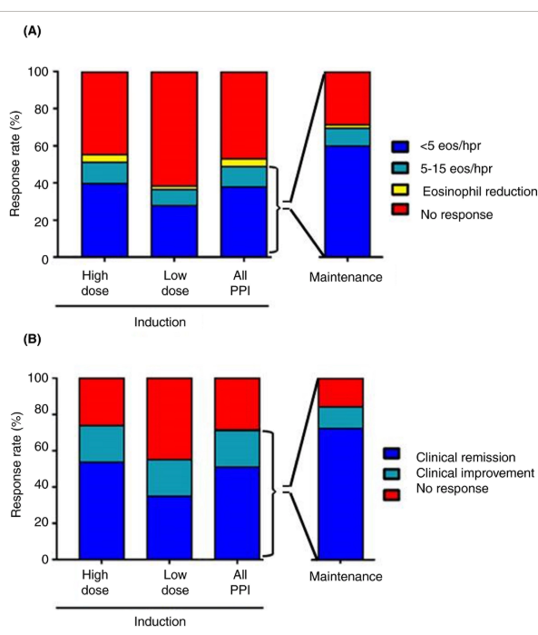
  • PPI therapy achieved eosinophil density below 15 eosinophils per high‐power field in 48.8% and a decreased symptom score ≥50% from baseline in 71.0% of patients.
  • More EoE patients with an inflammatory rather than stricturing phenotype accomplished clinico‐histological remission after PPI therapy (OR 3.7; 95% CI, 1.4‐9.5)
  • PPI treatment is more effective in achieving clinico‐histological remission of the disease when used in higher instead of standard or lower doses (50.8% vs 35.8%), and when the duration of therapy is prolonged from 8 to 12 weeks (50.4% vs. 65.2%)

My take: This study confirms previous studies which have generally found that PPIs are effective in 40-50% of patients with eosinophilic esophagitis.  Higher doses of PPIs are needed to achieve the highest response rates.

“Bar chart for histological (A) and symptomatic (B) responses for proton pump inhibitor (PPI) mono‐therapy to induce and maintain remission in patients with eosinophilic oesophagitis. For induction of remission, patients were classified according to the PPI dosage prescribed: high dose was double dosage or higher, and low dose was standard dosage or lower. For maintenance therapy, only patients with dosage reduction from that used for induction were included. eos/hpf: eosinophils per high power field”

Related blog posts:

IBD Update -August 2020

S Jansson et al JPGN 2020; 71: 40-5. This retrospective study (1998-2008) showed that pediatric patients with extraintestinal manifestations (EIM) had more severe IBD course than patients with IBD without an EIM.  EIM often had a temporal relationship with a relapse of IBD as well. Of 333 patients, 14 had an EIM at diagnosis and 47 had an EIM develop during followup.

PA Olivera, JS Lasa et al. Gastroenterol 2020; 158: 1554-73. This systematic review and meta-analysis ultimately included 82 studies with 66,159 patients (including those with IBD and other immune-mediated diseases) exposed to a JAK inhibitor; two-thirds of studies were randomized controlled trials.  Key findings:

  • Incidence rates of serious infections, herpes zoster infection, malignancy, and major cardiovascular events were 2.81, 2.67, 0.89, and 0.48 per 100 person year respectively. After meta-analysis, the authors conclude that there is an increased risk of herpes zoster (RR 1.57), but all other adverse events were not increased among patients treated with JAK inhibitors
  • Mortality was not increased in those receiving JAK inhibitors compared to placebo

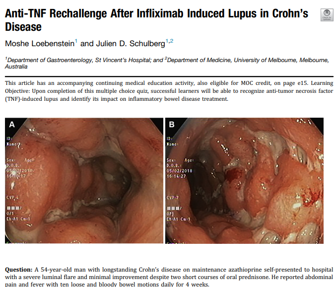
Loebenstein, JD Schulberg. Gastroenterol 2020; 158: 2069-71.  This case report describes a successful alternative anti-TNF rechallenge after infliximab induced Lupus in Crohn’s disease.  The authors note that in a previous study, 14 of 20 IBD patents with drug-induced lupus secondary to an anti-TNF agent were rechallenged with an alternative anti-TNF agent and 13/14 tolerated rechallenge without recurrent lupus (Inflamm Bowel Dise 2013; 19: 2778-86).

These images show active disease prior to intervention. The article provides f/u images showing endoscopic remission after re-starting a different anti-TNF agent.

Lingering Histologic Changes with Eosinophilic Esophagitis in Remission, Plus One

A recent study (KA Whelan et al. Clin Gastroenterol Hepatol 2020; 18: 1475-82) examined esophageal histology in 243 patients (mean age 16.9 years) in 3 groups: active eosinophilic esophagitis (EoE), inactive EoE (<15 eos/hpf), and a control non-EoE group.

Key findings:

Basal cell hyperplasia and spongiosis were present in 43 (29%) and 109 (74%) respectively of patients with inactive EoE. In comparison, these findings were present in 98% and 100% respectively of those with active EoE and in 6% and 33% of non-EoE patients

My take: This study provides some insight into the idea that esophageal damage may be ongoing in the absence of eosinophils.  These histologic findings could provide part of the reasons for symptoms in those who have had resolution of esophageal eosinophilia.

Related study: ES Dellon et al. Clin Gastroenterol Hepatol 2020; 18: 1483-92. This study showed rapid recurrence of eosinophilic esophagitis after discontinuation of topical steroids.  33/58  (57%) had symptom recurrence before 1 year (median time 244 for symptoms). At time of symptom recurrence, 78% had histologic relapse (≥15 eos/hpf).

Related blog posts:

Briefly Noted: Esophageal Stricture Dilatations

SJ Clark et al. JPGN 2020; 71: e1-e5. Examining the “Rule of 3” for Esophageal Dilations in Pediatric Stricture Patients.  In this retrospective study with 284 patients and 1384 balloon dilatations, the authors examined the perforation rate compared to the delta dilation diameter (ΔDD).  They note that the “rule of 3” is a 40-year-old expert opinion that suggests dilating an esophageal stricture more than 3 mm is unsafe. In this cohort, the perforation rate was 1.66% overall; there were 8 perforations in 1075 dilations with ΔDD ≤5 mm (0.7%) and 15 perforations in 309 dilations with ΔDD >5 mm (4.9%). The authors indicate that dilatations ≤5 mm may be acceptable.