Why Celiac Serology Needs To Be Looked At Differently in Children with Type 1 Diabetes

A recent study (M Wessels et al. J Pediatr 2020; 223: 87-92. Raising the Cut-Off Level of Anti-Tissue Transglutaminase Antibodies to Detect Celiac Disease Reduces the Number of Small Bowel Biopsies in Children with Type 1 Diabetes: A Retrospective Study) recommends changing the approach to celiac disease (CD) diagnosis in children with Type 1 Diabetes Mellitus (T1DM).

Background: The prevalence of CD among patients with T1DM is between 3-10%

Using a retrospective observational cohort with 63 children, the authors recommend raising the cut-off from performing biopsies from 3 times the ULN to 11 times ULN.

Here’s why:

  • This change in increases the specificity from 36% to 73% while reducing sensitivity from 96% to 87%.  In addition, this improves the positive predictive value from 88% to 94%, but lowered negative predictive value from 67% to 53%.  Overall, this leads to a reduction in “unnecessary biopsies.”
  • The authors note that while the serology sensitivity is reduced, it is still acceptable and justified because “normalization of elevated TG2A  can occur in up to one-third of patients.”
  • Another finding from this cohort was that 55% of children with Marsh 0 or 1 histology were symptomatic, indicating that symptoms are not specific for CD.

While the authors have recommended a higher threshold and advocated for repeating serology ~3 months later in those with lower titers, the associated editorial by Stefano Guandalini makes the following points:

  1. Raising the titer threshold would leave 13% of patients with celiac disease undiagnosed (or at least with a delay in diagnosis)
  2. “For lower titers, the physician will have to apply his or her knowledge and conscience in each individual case…we must be mindful of the serious risk of missing too many patients with celiac disease by applying a high threshold, a risk probably outweighing that of an unnecessary biopsy.”

My take: This study shows that in children with T1DM who have abnormal lower-value celiac serology, a careful discussion with parents is needed about the merits of endoscopy or deferring until persistent positivity.

Related blog posts:

Taken near Hunley bridge, Isle of Palms, SC

IBD Update -September 2020

EM Kim et al. Inflamm Bowel Dis 2020; 26: 1232-38. Mucosal Eosinophilia Is an Independent Predictor of Vedolizumab Efficacy in Inflammatory Bowel Diseases n=65 patients. In IBD cohort, colonic eosinophilia (340 +/- 156 vs 236 +/- 124) was associated with clinical non-response to vedolizumab (as was prior anti-TNF treatment). In those with ulcerative colitis, mean eosinophil count was 438 in nonresponders compared to 299 in responders. In those with Crohn’s disease, colonic biopsies showed a non-significant increase in eosinophil count in non-responders compared to responders: 352 vs. 232.

MA Sofia et al. Inflamm Bowel Dis 2020; 26: 1251-9. Poor Sleep Quality in Crohn’s Disease Is Associated With Disease Activity and Risk for Hospitalization or Surgery

  • Ninety-two CD and 82 control subjects
  • Crohn’s disease subjects with Pittsburgh Sleep Quality Index (PSQI) >5 more often had inflammatory phenotypes and reported increased benzodiazepine and psychiatric medication use. Crohn’s disease subjects with PSQI >5 also reported more night awakenings due to pain and bathroom use.
  • The PSQI correlated with HBI
  • PSQI >8 was predictive of surgery or hospitalization (hazards ratio 5.37; 95% confidence interval, 1.39-27.54).

My take: This study indicates that poor sleep is a marker for increased adverse outcomes/disease activity.  It may be that sleep disturbance is due to increased disease activity or this may be a bidirectional issue in which poor sleep triggers more disease activity as well.

A Ricciuto et al. Clin Gastroenterol Hepatol 2020; 18: 1509-1517. Primary Sclerosing Cholangitis in Children With Inflammatory Bowel Diseases Is Associated With Milder Clinical Activity But More Frequent Subclinical Inflammation and Growth Impairment

This retrospective study provides additional information on the observation that children with PSC often have subclinical disease; it is similar to a prospective study by the same group in 2018 (n=37):  (prior blog post: Active Colitis More Likely in Children in Clinical Remission Who Have IBD and PSC) Key finding: Higher proportions of children with PSC-IBD had backwash ileitis, pancolitis, and rectal sparing, and more severe right-sided disease, than controls (P < .05). Conclusions: “Despite the mild clinical activity of IBD in patients with PSC, lack of symptoms does not always indicate lack of mucosal inflammation. Children with PSC-IBD have greater growth impairments compared with children with ulcerative colitis or IBD-unclassified.”

“Implementing psychological therapies for gastrointestinal disorders in pediatrics”

Bonney Reed, Jessica Buzenski & Miranda A.L van Tilburg.  Expert Review of Gastroenterology & Hepatology (2020), DOI: 10.1080/17474124.2020.1806055 Full Text: Implementing psychological therapies for gastrointestinal disorders in pediatrics

This article is a useful and up-to-date review on the role of psychology to treat children with gastrointestinal disorders, particularly targeting functional GI disorders as well as children with inflammatory bowel disease. Also, I want to recognize Bonney and Jessica who have been so helpful for so many of our patients.

Areas covered:

  • Cognitive behavioral therapy (CBT)
  • Gut-directed hypnotherapy
  • Biofeedback-assisted relaxation training
  • E-treatment/telemedicine
  • Emerging therapies: Mindfulness, and Acceptance and Commitment therapy

Resources/Referrals:

Related blog posts:

Disclaimer: This blog, gutsandgrowth, assumes no responsibility for any use or operation of any method, product, instruction, concept or idea contained in the material herein or for any injury or damage to persons or property (whether products liability, negligence or otherwise) resulting from such use or operation. These blog posts are for educational purposes only. Specific dosing of medications (along with potential adverse effects) should be confirmed by prescribing physician.  Because of rapid advances in the medical sciences, the gutsandgrowth blog cautions that independent verification should be made of diagnosis and drug dosages. The reader is solely responsible for the conduct of any suggested test or procedure.  This content is not a substitute for medical advice, diagnosis or treatment provided by a qualified healthcare provider. Always seek the advice of your physician or other qualified health provider with any questions you may have regarding a condition.

Proactive Therapeutic Drug Monitoring in Pediatric Crohn’s disease -Better Outcomes

Y Gofin et al. Inflamm Bowel Dis 2020; 26: 1276-82.  Therapeutic Drug Monitoring Increases Drug Retention of Anti–Tumor Necrosis Factor Alpha Agents in Pediatric Patients With Crohn’s Disease

Retrospective study with 197 pediatric participants (2007-2018)

Key findings:

  • Compared with the TDM- group (n=98), the TDM+ group (n=99)
    • longer drug retention time (mean ± SE, 45.0 ± 2.7 vs 33.5 ± 2.4 months; P = 0.001)
    • lower hospitalization rate per patient per year (mean ± SE, 0.51 ± 0.7 vs 0.92 ± 0.81; P < 0.001)
    • higher treatment intensification rate (70% vs 18%; P < 0.001).
  • Analysis of the entire cohort showed a longer retention time for adalimumab vs infliximab (45.3 ± 2.8 vs 34.8 ± 2.5 months; P = 0.007)

My take: This is another study showing utility of proactive therapeutic drug monitoring

Related blog posts:

Which Crohn’s Disease Ulcerations Are Harder to Treat — Small Bowel or Colon?

K Takenaka et al. Clin Gastroenterol Hepatol 2020; 18: 1545-1552. Small Bowel Healing Detected by Endoscopy in Patients With Crohn’s Disease After Treatment With Antibodies Against Tumor Necrosis Factor

Methods: This was a post-hoc analysis of data from a clinical trial from 116 patients with CD (46 with ileal and 70 with ileocolonic type) who received induction and then maintenance therapy with anti-TNF agents (2013-18). Median age 29 years.

Key findings (based on findings from balloon-assisted enteroscopy )

  • Before treatment, small bowel ulcerations were present in 114 patients (98%); 42 patients (60%) with ileocolonic disease had colon ulcerations.
  • During maintenance therapy, 41/114 patients (36%) had small bowel endoscopic healing; all the patients with small bowel endoscopic healing also had colonic endoscopic healing.
  • Failure to achieve small bowel endoscopic healing was significantly associated with stricturing or penetrating disease (P = .014), lack of concomitant treatment with immunomodulators (P = .015), and having received previous treatment with an anti-TNF agents (P = .018).
  • The authors found that endoscopic healing was only 35% (36% for small bowel and 79% for colonic inflammation)

My take: Small bowel inflammation did not respond to treatment as well as colonic inflammation.  The implication of this study is that even in patients who are doing well clinically with treatment, disease progression especially in the small bowel may be ongoing.

Briefly noted: M Kayal et al. Inflamm Bowel Dis 2020; 26: 1079-1086.  Inflammatory Pouch Conditions Are Common After Ileal Pouch Anal Anastomosis in Ulcerative Colitis Patients.

  • In this retrospective study of adults with ulcerative colitis who had undergone total proctocolectomy (TPC) with ileal pouch anal anastomosis (IPAA). Acute pouchitis occurred in 205 patients (53%), 60 of whom (30%) progressed to chronic pouchitis.
  • Cuffitis and Crohn’s disease-like condition (CDLC) of the pouch occurred in 119 (30%) patients and 46 (12%) patients
  • Pouch failure was noted in 6.7%
  • Only one-third of patients with chronic pouchitiis, cuffitis and CDLC responded to biologic therapy

Related blog posts:

Does Clostridium difficile Increase the Risk of Surgical Resection in Pediatric Crohn’s Disease?

A recent retrospective & prospective study (J Hellmann et al. Inflamm Bowel Dis 2020; 26: 1212-21. Microbial Shifts and Shorter Time to Bowel Resection Surgery Associated with C. difficile in Pediatric Crohn’s Disease. Associated editorial 1222-3) suggests that C difficile infection (CDI) is associated with an increased risk of bowel resection surgery.

In the retrospective arm with 75 pediatric patients (<22 years): Key findings:

  • 14 of 75 had positive C difficile testing (mainly PCR, especially after 2009).
  • The rate of bowel resection surgery increased from 21% in those without C. difficile to 67% in those with (P = 0.003).
  • From a Kaplan-Meier survival model, the hazard ratio for time to first surgery was 4.4 (95% CI, 1.2–16.2; P = 0.00) in patients with positive C. difficile testing in the first year after diagnosis.

Importantly, the study was unable to distinguish between C difficile colonization versus infection.

In the prospective arm with 70 patients, patients underwent meatgenomic sequencing. Those with a positive PCR assay (irregardless of symptoms or calprotectin) were considered to have CDI.

  • 10 of 70 (14%) tested positive for CDI
  • 40% of those with CD and positive CDI had a history of surgery vs 15% with negative C difficile testing
  • Fecal calprotectin levels were elevated (>250) in 40% of both those testing positive for CDI and those testing negative
  • The overall fecal microbiome composition was not statistically significantly different between CDI-positive and CDI-negative
  • There were significant differences in the fecal microbiome composition between those with prior surgery and those without prior surgery.  Depletion of Alistipes and Ruminococcus species and reduction in methionine biosynthesis were noted in patients with both C. difficile carriage and past surgery

My takes:

  1. Based on my reading, the authors assert an association of shorter time to surgery associated with CDI in the retrospective cohort.  Because testing for CDI is common in those with flare-ups, it is unclear if this is a temporal phenomenon or is a causal relationship.
  2. It is interesting that their prospective cohort did not have an increased calprotectin level in those with CDI-positivity or overall composition change in microbiome in the CDI-positive group.  It would be of interest if these studies were confined to those with cytotoxin-assay positivity which has been shown to correlate with clinical outcomes.  In previous studies, individuals with PCR-positive CDI & cytotoxin-assay negative had similar outcomes to those with negative PCR assays.

Related blog posts:

COVID-19 -New Infection Fatality Data & How to Fix the Testing Mess

From Annals of Internal Medicine 2020 https://doi.org/10.7326/M20-5352: J Blackburn et al. Full Text: Infection Fatality Ratios for COVID-19 Among Noninstitutionalized Persons 12 and Older: Results of a Random-Sample Prevalence Study

Background: Mortality rates have been calculated from confirmed cases, which overestimates the infection fatality ratio (IFR). To calculate a true IFR, population prevalence data are needed from large geographic areas where reliable death data also exist.

Results: The Table below suggests IFR of 0.01% for those <40, 0.12% for those 40-59, and 1.71% for those ≥60 in noninstitutionalized persons.  The Table indicates nearly a 3-fold increase risk in Non-White persons. Whites had an IFR of 0.18%; non-Whites had an IFR of 0.59%. Also, I think the Table incorrectly suggests that Females have a higher IFR than Males (but the numbers suggest that they are equivalent).

From The New Yorker, Atul Gawande: We Can Solve the Coronavirus-Test Mess Now—If We Want To

This is a lengthy article which describes some of the mistakes that we’ve made with testing, some of the technical details with various tests, pooled testing, at-home testing, wastewater testing, and how to fix testing (including assurance testing) to gain control of this pandemic.

An excerpt:

We could have the testing capacity we need within weeks. The reason we don’t is not simply that our national leadership is unfit but also that our health-care system is dysfunctional….

In the United States, getting a test is anything but easy…[And] through early August, results routinely took four days or more, making the tests essentially useless. 

“Assurance testing” has been required by countries such as Iceland, France, and Germany for travellers from abroad in order to avoid a mandatory two-week quarantine

Intestinal Barrier Function and Risk of Crohn’s Disease

Several recent studies have examined biomarkers to predict Crohn’s disease.  A recent prospective study (W Turpin et al. Gastroenterol 2020; DOI: https://doi.org/10.1053/j.gastro.2020.08.005. Increased Intestinal Permeability is Associated with Later Development of Crohn’s Disease) sought to determine whether increased intestinal permeability, as measured by urinary fractional excretion of lactulose to mannitol ratio (LMR), is associated with future development of CD.

Methods: 1420 asymptomatic first-degree relatives (6–35 years old) of patients with CD (collected from 2008 through 2015) had LMR measured and were then followed for a diagnosis of CD from 2008 to 2017, with a median follow up time of 7.8 years. We analyzed data from 50 participants who developed CD after a median of 2.7 years during the study period, along with 1370 individuals who remained asymptomatic until October 2017

Key findings:

  • An abnormal LMR (> 0.03) was associated with diagnosis of CD during the follow-up period (hazard ratio, 3.03; 95% CI, 1.64–5.63; P=3.97×10 -4).
  • This association remained significant even when the test was performed more than 3 years before the diagnosis of CD (hazard ratio, 1.62, 95% CI, 1.051–2.50; P=.029).

My take:  It remains unclear whether abnormal barrier function primarily precedes or follows CD development.  The authors state that these findings support a model in which altered intestinal barrier function contributes to pathogenesis.

More Data on Magnetic Sphincter Augmentation for Gastroesophageal Reflux

In adults with gastroesophageal reflux, there is more data that magnetic sphincter augmentation (MSA) is a good option for many.  R Bell et al. Clin Gastroenterol Hepatol 2020; 18: 1736-1743. Full Text: Magnetic Sphincter Augmentation Superior to Proton Pump Inhibitors for Regurgitation in a 1-Year Randomized Trial)

Methods: Patients with moderate to severe regurgitation (assessed by the foregut symptom questionnaire) despite once-daily PPI therapy (n = 152) were randomly assigned to groups given twice-daily PPIs (n = 102) or laparoscopic MSA (n = 50) at 20 sites.

Key findings:

  • MSA resulted in control of regurgitation in 72/75 patients (96%); regurgitation control was independent of preoperative response to PPIs. It is noted that none of the patients in this study had grade C or grade D esophagitis.
  • Only 8/43 patients receiving PPIs (19%) reported control of regurgitation.
  • In the MSA group, MSA, 61 (81%) had improvements in GERD health-related quality of life improvement scores (greater than 50%) and 68 patients (91%) discontinued daily PPI use. The Demeester scores improved from 33.4 to 3.5 at 6 months post implanation
  • Esophageal acid exposure time decreased from 10.7% to 1.3% (P < .001) from study entry to 1-year after MSA.
  • Safety: No serious perioperative adverse events occurred in any arm of the study. Although 19 (39.6%) MSA patients and 10 (33.3%) MSA crossover patients reported instances of dysphagia, MSA patients reported less dysphagia at 6 and 12 months than at baseline

Discussion points from authors:

  • Relatively limited duration of follow-up, though “other studies of MSA have documented little decrease in efficacy between 1 and 5 years of follow-up”
  • The American College of Gastroenterology guidelines indicate “that surgical therapy is not recommended for patients who do not respond to PPI therapy. However, the basis for these recommendations and the types of symptoms evaluated for response to PPI therapy is unclear. Three prospective cohort studies have compared the effectiveness of laparoscopic fundoplication between PPI responsive and  nonresponsive populations and found significant symptom improvement with laparoscopic fundoplication in PPI nonresponders, though not quite as much as in PPI responders”
  • Increased bloating which is common after laparoscopic fundoplication was not evident with MSA which found a decrease from baseline.  “. Continued ability to belch was reported in 99% (n = 74 of 75) of all patients who received MSA at 12 months.”

In the associated editorial, JE Richter (Clin Gastroenterol Hepatol 2020; 18: 1685-1687 Full text: Laparoscopic Magnetic Sphincter Augmentation: Potential Applications and Safety Are Becoming More Clear—But the Story Is Not Over) notes that “erosion and migration of the MSA device have been a rare event thus far. In a total of nearly 10,000 device placements, there were 29 reported cases of erosions…Smaller devices were associated with higher rates of erosions. The 12-bead device was responsible for 18 of 29 erosions (62%) and is no longer available for implantation…To an admitted skeptic about new antireflux treatments, the available data about the symptomatic and
physiological effectiveness, durability, and safety of MSA are very impressive. I believe this procedure now deserves to be routinely done as an alternative surgical procedure to traditional fundoplication for patients with mild-moderate GERD….patients with severe GERD and structural sphincter dysfunction still need the traditional fundoplication”

My take: This study and others shows that MSA (aka Linx) is an effective treatment option for many adult patients with medically-refractory regurgitative reflux.

Related blog posts:

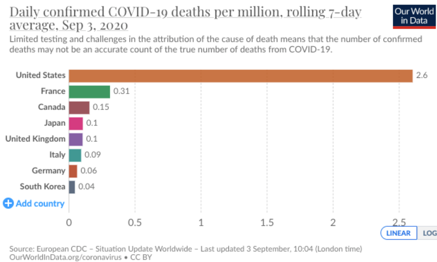
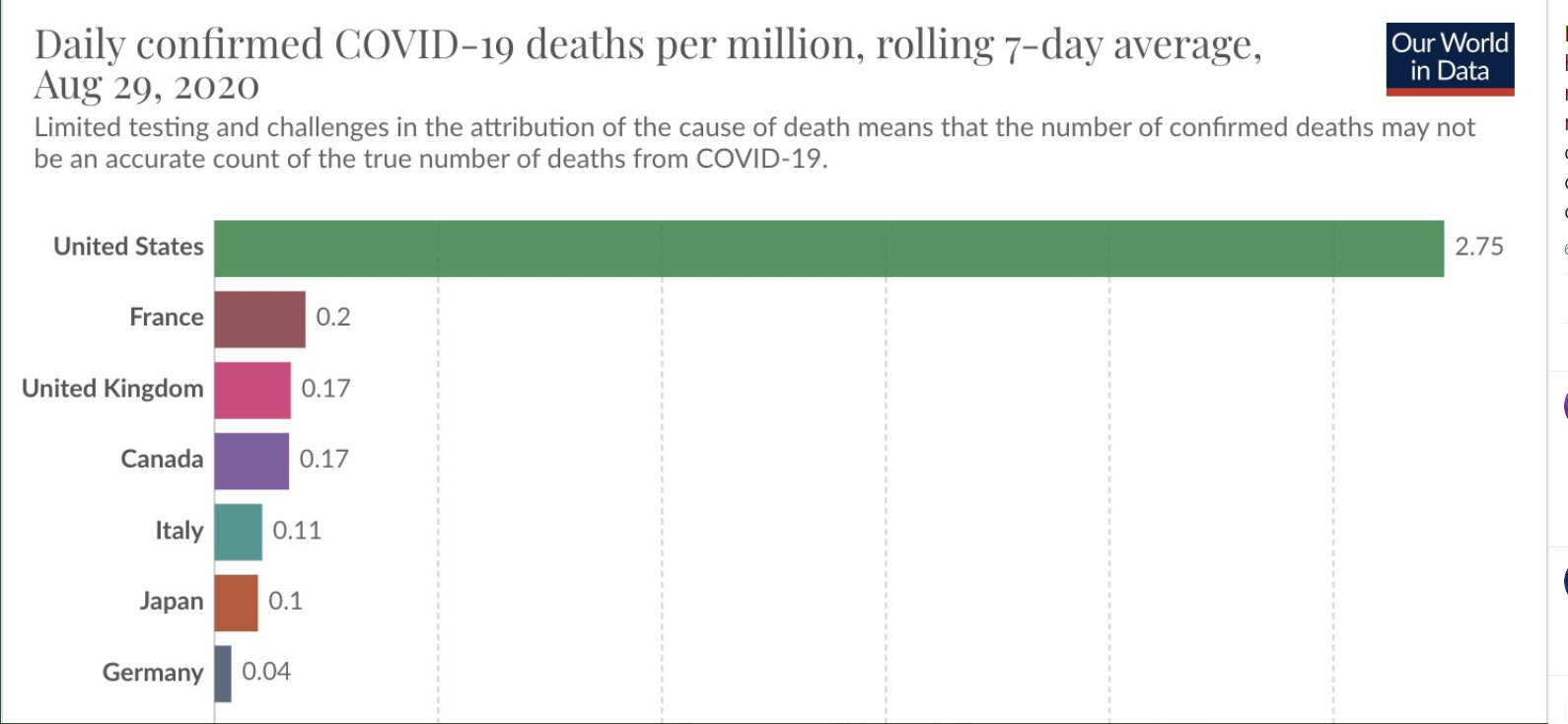
It is surprising to me that almost half of the U.S. thnk that we have handled the pandemic well

Are Your Geneticists Looking For Celiac Disease in Children With Down Syndrome?

Briefly noted: E Liu et al. JPGN 2020; 71: 252-6. Routine Screening for Celiac Disease in Children With Down Syndrome Improves Case Finding

  • Retrospective chart single center review of children with Down syndrome (2011 to 2017).
  • Prevalence of celiac disease in our population of children with Down syndrome ages 3 years or older was 9.8%.
  • 90 with celiac disease diagnosis:
    • 58 biopsy-confirmed
    • 17 with diagnosis via serology threshold in accordance with ESPGHAN
    • 9 diagnosis at outside center
    • 6 with serology but not meeting definitive criteria
  • 82% were identified through screening rather than clinical symptoms

My take: To identify celiac disease in children with Down syndrome, routine screening is needed.

Pictures from Sullivan’s Island, SC -This first picture looks out on Charleston Harbor and Ft Sumter is in the distance