How Genetics Influence Response to PPIs in Eosinophilic Esophagitis

About two years ago, James Franciosi presented research at NASPGHAN meeting indicating that the main difference between children with eosiniophilic esophagitis (EoE) who respond to proton pump inhibitiors (PPIs) compared to those who do not was related to their metabolism of PPIs and not related to the nature of their underlying EoE.

Related blog: #NASPGHAN17 Eosionophilic Esophagitis Session

Now, more has been published on this topic: EB Mougey et al. JPGN 2019; 69: 581-7.

In this study with 92 patients, data was collected from participants in a prospective clinical trial of high-dose PPI for EoE.

Key findings:

  • 57 (62%) were responsive to PPIs and 35 (38%) were not responsive to PPIs
  • Carriage of STAT6 allele variant rs1059513 predicted responsiveness to PPIs with OR of 6.16
  • Carriage of STAT6 rs324011 synergizes with CYP2C19*17 to predict PPI-nonresponsive EoE

Discussion: 

  • Carriers of CYP2C19*17 are more likely to fail PPIs for EoE.  Children with CYP2C19*17 gain of function “have a 7.7 fold better odds of failing PPI therapy” than noncarriers.
  • CYP2C19*17 effects “appears to be exerted within a specific range of PPI doses…and does not appear to exert influence at the low and high ends of this dose range.”
  • STAT6, which in this study is a cofactor, “upregulates transcription of CCL26 (eostaxin-3) 53-fold in esophageal eosinophilia relative to levels in peptic esophagitis and 490-fold over levels found in normal esophageal biopsies.”
  • PPIs effectiveness “does not correlate with esophageal” acid exposure; thus, its effects are mediated via an anti-inflammatory mechanism.

My take: This study indicates that genotype-guided dosing of PPIs for the treatment of EoE is likely to be worthwhile.

 

View from Yonah Mountain, GA

How Bad is Reflux in Children with Esophageal Atresia?

A recent retrospective study (FWT Vergouwe et al. JPGN 2019; 69: 515-22) with 57 children with esophageal atresia (EA) found most children have a normal reflux index.

This study, analyzing data between 2012-2017, reviewed all 24-hour pH-impedance (MII) studies in children at ≤18 months and 8 year olds with EA.  “All children with EA born in our hospital are offered a 24-hour pH-MII study at the age of 0.5 years and 8 years.”  In this institution, PPI treatment is given for at least 6 months after surgery. Of the 57 in the cohort, 20 had completed pH-MII at <18 months of age and 32 at age 8 years.

Key findings:

  • In children ≤18 months of age, median reflux index was 2.6% (abnormal in 2), median number of retrograde boluses was 61 (62% nonacid, 58% mixed)
  • In the older cohort (~8 years of age), median reflux index was 0.3% (abnormal in 4) and median number of retrograde boluses was 21 (64% nonacid, 75% mixed)
  • Overall, 10 of 57 children (17.5%) had GERD with reflux index >7% (n=6) or positive SI/SAP (n=4).  The authors note that much higher rates of GERD have been found in prior studies.  If they included children with fundoplication who were considered as having GERD (prior to fundoplication), then the GERD rate was 32%.

My take: This study showed that reflux in this cohort of children with EA was similar to the general population and likely indicates that a substantial portion of patients with EA do not need indefinite PPI therapy.  In children with more complex EA, PPI therapy is likely to be more beneficial.

Related blog posts:

Recent (November 4th) GI-Related Tweets:

Diabetes Mellitus Associated with Acute Recurrent and Chronic Pancreatitis

Briefly noted: MD Bellin et al. JPGN 2019; 69: 599-606.

Using the INSPPIRE database with 397 children with either acute recurrent pancreatitis or chronic pancreatitis, the authors examined the frequency of diabetes mellitus (DM).

Key findings:

  •  6% (n=24) had a diagnosis of DM. This is 30-fold higher than the general pediatric population
  • The group with DM was more likely to have elevated triglycerides (OR 5.21) coexisting autoimmune disease (OR 3.94) or pancreatic atrophy (OR 3.64)
  • The group with DM tended to be older with a mean at first diagnosis of acute pancreatitis of 12.9 years compared to 8.7 years in those who did not develop DM

Related blog posts:


Frontenac Hotel, Quebec City

Is CMV a Trigger for Necrotizing Enterocolitis?

Previously, it has been recognized that increased rates of necrotizing enterocolitis (NEC) have been associated with viral outbreaks (eg. rotavirus, norovirus) (J Pediatr Surg. 2004;39:453–7 , 2010l;29: 644-7.).

A more recent study (S Panesso-Gomez et al. J Pediatr 2019; 214: 34-40) examines the association between cytomegalovirus (CMV) and NEC.

Key Findings:

  • In this retrospective cohort (2000-2016) with paraffin-embedded samples, the authors detected CMV by PCR or immunohistochemistry (IHC) in 7 (4%) of 178 infants with either NEC (n=143) or spontaneous intestinal perforation (n=35).
  • PCR was more sensitive and was positive in all 7 detected cases whereas IHC identified CMV in 4 of the cases.

One of the limitations of the study was the lack of a control group, though previous reports have found congenital CMV in 0.4% of preterm infants with very low birth weight (<1500 g) and in 0.5-1% of all liver births.

My take: This study shows an association between the presence of CMV and NEC; hence, CMV may be one of many factors which increase susceptibility to NEC.

Related blog posts:


Yonah Mountain

Esophageal Disorders: POEM in Kids, Mitomycin C for Refractory Strictures

At our recent national meeting, Dr. Peter Kahrilas indicated that POEM (Per-oral Endoscopic Myotomy) was now the treatment of choice for most adults with achalasia (#NASPGAN19 Postgraduate Course -Part 3).

A Chone et al (JPGN 2019; 69: 523-7) provide recent multicenter retrospective data on POEM in the pediatric age group (mean age 14 years), n=117.

Key findings:

  • Clinical success, defined as Eckardt score ≤3 during followup, was achieved in 90.6% of cases. The Eckardt score was >3 in 5 (4.3%) and data was missing in 6 (5.1%)
  • Adverse events included 1 case with significant bleeding, 2 cases of aspiration pneumonia (related to anesthesia), 1 esopleural fistula (managed endoscopically), and 6 mild AEs (4 mucosomtomies, 2 subcutaneous emphysema)

Additional related blog posts:

D Ley et al (JPGN 2019; 69: 528-32) provide retrospective data on 39 patients, median age 19 months, with refractory esophageal strictures which were treated with mitomycin C.  The authors considered mitomycin C after a minimum of two previous dilatations.

Key findings:

  • Etiology: The majority had strictures/stenosis associated with esophageal atresia (n=25) followed by caustic ingestion in 9.
  • Number of stenosis: The majority (n=35) had a single stenosis.
  • In 26 patients (67%), topical application of mitomycin C was considered a success based on a reduction in the number of dilatations.  In this group, the number of dilatations dropped from 102 to 17 over a comparable period.
  • 16 (41%) never required further dilatation following mitomycin C application

My take: This study provides some of the best evidence that mitomycin C may be helpful.  Long-term followup and more studies are needed.

Related blog posts:

Disclaimer: This blog, gutsandgrowth, assumes no responsibility for any use or operation of any method, product, instruction, concept or idea contained in the material herein or for any injury or damage to persons or property (whether products liability, negligence or otherwise) resulting from such use or operation. These blog posts are for educational purposes only. Specific dosing of medications (along with potential adverse effects) should be confirmed by prescribing physician.  Because of rapid advances in the medical sciences, the gutsandgrowth blog cautions that independent verification should be made of diagnosis and drug dosages. The reader is solely responsible for the conduct of any suggested test or procedure.  This content is not a substitute for medical advice, diagnosis or treatment provided by a qualified healthcare provider. Always seek the advice of your physician or other qualified health provider with any questions you may have regarding a condition.

Lincoln Park, Chicago

China Is Catching and Passing U.S. with NAFLD Plus Updates

F Zhou et al. Hepatology 2019; 70: 1119-33. The authors performed a systematic review (n=392 studies, more than 2 million subjects) and found that NAFLD in China increased from 25.4% in 2008-2010 to 32.3% in 2015-2018. The pooled prevalence across all studies was 29.2%. The associated editorial speculates that some of this increase is related to diet changes as well as PNPLA3 gene.  This allele “is more common among East Asians than Caucasians”  It is lower in African Americans in the U.S. which helps explain why this population is at reduced risk.

JB Schwimmer, JS Johnson et al Gastroenterol 2019; 157: 1109-22.  In this prospective study with 87 children (89% Hispanic), the authors associated fecal microbiomes with NAFLD and NASH.  Both NAFLD and NASH were associated with intestinal dysbiosis with lower diversity and high abundance of Prevotella copri. Full text link: Microbiome Signatures Associated With Steatohepatitis and Moderate to Severe Fibrosis in Children With Nonalcoholic Fatty Liver Disease

S Pelusi et al. Clin Gastroenterol Hepatol 2019; 17: 2310-9.  This study analyzed data from 1738 subjects (45% with severe obesity) who had undergone liver biopsy.  132 of 389 (33.9%) with significant fibrosis did NOT have nonalcoholic steatohepatitis (NASH) and 39 patients (10%) had no inflammation. NASH diagnosis required steatosis (≥5% of hepatocytes), hepatocellular ballooning, and lobular inflammation. Factors associated with significant fibrosis in the absence of NASH, included fasting hyperglycemia, severe steatosis, mild inflammation or ballooning, and PNPLA3 1148M variant.  My take: this study shows that the finding of NASH on liver biopsy is NOT required for the development of severe liver disease related to NAFLD.

D Linden et al. Molecular Metabolism 2019; 22: 49-61. This study, summarized in Gastroenterol 2019; 157: 1156-9) showed that PNPLA3 silencing with antisense oligonucleotides ameliorates NASH in PNPLA3 1148M knock-in mice.  The summary notes that the mutated 1148 M PNPLA3 protein variant accumulates on lipid droplets altering clearance and affecting triglycerides and phospholipid turnover.

Genetic Risk for Failing Infliximab

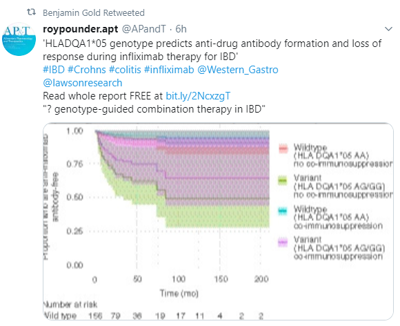
A recent study (A Wilson et al. AP&T 2019; https://doi.org/10.1111/apt.15563) noted that a HLADQA1*05A>G genotype predicts a high risk of developing anti-drug antibodies in patients receiving infliximab.

Here’s a link to the full article:  HLADQA1*05 genotype predicts anti‐drug antibody formation and loss of response during infliximab therapy for inflammatory bowel disease

Here’s the abstract (my highlights in bold):

Background

Anti‐drug antibodies (ADAs) are a leading contributor to infliximab loss of response and adverse drug events. It is not feasible to identify patients at risk of antibody formation before initiating infliximab. The genetic variation HLADQA1*05 (rs2097432) has been linked to infliximab antibody formation in Crohn’s disease (CD).

Aims

To evaluate the association between HLADQA1*05 and infliximab antibody formation, infliximab loss of response, treatment discontinuation and adverse drug events in patients with inflammatory bowel disease (IBD)

Methods

In a retrospective cohort study, infliximab‐exposed patients with IBD (n = 262) were screened for the genetic variation, HLADQA1*05A>G (rs2097432). Risk of infliximab ADA formation, infliximab loss of response, adverse events and discontinuation were assessed in wild‐type (GG) and variant‐carrying (AG or AA) individuals.

Results

Forty per cent of all participants were HLADQA1*05A>G variant carriers, with 79% of participants with infliximab antibodies carrying at least one variant allele. The risk of infliximab antibody formation was higher in HLADQA1*05A>G variant carriers (adjusted HR = 7.29, 95% confidence interval (CI) = 2.97‐17.191, P = 1.46 × 10−5) independent of age, sex, weight, dose and co‐immunosuppression with an immunomodulator. Variant carrier status was associated with an increased risk of infliximab loss of response (adjusted HR = 2.34, 95% CI = 1.41‐3.88, P = .001) and discontinuation (adjusted HR = 2.27, 95% CI = 1.46‐3.43, P = 2.53 × 10−4) although not with infliximab‐associated adverse drug events.

Conclusions

HLADQA1*05 is independently associated with a high risk of infliximab antibody formation in addition to infliximab loss of response and treatment discontinuation. There may be a role for genotype‐guided application of combination therapy in IBD.

Related blog posts:

Grading Treatment Response in Eosinophilic Esophagitis

Full Text Link: A Conceptual Approach to Understanding Treatment Response in Eosinophilic Esophagitis

Also, related articles:

  1. D Bushyhead et al. Gastroenterology 2019; 157: 944-5. This practical teaching case report noted that oral immunotherapy (OIT) has been shown to trigger new onset EoE in 2.7% (AJ Lucendo et al. Ann Allergy Asthma Immunol 2014; 113: 624-9).
  2. R Alexander et al. Clin Gastroenterol Hepatol 2019; 17: 2371-3. This study compared eating behaviors of adults with active EoE (n=10), inactive EoE (n=10) and control patients (n=10).  Not surprisingly, those with active EoE took longer to eat (18.3 min compared to 12.4 min, and 13.0 min respectively) and had more drinks after a single bite (11.6 compared with 5.1 and 2.5 respectively)

Related blog posts:

IBD Updates November 2019

M Lowenberg et al. Gastroenterol 2019; 157: 997-1006. This LOVE-CD study, a prospective study with 110 patients with active Crohn’s disease, found that treatment with vedolizumab resulted in 29% and 31% corticosteroid-free clinical remission at weeks 26 and 52 respectively (CDAI <150).  Endoscopic remission, based on intent-to-treat analysis, was 33% and 36% at weeks 26 and 52.  Serum vedolizumab levels above 10 mg/L at week 22 were associated with endoscopic remission at week 26.

S Danese et al. Gastroenterol 2019; 157: 1007-18.  This VERSIFY trial, a phase 3b, open-label, single-group study of 101 patients with Crohn’s disease, found that treatment with vedolizumab resulted in 11.9% and 17.9%  endoscopic remission at week 26 and 52 respectively. Remission by MRE was 21.9% and 38.1% at those respective time points. No notable safety issues were reported.

N Khan et al. Clin Gastroenterol 2019; 17: 2262-8. Using a retrospective cohort of 54,919 patients with IBD followed by the VA System (2000-2018), the authors identified 467 patients with incident squamous cell cancer (SCC); median age ~70 years.  11 patients with SCC died from related-complications.  In this group, 8 had been exposed to thiopurines.   Thus, exposure to thiopurines increased mortality related to SCC compared to those exposed to mesalamine therapy, though the absolute risk among the entire cohort was less than 1 in 5000.  My take: Long-term use of thiopurines should be paired with dermatology evaluation and good skin care.

Pics from Mechandise Mart Light Show, Chicago

Waiting for the String Test for Eosinophilic Esophagitis

A recent study (SJ Ackerman et al. American Journal of Gastroenterology: October 2019 – Volume 114 – Issue 10 – p 1614–1625) provides additional data supporting the ‘string’ test to determine whether eosinophilic esophagitis is active or inactive.  Thanks to Ben Gold for sharing this reference.

My take: The string test could be a useful test for monitoring response to treatment, especially if it could garner insurance coverage. When/if will it ever become available clinically? (prior publication as early as 2012: String test for EoE)

Here’s the link to the full-text open-access article: One-Hour Esophageal String Test: A Nonendoscopic Minimally Invasive Test That Accurately Detects Disease Activity in Eosinophilic Esophagitis

Abstract:

OBJECTIVES: Eosinophilic esophagitis (EoE), a chronic food allergic disease, lacks sensitive and specific peripheral biomarkers. We hypothesized that levels of EoE-related biomarkers captured using a 1-hour minimally invasive Esophageal String Test (EST) would correlate with mucosal eosinophil counts and tissue concentrations of these same biomarkers. We aimed to determine whether a 1-hour EST accurately
distinguishes active from inactive EoE or a normal esophagus.

METHODS: In a prospective, multisite study, children and adults (ages 7–55 years) undergoing a clinically indicated esophagogastroduodenoscopy performed an EST with an esophageal dwell time of 1 hour. Subjects were divided into 3 groups: active EoE, inactive EoE, and normal esophageal mucosa. Eosinophil-associated protein levels were compared between EST effluents and esophageal biopsy extracts. Statistical modeling was performed to select biomarkers that best correlated with and
predicted eosinophilic inflammation.

RESULTS: One hundred thirty-four subjects (74 children, 60 adults) with active EoE (n 5 62), inactive EoE (n 5 37), and patient controls with a normal esophagus (n 5 35) completed the study. EST-captured eosinophil-associated biomarkers correlated significantly with peak eosinophils/high-power field, endoscopic visual scoring, and the same proteins extracted from mucosal biopsies. Statistical modeling, using combined eotaxin-3 and major basic protein-1 concentrations, led to the development of EoE scores that distinguished subjects with active EoE from inactive EoE or normal esophagi. Eightyseven percent of children, 95% of parents, and 92% of adults preferred the EST over endoscopy if it provided similar information.

DISCUSSION: The 1-hour EST accurately distinguishes active from inactive EoE in children and adults and may facilitate monitoring of disease activity in a safe and minimally invasive fashion.

First Floor of Hancock Building, Chicago