Gluten Intake and Development of Celiac Disease -Two Studies

  • NA Lund-Blix et al. Am J Gastroenterol 2019; 114: 1299-1306.
  • K Marild et al. Am J Gastroenterol 2019; 114: 1307-14.

Thanks to Ben Gold for these references.

In the first study, the authors used an observational prospective nationwide cohort study, the Norwegian Mother and Child Cohort Study (MoBa) with 67,608 children born between 2000-2009 and with a mean followup of 11.5 years.

Key findings:

  • Celiac disease (CD) was diagnosed in 738 children (1.1%)
  • The adjusted relative risk of CD was 1.1 per standard deviation increase in daily gluten amount at age 18 months.
  • Compared to children in the lowest quartile of gluten ingestion, those in the upper quartile had an adjusted relative risk of 1.29.
  • Timing of gluten introduction, ≥6 months or before 4 months, was also an independent risk factor for CD. In those before 4 months the aRR was 1.45 and for those ≥6 months the aRR was 1.34

In the second study, the authors used the prospective Diabetes Autoimmunity Study in the Young cohort with 1875 at-risk children.

Key findings:

  • Children in the highest tertile of gluten intake between ages of 1 and 2 had a 2-fold greater hazard of developing CD autoimmunity (positive tTG antibodies) (aHR 2.17) than those in the lowest tertile.
  • The risk of CDA increased by 5% per daily gram increase in gluten intake in 1 year olds.

My take: Taken together, these studies indicate that higher gluten exposure (between 1-2 years) is associated with a modestly-higher risk of CD; in addition, early (<4 months) and late exposure (>6 months) may increase the risk as well.

Related blog posts:

Spoiler alert: This case study by A Fasano et al. NEJM 2020; 382: 180-9. describes a presentation of celiac disease and Addision’s disease. I recently had a teenager present with similar symptoms who had Addison’s alone (clues were fatigue, low sodium and hyperpigmentation)

ESPGHAN Guidelines for Diagnosing Coeliac Disease 2020

Briefly noted: S Husby et al. JPGN 2020; 70: 141-56.

Link to document: ESPGHAN Guidelines for Diagnosing Coeliac Disease 2020

Key recommendations for diagnosing celiac disease (CD):

  • If CD is suspected, measurement of total serum IgA and IgA-antibodies against transglutaminase 2 (TGA-IgA) is superior to other combinations
  • We recommend against deamidated gliadin peptide antibodies (DGP-IgG/IgA) for initial testing
  • Only if total IgA is low/undetectable, an IgG-based test is indicated
  • If TGA-IgA is ≥10 times the upper limit of normal (10× ULN) and the family agrees, the no-biopsy diagnosis may be applied, provided endomysial antibodies (EMA-IgA) will test positive in a second blood sample. HLA DQ2-/DQ8 determination and symptoms are not obligatory criteria
  • In children with positive TGA-IgA <10× ULN at least 4 biopsies from the distal duodenum and at least 1 from the bulb should be taken

Related blog posts:


P’tit Train du Nord Linear Park

Celiac Disease: “”80 percent of success is just showing up”

“80 percent of success is just showing up” —Woody Allen

Reading a recent (brief) study (BA Blansky et al. Clin Gastroenterol Hepatol 2019; 17: 2503-4) reminded me of the quote from Woody Allen.  This study of children with Celiac disease (CD) demonstrates a high rate of children who were lost to follow-up at a leading Children’s hospital.

Key findings:

  • From a randomly selected retrospective cohort (2010-2014) with 241 eligible subjects, one-fourth of children were lost to follow-up within a year of diagnosis. 22 (9%) had NO GI visits after their diagnostic procedure.
  • Risk factors for loss of follow-up: sibling with CD (HR 1.90), Medicaid insurance (HR 2.19), and older age at diagnosis; those with adherence had median age at diagnosis of 8.7 years compared with 11.4 years for those lost to follow-up.
  • Median time to tissue transglutaminase (TTG) IgA normalization was 17 months.  Of 141 who had recommended follow-up, 25% had elevated TTG IgA at last GI visit.

My take: These numbers should not be surprising to most clinicians.  If clinicians want to improve follow-up and outcomes, then families will need more nudging; EMRs can be configured to help in this task.

Related blog posts:

Quebec City

#NASPGHAN19 Selected Abstracts (Part 1)

Link to full NASPGHAN 2019 Abstracts.

Here are some abstracts that I found interesting at this year’s NASPGHAN meeting:

NAFLD:

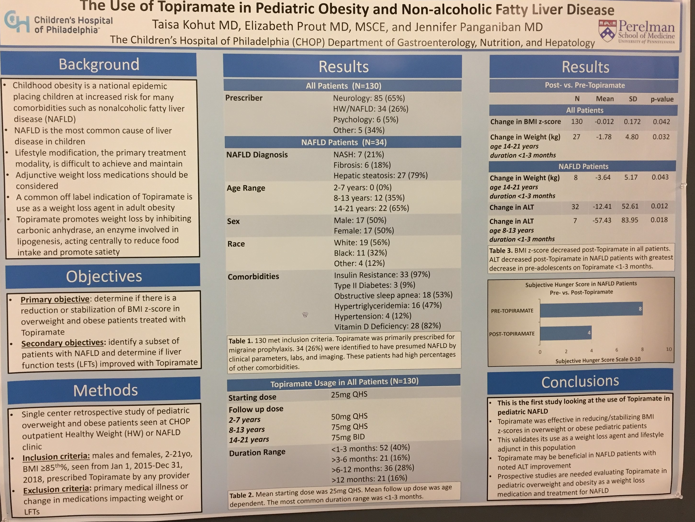
  1. Off-label use of topiramate may be helpful in stabilizing weight and improving NAFLD
  2. Socioeconomic barriers are frequent in NAFLD patients (the 2nd poster did not appear to show a control population):

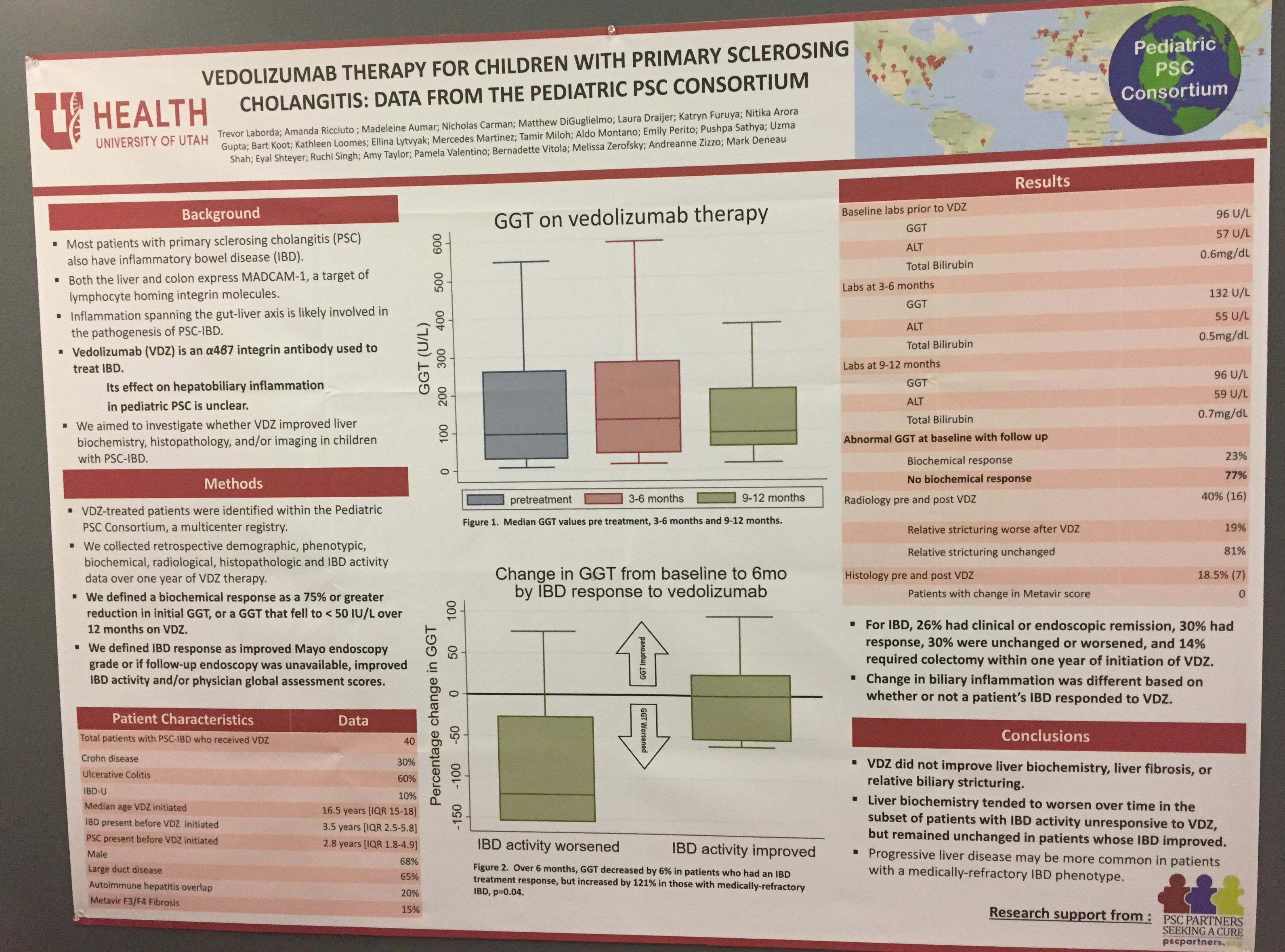
Primary Sclerosing Cholangitis -Use of Vedolizumab for PSC did not appear to help

Eosinophilic Esophagitis

  1. EoE is four times more likely in this cohort with inflammatory bowel disease
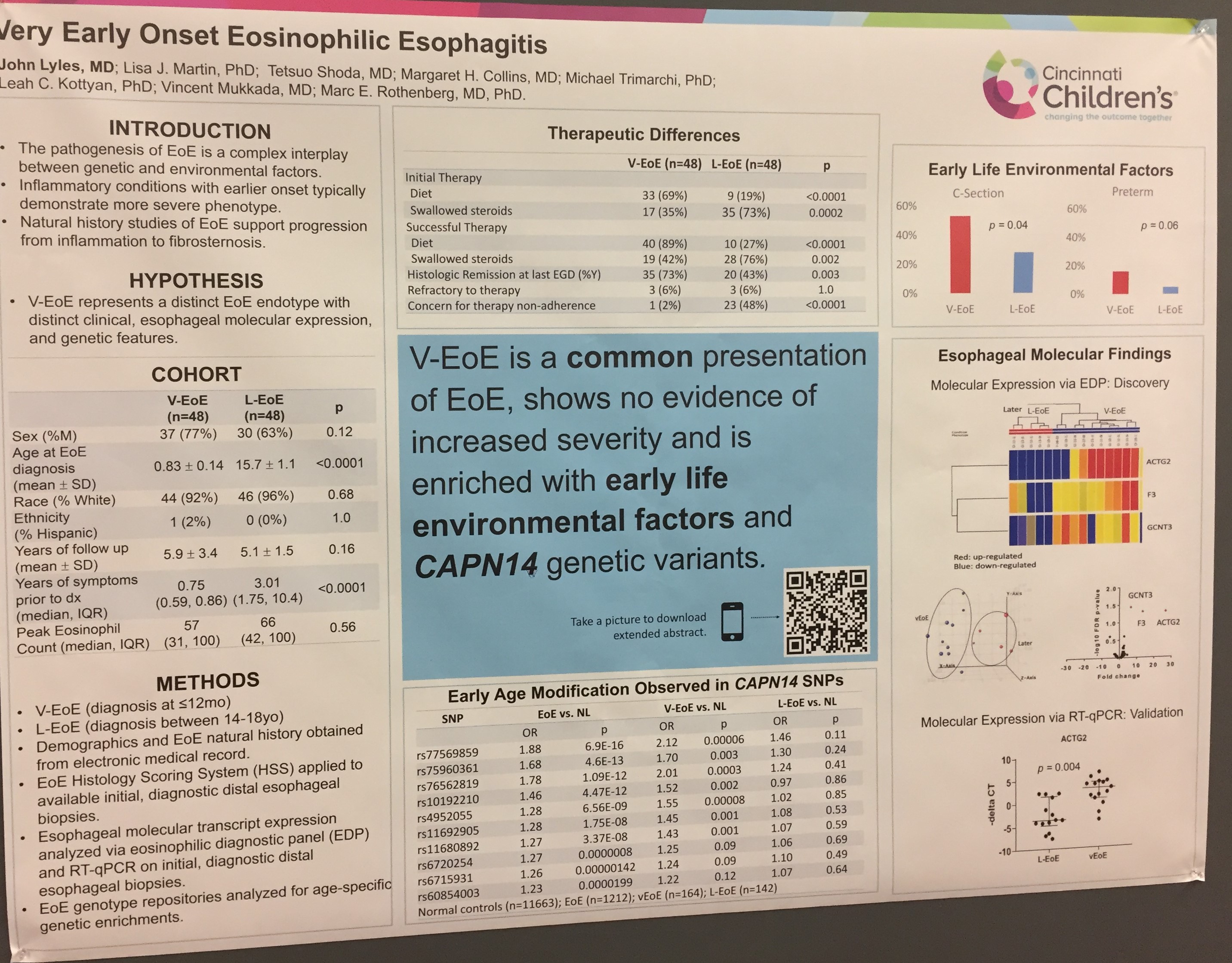
  2. 2nd poster describes very early-onset EoE

Inflammatory Bowel Disease:

  1. Use of infliximab in VEO IBD.  Used in 46/122 (38% of patients) and 50% had persistent use 3 years later

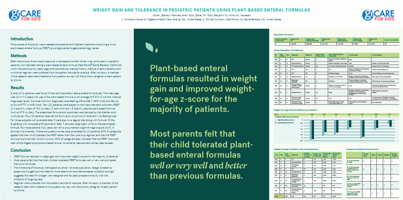
Enteral nutrition –poster from our group describing good tolerance of plant-based formula (with Ana Ramirez).
Celiac disease.  This poster indicates low yield of additional serology for celiac disease besides TTG IgA and serum IgA. This includes testing in young patients (< 2 years) with celiac disease.

#NASPGHAN19 Postgraduate Course (Part 2)

Here are some selected slides and notes from this year’s NASPGHAN’s postrgraduate course. My notes from these lectures may contain errors of omission or transcription.

Link to the full NASPGHAN PG Syllabus 2019 (Borrowed with permission)

9:00 – 10:20 “Potpourri”

46 Alessio Fasano, MD, MassGeneral Hospital for Children  Celiac disease: Beyond diagnosis

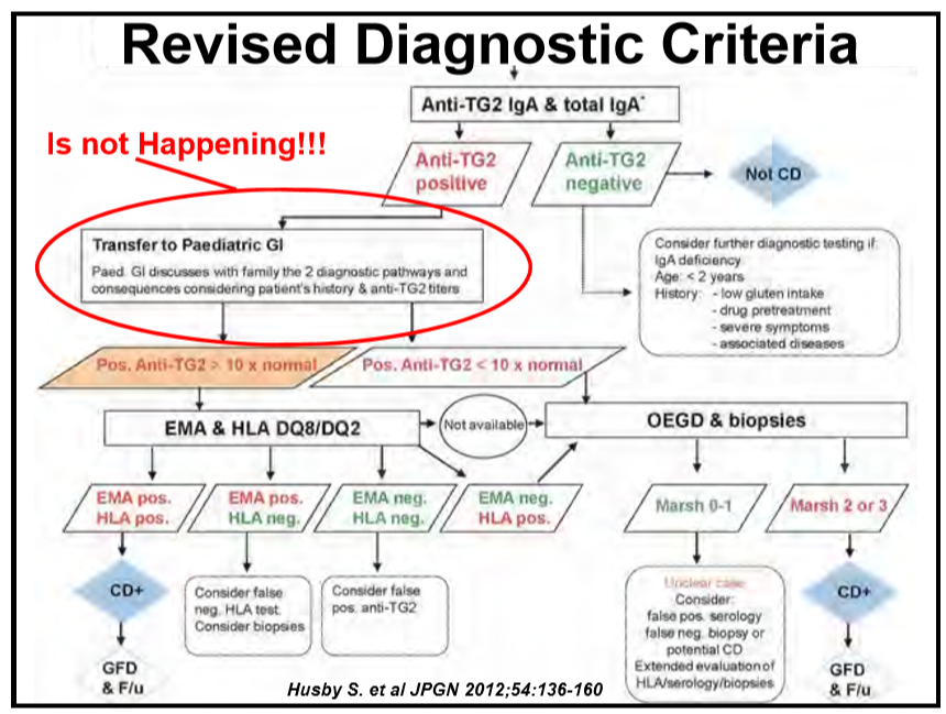
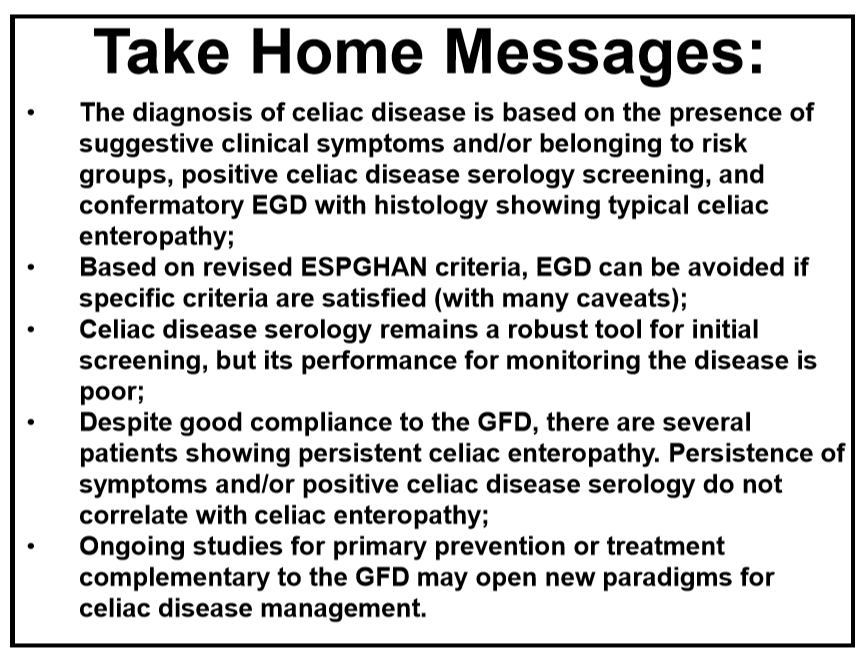
  • Reviewed potential non-biopsy option for diagnosis if anti-TG2 >10 x normal. Pediatricians are not following recommendations –>many children placed on gluten-free diet at lower titer antibody-positivity.
  • Recommends checking Hepatitis B antibody because many children with celiac disease do not seroconvert.
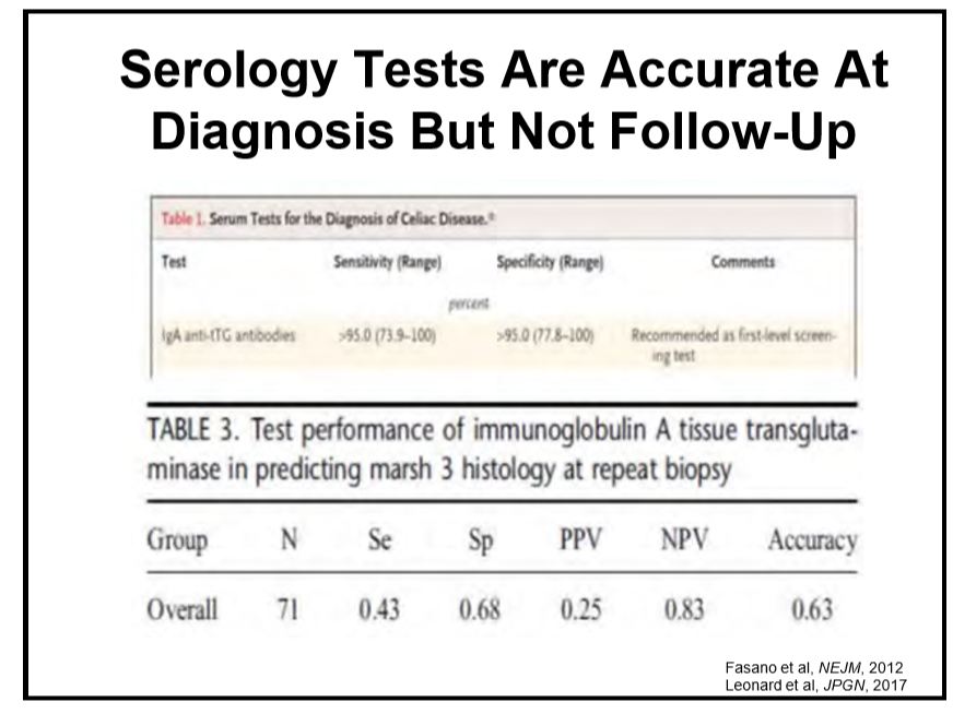
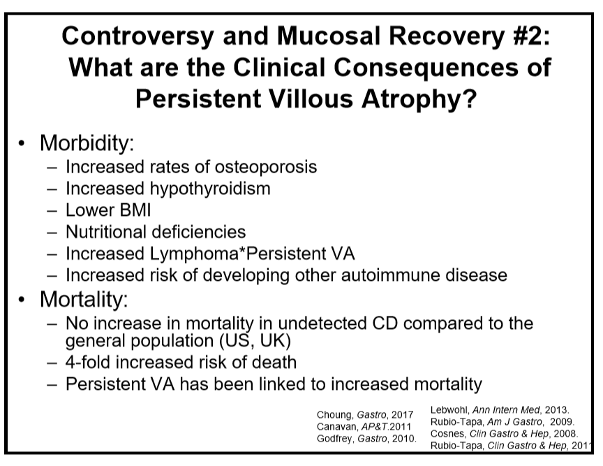
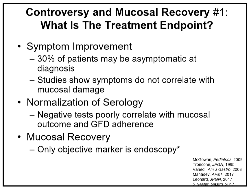
  • TTG levels are good for diagnosis but not as helpful for monitoring after diagnosis.
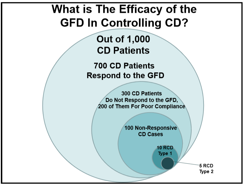
  • Only 10 out of 1000 are true refractory, about 100 out of 1000 are exquisitely sensitive to gluten

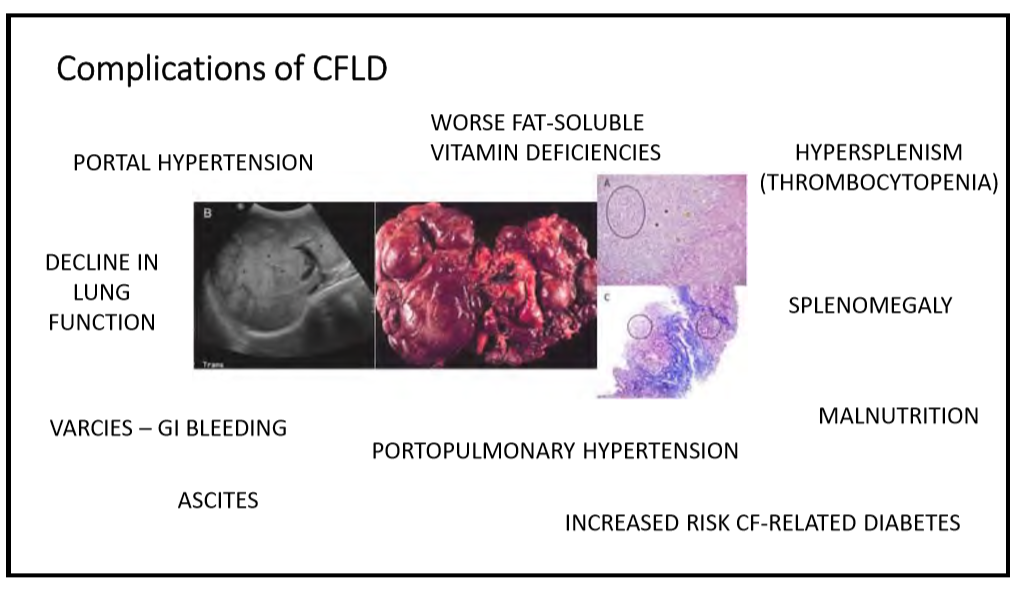
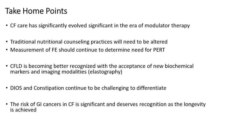
56 Meghana Sathe, MD, UT Southwestern Medical Center The role of the gastroenterologist and hepatologist in Cystic Fibrosis (CF) care today

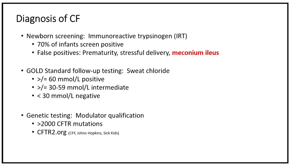
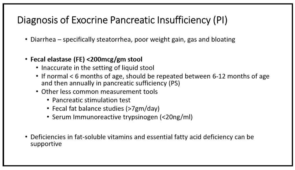
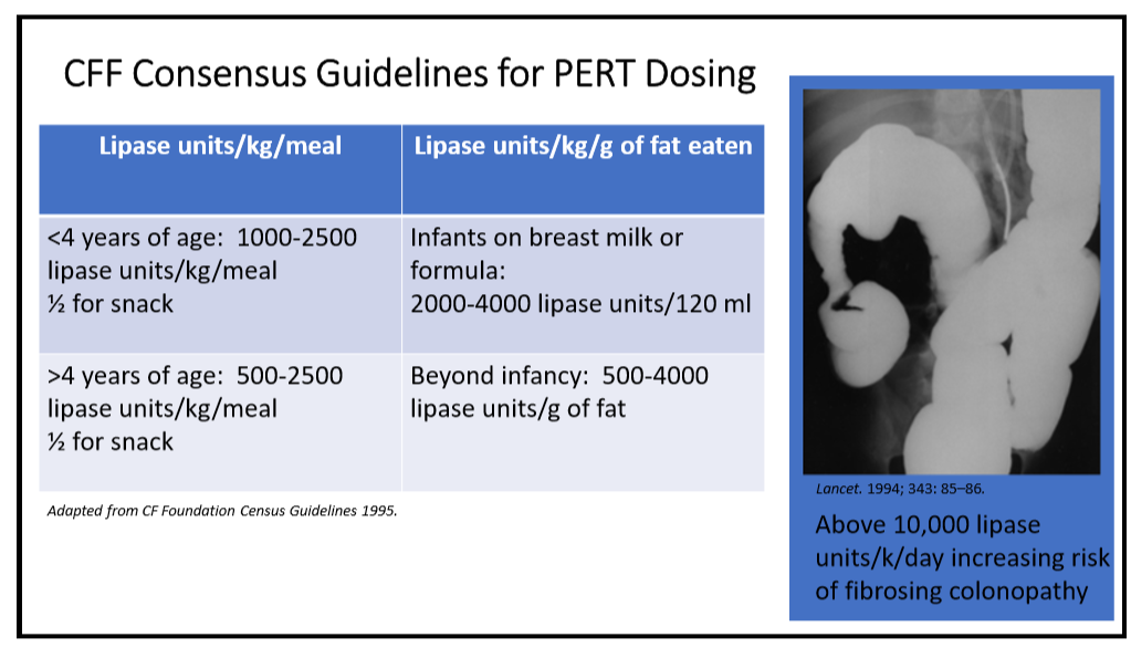
  • Fecal elastase monitoring useful for determining need for PERT.
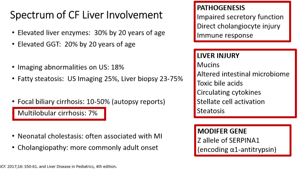
  • Discussed CF liver involvement.  Multilobular cirrhosis, 7% of individuals, is most important liver disease in CF.
  • Modulator therapy can elevate liver enzymes and may need to hold if ALT >5 ULN or lower elevation if elevated bilirubin (see Stop Rules -Practical Advice on DILI)
  • DIOS -for partial obstruction, polyethylene glycol and/or gastrogastrin enemas could be used.
  • Consider treatment of SBBO as well which is frequent with CF.

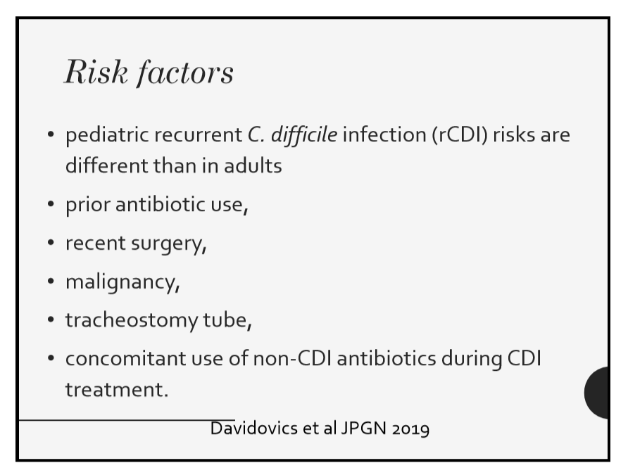
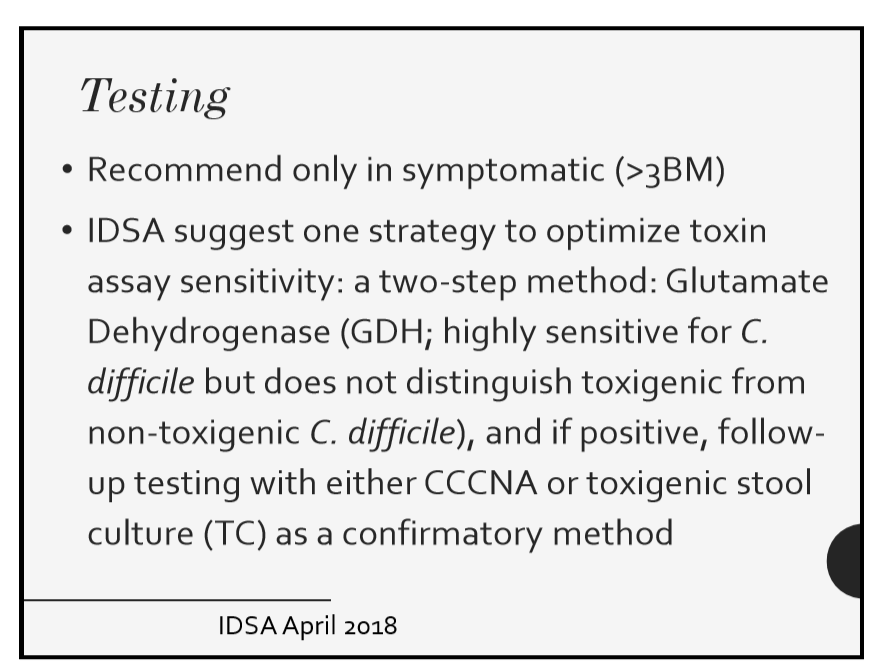
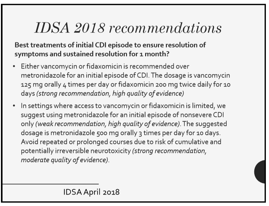
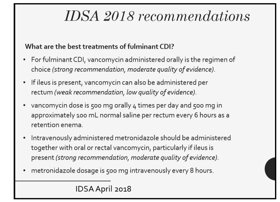
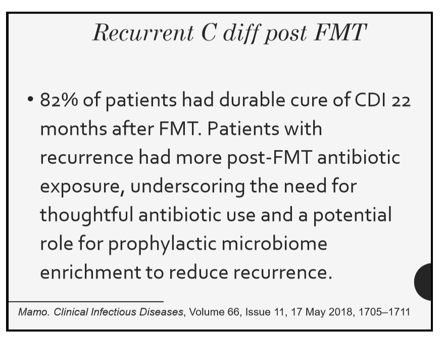
67 Sonia Michail, MD, Children’s Hospital Los Angeles Update on C. difficile

The slide I liked the best was showing a change in microbiome after FMT which is not in syllabus.

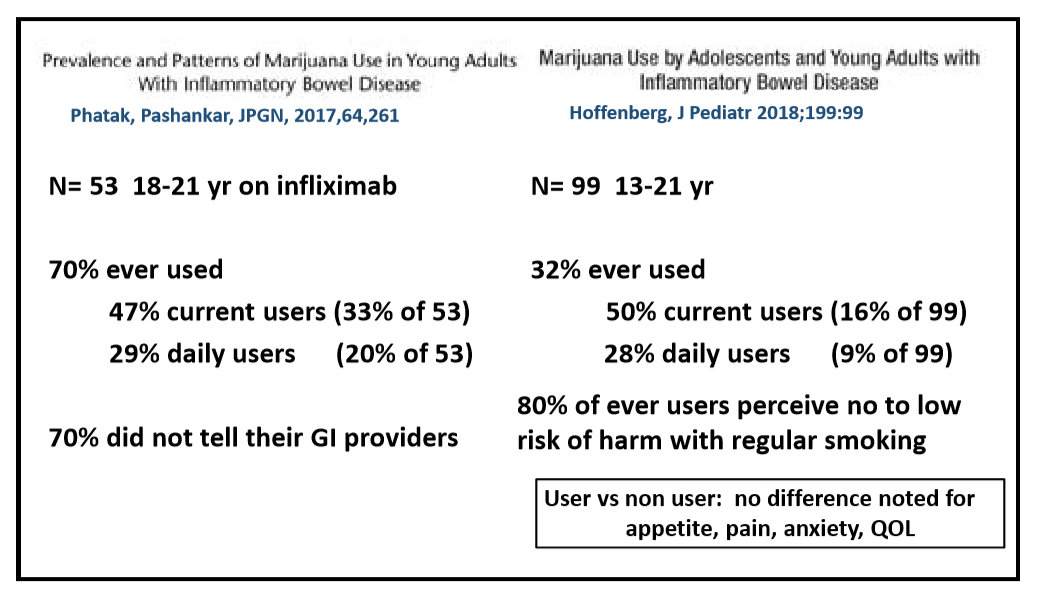
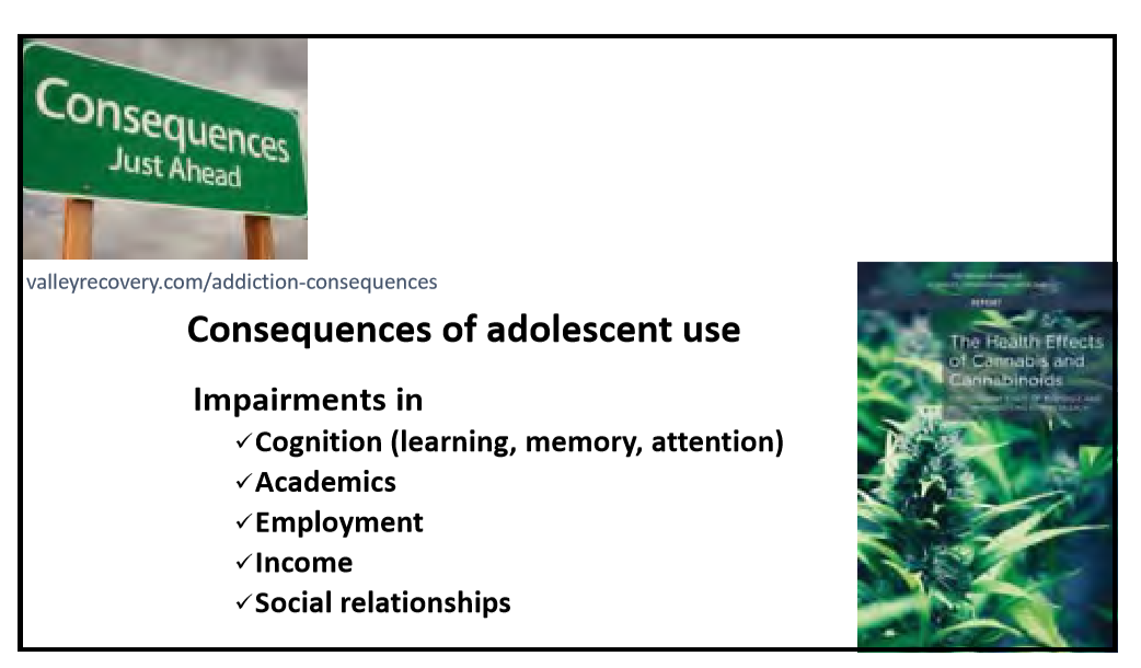
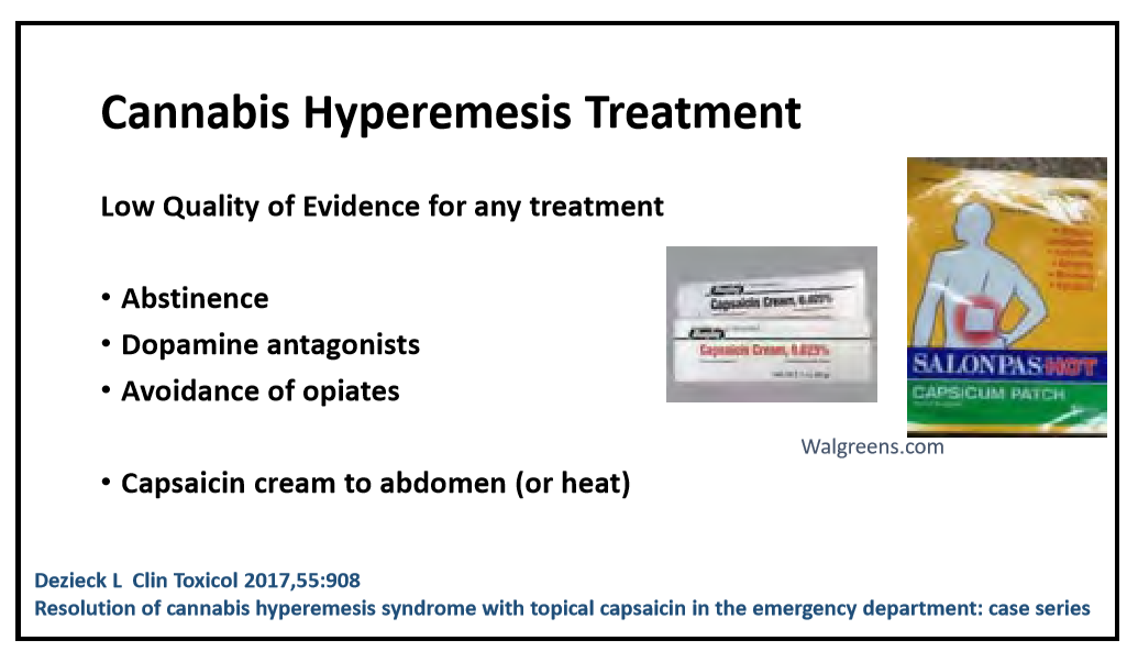
82 Ed Hoffenberg, MD, Children’s Hospital Colorado  What the pediatric GI provider needs to know about cannabis

Disclaimer: NASPGHAN/gutsandgrowth assumes no responsibility for any use or operation of any method, product, instruction, concept or idea contained in the material herein or for any injury or damage to persons or property (whether products liability, negligence or otherwise) resulting from such use or operation. The discussion, views, and recommendations as to medical procedures, choice of drugs and drug dosages herein are the sole responsibility of the authors. Because of rapid advances in the medical sciences, the Society cautions that independent verification should be made of diagnosis and drug dosages. The reader is solely responsible for the conduct of any suggested test or procedure. Some of the slides reproduced in this syllabus contain animation in the power point version. This cannot be seen in the printed version.

#NASPGHAN19 Liver Symposium Notes (Part 1)

Although I was unable to attend this year’s liver symposium at NASPGHAN19, I reviewed the lecture notes.  There is some terrific content.  Here are some of the slides (borrowed with permission from NASPGHAN).

Link to complete NASPGHAN Chronic Liver Disease Symposium 2019

Session I

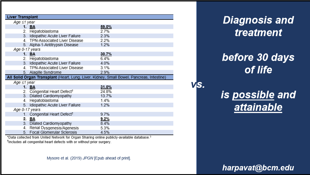
How do I best evaluate a cholestatic infant? Sanjiv Harpavat MD Texas Children’s Hospital 

Related blog post: What is the evidence that biliary atresia starts in utero?

As for this algorithm, in my opinion, the 1st step needs to be to exclude emergencies associated with infantile cholestasis: coagulopathy, hypoglycemia, sepsis, and checking urine for reducing substances (cow’s milk formula can worsen liver disease if galactosemia is present). Subsequently, evaluation needs to proceed quickly to determine the etiology.

How do I interpret genetic results?  Saul J. Karpen MD, PhD, Emory University School of Medicine/Children’s Healthcare of Atlanta

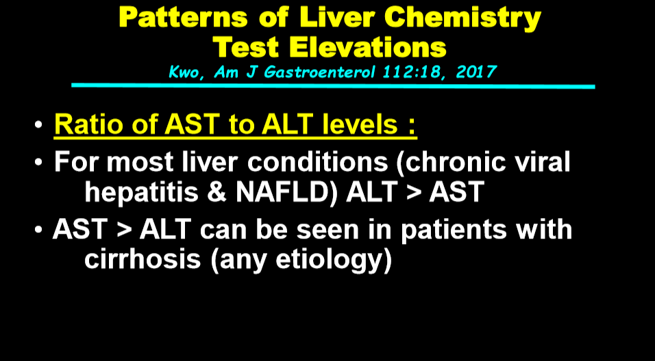
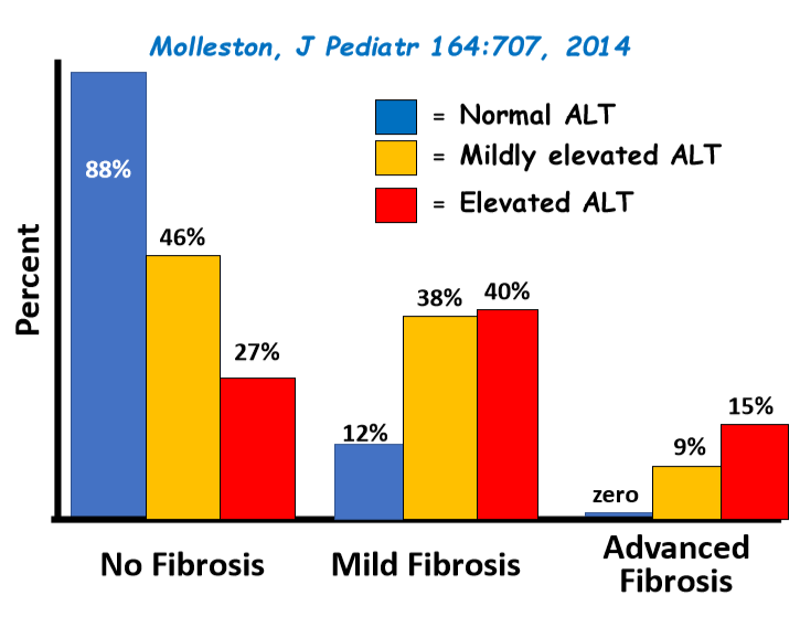
What do abnormal liver enzyme levels mean in a tween?  William F. Balistreri MD, Cincinnati Children’s Hospital Medical Center

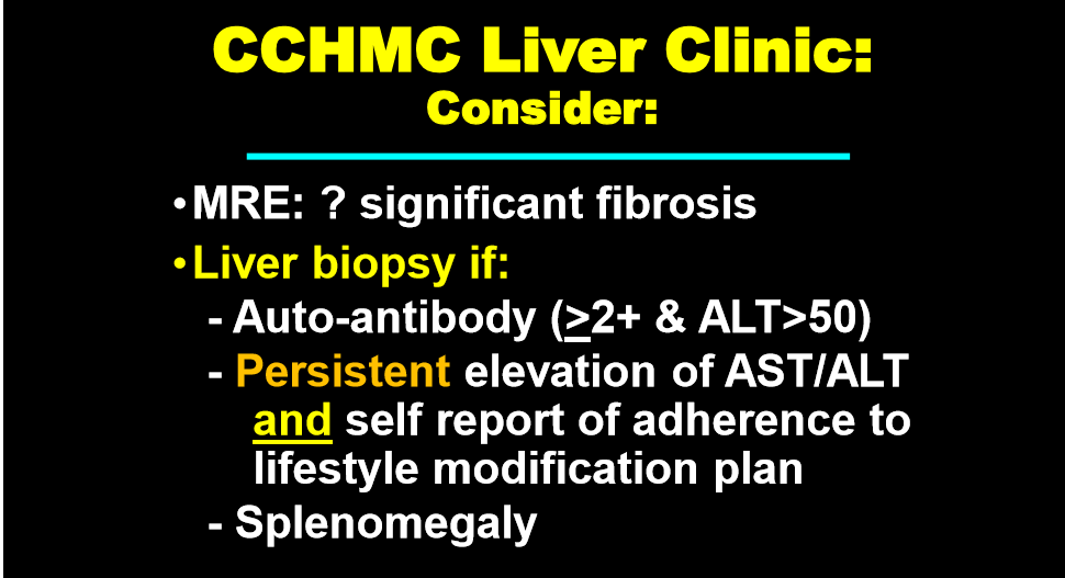
What do I do with this abnormal radiology finding? Jean Molleston MD, Riley Children’s Hospital

I have not selected slides from Dr. Molleston’s handout –the images are terrific.  For most of the problems that are presented, the lecture notes do not provide specific recommendations for management.

Disclaimer: NASPGHAN/gutsandgrowth assumes no responsibility for any use or operation of any method, product, instruction, concept or idea contained in the material herein or for any injury or damage to persons or property (whether products liability, negligence or otherwise) resulting from such use or operation. The discussion, views, and recommendations as to medical procedures, choice of drugs and drug dosages herein are the sole responsibility of the authors. Because of rapid advances in the medical sciences, the Society cautions that independent verification should be made of diagnosis and drug dosages. The reader is solely responsible for the conduct of any suggested test or procedure. Some of the slides reproduced in this syllabus contain animation in the power point version. This cannot be seen in the printed version.


Quantifying the Risk of Autoimmunity for Celiac Disease

A recent study (MR Khan et al. JPGN 2019; 69: 438-42) examined the rates of autoimmune disorders (AD) among patients with celiac disease (CD) (n=249) compared to a control group (n=498) over an 18 year period (1997-2015). The authors utilized the  a database of medical records via the Rochester Epidemiology Project (Mayo Clinic/Olmstead County).

Key findings:

  • Five years after the index date, 5.0% of CD patients and 1.3% of controls had a de novo AD diagnosis
  • In the pediatric age group, there was an increased risk of AD: 5/83 (7.3%) of CD patients and 0/179 (0%) developed a AD diagnosis at the 5-year mark
  • The authors note that they observed a lower rate of Hashimoto thyroiditis after the diagnosis of CD, likely indicating a protective role of a gluten-free diet
  • Thyroid disorders, type 1 DM, psoriasis/psoriatic arthritis and rheumatoid arthritis were the most common AD identified in patients with CD

Limitations:

  • Retrospective study
  • Adherence with GFD was not assessed

My take: Screening for AD periodically is worthwhile in patients with CD, particularly thyroid disorders and type 1 diabetes which accounted for ~80% of the autoimmune conditions identified.

Briefly noted: R Ahawat et al. JPGN 2019; 69: 449-54. In this study with 38 newly-diagnosed CD, the authors found a high prevalence of low vitamin D (25OHD) levels (65.8%) -defined as <30 ng/mL; however, the control population had a higher rate of 79.3%.  While the authors advocate checking vitamin D levels due to the risk of bone disease, it is noted that bone mineral density and vitamin deficiencies frequently improve with a gluten-free diet (Related post: Celiac studies)

Related posts:

Barcelona

“When is Celiac Disease Celiac Disease?”

A recent study (R Auricchio et al. Gastroenterol 2019; 157: 413-20, and editorial PR Green S Guandalini, pg 293-4) provides insight into the topic of “potential celiac disease.”

It is difficult trying to explain the concept of potential celiac disease (CD) to families.  Potential CD refers to the situation of having positive celiac serology but normal duodenal mucosa. In this study, the authors prospectively followed 280 children (age 2-18 yrs) with 2 consecutive abnormal serological tests (anit-TG2, EMA) along with normal duodenal architecture who continued a diet containing gluten.

Key findings:

  • 42 (15%) developed villous atrophy at median followup of 60 months
  • 89 (32%) became serologically-negative for CD
  • Cumulative incidence of progression to villous atrophy was 43% at 12 years.
  • The strongest predictive factor for villous atrophy was age: 7% of children less than 3 years developed flat mucosa, compared with 51% for age 3-10 and 55% for those older than 10 years

Advice on potential CD from editorial –titled “When is Celiac Disease Celiac Disease?”

  • Review the biopsies: were there adequate biopsy specimens? ≥4 from descending duodenum and ≥1 from duodenal bulb
  • Have a second specialist pathologist review specimens
  • If a patient with potential CD is symptomatic, institute a gluten-free diet and then follow for clinical and serologic response
  • If asymptomatic, “a wait and see approach is appropriate with interval biopsies every 2 years, if the elevated antibodies persist”

The editorial also note that none of the patients in this cohort would have been mislabeled with a diagnosis of CD using the non-biopsy approach as none of them had tTG antibodies >10 times the upper level of normal.

My take: This useful study should help with counseling parents about the likelihood of developing celiac disease in those with the “potential” label.  Younger children (<3 yrs), compared to older children, are less likely to convert from potential celiac disease to actual celiac disease..

Related blog posts:

Crater Lake, OR

Dust Mites and Eosinophilic Esophagitis

Given seasonal fluctuation in the activity of eosinophilic esophagitis (EoE), aeroallergens have been considered a trigger in some patients.

Briefly noted: A recent study (A Ravi et al. Gastroenterol 2019; 157: 255-6, editorial 17) showed that dust mite antigen was present in esophageal biopsy specimens at a greater level in adult patients with EoE compared to controls.  With active EoE, patients had dust mite staining in 1.6% of the field which was significantly greater than patients with inactive EoE (0.7). The control group had a complete absence of epithelial dust mite staining.

The editorial (Seena Aceves) notes that these investigators have also shown gluten accumulation in the EoE esophagus.  Whether dust mite antigens or other specific postulated aeroallergens plays a causative role is unclear.  This study shows the presence of these antigens in the esophagus but does not show whether this is an epiphenomenon due to increased permeability or whether these antigens activate the local immune system.

A second study (T Patton et al. JPGN 2019; 69: e43-e48) describes the outcome of coexisting celiac disease and eosinophilic esophagitis in 22 children (from a cohort of 350 children with celiac disease. 17 had repeat biopsies.  Four of 17 (23.5%) had resolution of EoE with a gluten-free diet.  Related blog post: Is there a Link Between Eosinophilic Esophagitis and Celiac Disease?

Sagrada Familia, Barcelona

Should Pediatric Patients with Celiac Disease Be Screened for Low Bone Mineral Density?

A recent retrospective study (J Webster et al. Clin Gastroenterol Hepatol 2019; 17: 1509-14) with 673 children with newly diagnosed (biopsy-proven) celiac disease (CD) (median age 10.6 y) evaluated DXA studies at time of diagnosis.

Key findings:

  • Approximately 7% (n=46) had a low lumbar spine areal bone mineral density (aBMD) z-score (less than -2)
  • Of those with abnormal aBMDs, 18 had repeat studies.  11 of 18 normalized after institution of dietary management. Mean time for repeat DXA was 2.3 years
  • Of note, mean BMI z-score at time of repeat DXA was 0.005 (this includes 90 who had followup studies after a normal baseline DXA).
  • Low body mass index (BMI) with z-score of -0.4 identified a >10% risk of an abnormal aBMD

The authors acknowledge than DXA screening is controversial.  The current study’s strength is its large size.  Limitations include the inability to correlate with clinical factors including adherence to a gluten-free diet.

My take:

  1. Based on this study, it is likely that only 2-3% of pediatric patients with celiac disease will have a persistently abnormal DXA after institution of a gluten free diet for 2 years; it is likely that even more will improve with time if receiving appropriate dietary treatment.
  2. I am not likely to recommend obtaining a baseline DXA study in pediatric patients with newly diagnosed celiac disease; the treatment for low bone mineral density in the setting of celiac disease is the same as for all children with celiac disease.  If one were inclined to look for low BMD, optimal timing would likely be AFTER being adherent on a gluten free diet for at least two years particularly in those who had low BMI at presentation.

Related blog posts:

Columbus Monument, Barcelona

Disclaimer: These blog posts are for educational purposes only. Specific dosing of medications/diets (along with potential adverse effects) should be confirmed by prescribing physician/nutritionist.  This content is not a substitute for medical advice, diagnosis or treatment provided by a qualified healthcare provider. Always seek the advice of your physician or other qualified health provider with any questions you may have regarding a condition