Must-Read: How to Handle Post-Procedure Fevers

A recent study (JM Boster, M Iwanowski, RE Kramer. JPGN 2021; 72: 250-254. Management of Pediatric Postendoscopy Fever: Reducing Unnecessary Health Care Utilization With a Clinical Care Guideline) provides clear guidance for how to handle post-procedure fevers.

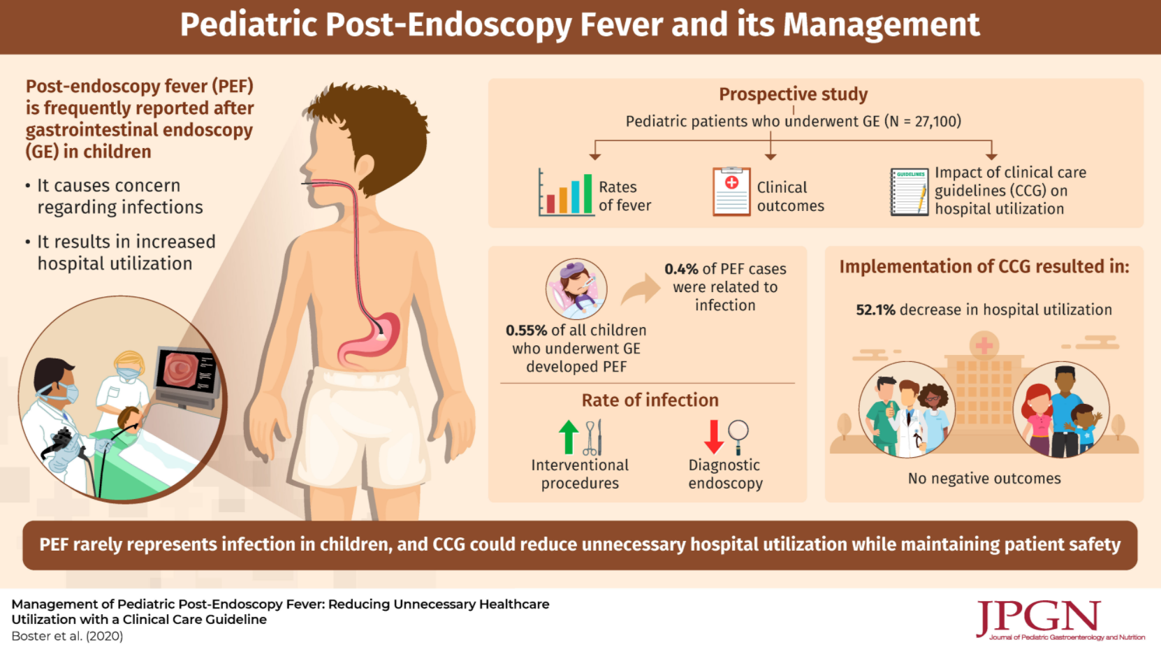
Using a prospective database with 27,100 endoscopies, the authors evaluated a clinical care guideline to reduce unnecessary medical care following endoscopy.

Key findings:

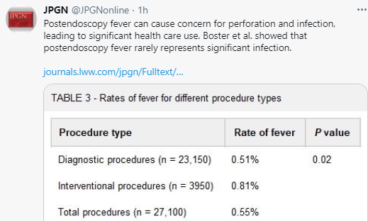
  • Post-endoscopy fever (PEF) occurred in 0.55% (n=150)
  • ONLY 6 of these 150 PEFs (0.4%) were attributed to a procedure complication: 3 had perforations (all with abdominal pain), 2 had aspiration (both had emesis at time of endoscopy) and 1 had a positive blood culture (though had undergone a liver biopsy as well as endoscopy)
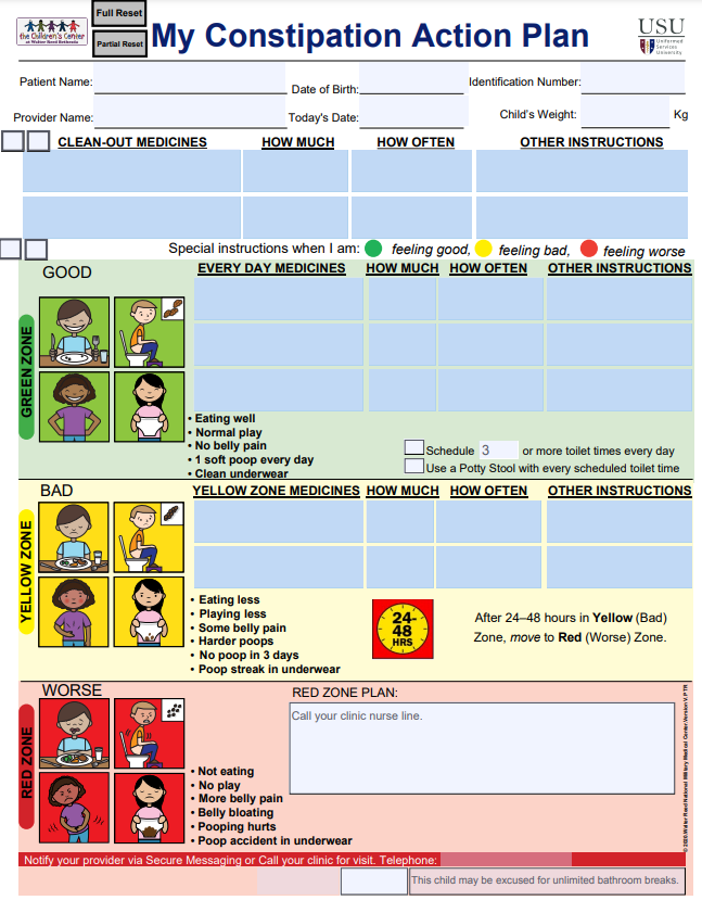
  • The authors published their care guideline (Figure 1) which stratifies risk based on whether the procedure was an interventional (high risk) vs diagnostic (low risk), ASA class, duration of fever, concomitant immunosuppression (eg. steroids), and associated symptoms including vomiting, diarrhea, bleeding, new abdominal pain, impact on activities of daily living, and hydration
  • Interestingly, the authors note that their cohort had a total of 23 perforations, but only 3 presented with fever
  • Using the care guideline resulted in a “significant shift in the prevalence of Grade 2 and above (requiring hospital use) to Grade 1 (clinical observation and reassurance) adverse events, dropping ED visits and admissions by 43.6% and 76.4% respectively for the post-endoscopy fever patients.” This shift was not associated with any observed negative patient outcomes or missed diagnoses.

My take: The authors note that fever is often related to release of inflammatory cytokines which can occur with endoscopy in the absence of complications. The authors methodical guideline to post-procedure fever provides a logical approach to this common problem.

Related blog posts:

Disclaimer: This blog, gutsandgrowth, assumes no responsibility for any use or operation of any method, product, instruction, concept or idea contained in the material herein or for any injury or damage to persons or property (whether products liability, negligence or otherwise) resulting from such use or operation. These blog posts are for educational purposes only. Specific dosing of medications (along with potential adverse effects) should be confirmed by prescribing physician.  Because of rapid advances in the medical sciences, the gutsandgrowth blog cautions that independent verification should be made of diagnosis and drug dosages. The reader is solely responsible for the conduct of any suggested test or procedure.  This content is not a substitute for medical advice, diagnosis or treatment provided by a qualified healthcare provider. Always seek the advice of your physician or other qualified health provider with any questions you may have regarding a condition.

Short Bowel Syndrome -YouTube Information

Some good patient educational videos for short bowel syndrome:

Related article: AE Wiskin et al. Clin Nutrition 2021; https://doi.org/10.1016/j.clnesp.2020.12.029. Prevalence of Home Parenteral Nutrition in children Key finding: In 2019, 389 children received HPN (home parenteral nutrition) in the UK; this is nearly double the number last reported in 2012 and is a prevalence of 30 per million children (Thanks to Kipp Ellsworth for this reference)

Other resources:

Related blog posts for Short Bowel Syndrome:

Should We Worry About Osteopenia in Children with Crohn’s Disease?

A recent retrospective study (N Ronel, et al. JPGN 2021; 72: 270-275. Clinical Criteria Can Identify Children With Osteopenia in Newly Diagnosed Crohn Disease) included 116 children (mean age 13 years)

Key finding:

  • In total, 59% of children with BMI z-score <−0.5 had moderate-severe osteopenia and only 18% of those with higher z-scores. 
  • Osteopenia was associated with lower BMI z-score (−0.8 ± 1.2 vs −1.8 ± 1.1, P < 0.001) and higher PCDAI  (33.7 ± 15.2 vs 25.7 ± 16.5; P = 0.009)
  • None of the higher risk patients were receiving long-term corticosteroids 
  • Limitations: retrospective study with relatively small sample size, pubertal stage not recorded, variability in DXA studies, and lack of followup information

My take: The authors have NOT shown that identification of osteopenia at the time of diagnosis improves outcome of Crohn’s disease or bone disease. This is why I disagree with their recommendation to routinely screen children with BMI z-score <−0.5. In those in which finding osteopenia may influence treatment, then a DXA study would be worthwhile.

Related blog posts:

Chicago 2021

Seronegative Villous Atrophy

A recent large retrospective study (R Mandile et al. JPGN 2021; 72: 282-287. Seronegative Villous Atrophy in Children: Clinical and Immunohistochemical Features) provides information about conditions, besides celiac disease (CD) which present with villous atrophy. 64 of 1282 pediatric patients were seronegative with villous atrophy; seronegative was defined as testing negative twice for serology (TTG IgA/EMA or if IgA-deficient, IgG antibody serology).

Key findings:

  • Diagnoses were: inflammatory bowel diseases (IBD) (21/64), food allergy (8/64), infections (7/64, of which 3 HIV infections), immune deficiency (3/64), short bowel syndrome (3/64), congenital diarrhea (2/64), other/inconclusive diagnosis (8/64). In addition, there were 12 with Gastro-Esophageal Reflux Disease (GERD) & the authors speculate that perhaps hyperacidity could play a role in some of these cases.
  • Only one quarter of the seronegative patients had an increased number of intraepithelial lymphocytosis (IELs)
  • Among those with villous atrophy attributed to IBD, this was nearly equally-split between Crohn’s disease and ulcerative colitis, 10 and 11 patients respectively (according to Table 1)
  • The authors note that the ~5% of patients with seronegative villous atrophy with alternative diagnosis than Celiac disease may be an overestimation as more individuals are being diagnosed without biopsy based on serology
  • Despite the large cohort, there are still other rare conditions that were not identified in this study (eg. autoimmune enteropathy, CTLA4B deficiency,drug-induced enteropathy, and tropical sprue)

My take: This article provides a good starting point for patients with villous atrophy and negative serology.

Related article: J Devara et al. JPGN 2021; 72: 288-293. The Significance and Clinical Outcome of Lymphocytic Duodenosis in Children: Mayo Clinic Experience and Systematic Review Background: Lymphocytic duodenosis (LD) defined as increased intraepithelial lymphocytes >25 intraepithelial lymphocytes (IELs) per 100 epithelial cells with normal villous architecture is associated with many gastrointestinal (GI) disorders.

Key findings:

  • During the study period 12,744 children underwent an EGD with biopsies. Of those, we identified 426 children with LD (3%).
  • Among the LD (compared to control group), 5% had celiac disease (vs 0%, P < 0.001), 9% had Crohn disease (3%, P = 0.003) and 3% had Helicobacter pylori gastritis (1%, P = 0.021).

Related blog post: @AmyOxentenkoMD: Celiac Disease and Mimics

Pictographic Constipation Action Plan

A recent study (PT Reeves et al. J Pediatr 2021; 229: 118-126. Full text link: Development and Assessment of a Pictographic Pediatric Constipation Action Plan) highlighted patient education efforts. “This study focused on the design and assessment of a low literacy pictographic CAP for the care of functional constipation in children.”

My take: I agree with the authors that a simple plan like this has “the potential to become an important tool to be used in the care of children with functional constipation, improving both quality-of-care and clinical outcomes.”

Link to PDF: Constipation Action Plan

Related blog posts:

This QR code provides 9 minute explanation of constipation and action plan:

Disclaimer: This blog, gutsandgrowth, assumes no responsibility for any use or operation of any method, product, instruction, concept or idea contained in the material herein or for any injury or damage to persons or property (whether products liability, negligence or otherwise) resulting from such use or operation. These blog posts are for educational purposes only. Specific dosing of medications (along with potential adverse effects) should be confirmed by prescribing physician.  Because of rapid advances in the medical sciences, the gutsandgrowth blog cautions that independent verification should be made of diagnosis and drug dosages. The reader is solely responsible for the conduct of any suggested test or procedure.  This content is not a substitute for medical advice, diagnosis or treatment provided by a qualified healthcare provider. Always seek the advice of your physician or other qualified health provider with any questions you may have regarding a condition

Highly Prevalent Pediatric Feeding Disorders

A recent retrospective study (K Kovacic et al. J Pediatr 2021; 228: 126-131. Pediatric Feeding Disorder: A Nationwide Prevalence Study) provides epidemiologic data for pediatric feeding disorders.

The authors utilized three databases for children aged 2 months to 18 years: Medicaid Databases from Arizona (2009-2017) and Wisconsin (2005-2014) (public insurance databases) and The Truven Health Analytics MarketScan Commercial Claims and Encounters Database (2009-2015) (a nationwide private insurance database).

Key findings:

  • There were 126 002 and 367 256 children 5 years of age or younger with pediatric feeding disorders (PFD) with public and private insurance, respectively
  • In 2014, the annual prevalence of PFD was 1 in 23, 1 in 24, and 1 in 37 in children under 5 years in the publicly insured cohorts in Wisconsin, Arizona, and the privately insured cohort, respectively.
  • The prevalence of PFD in children <5 years (range: 27-44 per 1000 children) exceeds the prevalence of U.S. children with autism spectrum disorder (~17 per 1000 children at age 8 year) and eating disorders like anorexia nervosa and bulimia (8 and 13 per 100,000 persons per year).
  • In an associated editorial (pg 13-14), Rachel Rosen notes that “despite their high prevalence, the lack of studies funded by the National Institutes of Health…is striking.”

My take: This study provides useful data on PFD prevalence. PFD have a wide range of associated diseases, including prematurity, neurologic disorders/developmental delay, congenital heart disease, chronic lung disease, autism, and congenital bowel disorders. In some, PFD are related to poorly-understood feeding aversions.

Related blog posts:

Chicago, IL & Lake Michigan

FMT Research & The Shawshank Redemption

In The Shawshank Redemption, Andy Dufresne (Tim Robbins) manages to escape prison by crawling through 500 yards of a filthy sewage pipe. It seems like a similar effort will be needed to find out how to benefit from fecal transplantation when given for problems like irritable bowel syndrome and metabolic disease/obesity. Some recent studies and associated editorials are noted below.

T Holvoet et al. Gastroenterol 2021; 160: 145-157. Fecal Microbiota Transplantation Reduces Symptoms in Some Patients With Irritable Bowel Syndrome With Predominant Abdominal Bloating: Short- and Long-term Results From a Placebo-Controlled Randomized Trial

  • Key finding: At week 12, 56% of patients given donor stool reported improvement in both primary endpoints compared with 26% of patients given placebo (P = .03).
  • Commentary: PW O’Toole, F Flanahan. Gastroenterol 2021; 160: 15-17. Full Text: Transplanting Microbes for Irritable Bowels or Irritated Microbes or Both?
    • This editorial stresses that trials of FMT in IBS have had inconsistent results and risks are unclear. “How many clinicians inform patients receiving FMT that the donor microbiota might include components that increase (or decrease) one’s risk of colorectal cancer?” Part of the problem is “due, in part, because a normal microbiome has not been defined.”

E Rinott et al. Gastroenterol 2021; 160: 158-173. Full text Effects of Diet-Modulated Autologous Fecal Microbiota Transplantation on Weight Regain

Key findings:

  • In this randomized controlled trial with 90 participants, autologous FMT (aFMT) significantly attenuated weight regain in the green-Mediterranean group (aFMT, 17.1%, vs placebo, 50%; P = .02) and improved insulin resistance: insulin rebound (aFMT, –1.46 ± 3.6 μIU/mL vs placebo, 1.64 ± 4.7 μIU/mL; P = .04) (Graphical abstract below)
  • In mice, Mankai-modulated aFMT in the weight-loss phase compared with control diet aFMT, significantly prevented weight regain and resulted in better glucose tolerance during a high-fat diet–induced regain phase (all, P < .05).

Commentary: M Nieurdorp, K Madsen. Gastroenterol 2021; 160: 17-19. Full text The Promise of Maintaining Diet-Induced Weight Loss by Swallowing One’s Own Feces: Time to Provide a Do-It-Yourself Manual?

  • “These findings add support to the current body of evidence that the gut microbiota have a role in weight gain and metabolism. However, many questions remain. Indeed, although studies have shown varying degrees of effectiveness of FMT in the improvement of metabolic parameters in human participants, there has been no evidence yet that FMT can induce weight loss in obese patients.”
  • “The finding that maintenance of weight loss was only seen in the one dietary group consuming the Mediterranean diet plus green tea and Mankai supplement who received autologous FMT, would suggest that specific microbial profiles may be involved and that weight loss per se may not result in the required microbial profiles.”
Figure 1 from editorial: Challenges associated with the use of fecal microbial transplantation (FMT) as treatment

My take: Both of these studies show that modulation of the fecal microbiome may be helpful under the right set of circumstances to help with both irritable bowel syndrome and metabolic syndrome. However, ‘hundreds of yards’ of more research is needed to determine if this is really feasible and to assure that the benefits outweigh the potential risks.

Related blog posts:

Mechanisms of Postinfectious Irritable Bowel Syndrome & Functional Disorders

Aguilera-Lizarraga, J., Florens, M.V., Viola, M.F. et al. Local immune response to food antigens drives meal-induced abdominal painNature (2021). https://doi.org/10.1038/s41586-020-03118-2 (Thanks to Ben Gold’s twitter feed for this reference)

Background: “Up to 20% of people worldwide develop gastrointestinal symptoms following a meal, leading to decreased quality of life, substantial morbidity and high medical costs”

“Here we show that a bacterial infection and bacterial toxins can trigger an immune response that leads to the production of dietary-antigen-specific IgE antibodies in mice, which are limited to the intestine. Following subsequent oral ingestion of the respective dietary antigen, an IgE- and mast-cell-dependent mechanism induced increased visceral pain. This aberrant pain signaling resulted from histamine receptor H1-mediated sensitization of visceral afferents. Moreover, injection of food antigens (gluten, wheat, soy and milk) into the rectosigmoid mucosa of patients with irritable bowel syndrome induced local oedema and mast cell activation.”

My take: This study shows how innocuous food can trigger pain after an intestinal infection.

Related blog posts:

Ustekinumab Escalation in Patients with Crohn’s Disease & Healthy Lifestyle Choices for IBD Patients

JE Ollech et al. Clin Gastroenterol Hepatol 2021; 19: 104-110. Effectiveness of Ustekinumab Dose Escalation in Patients With Crohn’s Disease

In patients with Crohn’s disease, dose escalation of biologic therapy (eg. anti-TNF agents, vedolizumab) has been shown to be helpful in recapturing response to treatment. In a retrospective study with 110 patients, Ollech et al explore the outcomes in those who had their subcutaneous ustekinumab interval shortened to 4 weeks (from every 8 weeks).

Key findings:

  • Following dose interval shortening, the patients’ median Harvey Bradshaw Index (HBI) decreased from 4.5 to 3 ( P = .002), the median level of CRP decreased from 8 mg/L to 3 mg/L ( P = .031), and median level of fecal calprotectin decreased from 378 μg/g to 157 μg/g ( P = .57).
  •  Among patients with active disease (HBI >4, CRP ≥/=5mg/dL, fecal calprotectin >250ug/g, or endoscopic evidence for disease activity), dose interval shortening was associated with a 28% clinical remission (an HBI score ≤4), and 50% had reduced levels of fecal calprotectin; 36% achieved endoscopic remission.
  • The authors did not identify serious adverse events with dose shortening.

My take: Prospective studies are needed. This study indicates that more frequent dosing improves outcomes in a significant fraction of those with active disease.

Related blog posts:

Unrelated article: C-H Lo et al. Clin Gastroenterol Hepatol 2021; 19: 87-95. Healthy Lifestyle Is Associated With Reduced Mortality in Patients With Inflammatory Bowel Diseases In this study, using data from three large cohort studies, the authors assessed the impact of 5 healthy lifestyle factors: never smoking, body mass index 18.5–24.9 kg/m 2, vigorous physical activity in the highest 50% with non-zero value, alternate Mediterranean diet score ≥4, and light drinking [0.1–5.0 g/d]. Key finding:

  • Compared to patients with IBD with no healthy lifestyle factors, patients with IBD with 3–5 healthy lifestyle factors had a significant reduction in all-cause mortality (hazard ratio [HR], 0.29; 95% CI, 0.16–0.52; Ptrend < .0001). 

My take: Like the general population, healthy lifestyle choices are important in individuals with IBD; this study provides some data on the effects on outcomes.

Why Observational Studies Are Misleading & PPI Association with Kidney Stones

M Simonov et al. Clin Gastroenterol Hepatol 2021; 19: 72-79. Use of Proton Pump Inhibitors Increases Risk of Incident Kidney Stones

Commentary: P Moayyedi. Clin Gastroenterol Hepatol 2021; 19: 41-42. Full Text Link: Leaving No Stone Unturned in the Search for Adverse Events Associated With Use of Proton Pump Inhibitors

 The retrospective study by Simonov et al used data from the Women’s Veteran’s Cohort Study (1999-2017) with 465,891 patients. Key findings:

  • Overall, 2.4% of the cohort developed kidney stones. PPI use was associated with kidney stones in the unadjusted analysis, hazard ratio [HR], 1.74 (95% CI, 1.67–1.82), and persisted in the adjusted analysis with HR, 1.46 (CI, 1.38–1.55). The association was maintained in a propensity score-matched subset of PPI users and nonusers (adjusted HR, 1.25; CI 1.19–1.33).
  • H2RAs were also associated with increased risk with adjusted HR, 1.47

While this study is interesting, the editorial provides a great deal of insight into how this study and many others can be misleading. Key points:

  • “It seems that every few months a new issue arises, with the list of problems that PPIs might cause becoming ever longer, including pneumonia, fracture, heart disease, Clostridium difficile–associated diarrhea, dementia, chronic kidney disease, low B12 levels, gastric cancer, and even all-cause mortality.”
  • If the findings in the study are correct, with the unadjusted HR, “this translates to a number needed to harm (NNH) of 365 patients who need to take PPIs for 1 year to observe 1 extra episode of kidney stones…if the adjusted HR is used …the NNH was 1550.”

Limitations:

  • Confounding variables are hard to eliminate in an observational study. “Studies usually show that patients who are prescribed PPIs are, on average, sicker than those who are not taking these drugs.”
  • In the only large randomized controlled study (>17,000 patients over 3 years) of PPIs, there was no difference in pneumonia, Clostridium difficile infection, fracture, gastric atrophy, chronic kidney disease, dementia, cardiovascular disease, cancer, hospitalizations, and all-cause mortality in the PPI compared with the placebo arms.”Enteric infections, which were slightly more common in patients randomized to PPIs, but even there the NNH was more than 900 per year.”
  • Biases undermine the interpretation of observational studies. One example for PPIs is its association with pneumonias in prior observational studies.
    • “The effect was strongest within the first week of prescription when the odds ratio was approximately 4, although this was reduced to approximately 1.5 after 1 month. This marked reduction in risk over a relatively short period of time is not biologically plausible and a more likely explanation is that the association is the result of protopathic bias. Patients presenting to the clinician with a cough may be diagnosed with silent reflux and given a PPI. A few days later other symptoms develop, and pneumonia is made as the final diagnosis. This will not be apparent when simply interrogating a database where the researcher will observe that a PPI was prescribed before the onset of pneumonia and will imply the association is causal when this is not the case.”
  • This same type of bias could be present with the association between PPI and kidney stones.
    •  “Patients may present with abdominal pain and be given a PPI as a therapeutic trial, assuming the pain may be acid-related when subsequently it is found that the pain relates to kidney stones. Simonov et al reported that 3% were prescribed PPIs within 1 month of the diagnosis of kidney stones, but did not provide the analysis that would allow us to interpret whether protopathic bias may play a role in the associations observed”

My take: It is unlikely that PPIs cause kidney stones; however, if this is a risk factor, it is very rare. Understanding how PPIs have been incorrectly linked to a multitude of problems is a valuable lesson for any practitioner and emphasizes the need for randomized controlled studies to determine medication safety.

Related blog posts: