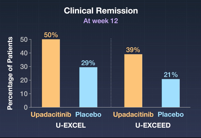
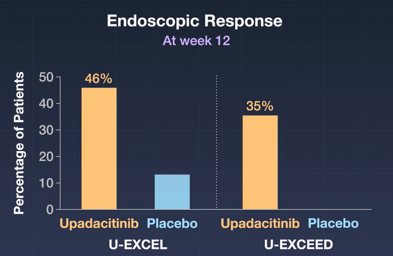
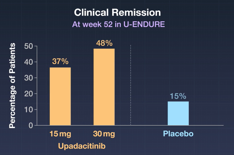
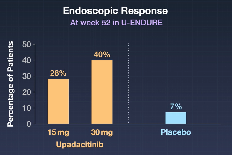
“Patients should start with 45 mg of Rinvoq once daily for 12 weeks. Following the 12-week period, the recommended maintenance dosage is 15 mg once a day. A maintenance dosage of 30 mg once daily can be considered for patients with refractory, severe, or extensive Crohn’s disease.”
“The most common side effects of Rinvoq as indicated for Crohn’s disease are upper respiratory tract infections, anemia, fever, acne, herpes zoster, and headache…. Serious infections, mortality, malignancy, major adverse cardiovascular events, and thrombosis have occurred with JAK inhibitors such as Rinvoq.”
Lately, there has been a lot of ‘buzz’ about the potential use of point-of-care bowel sonography (aka intestinal ultrasound). This study (2019-2021) prospectively enrolled patients with a high likelihood of Crohn’s disease (CD) and compared handheld bowel sonography (HHBS), MRE (all patients, n=85, had ileocolonoscopy)
Key findings:
Sensitivity, specificity, positive predictive values, and negative predictive values for CD diagnosis were 87.50%, 91.89%, 93.33%, and 85% for HHBS; and 91.67%, 94.59%, 95.65%, and 89.74% for MRE, without significant differences in terms of diagnostic accuracy (89.41% for HHBS vs 92.94% for MRE, P = NS)
Magnetic resonance enterography was superior to HHBS in defining CD extension (r = 0.67; P < .01) with a better diagnostic performance than HHBS for detecting location (k = 0.81; P < .01), strictures (k = 0.75; P < .01), abscesses (k = 0.68; P < .01), and fistulas (k = 0.65; P < .01).
My take: In this study, MRE was clearly superior at defining CD complications. This study suggests that HHBS could be an effective screening tool but is not likely a definitive imaging study. In terms of bedside monitoring, it would be helpful to see how clinical monitoring with HBSS compares with a highly sensitive marker like a calprotectin. I also worry that HBSS could perform more poorly with more widespread application due to potential increase in operator error.
The authors examined 29 RCTs (3772 patients). Key findings:
Only two medications (neither available in U.S.) were identified as being more effective than placebo for global symptoms of gastroparesis: clebopride (RR, 0.30) followed by domperidone (RR, 0.68)
Oral metoclopramide ranked first for nausea (RR 0.46), fullness (RR 0.67), and bloating. Though, use may result extrapyramidal adverse effects
I tend to abide by the recommendation proposed by Masaoka and Tack10 some years ago that one should use the term gastroparesis only “when persistently and severely delayed gastric emptying is found in the absence of mechanical obstruction.”
An obsession with gastroparesis as the basic issue among patients with “gastroparesis-like” symptoms has translated into a therapeutic fixation on the acceleration of gastric emptying. This too has led to frustration and disappointment. As already mentioned, symptoms are a poor predictor of the rate of gastric emptying, and a normalization of delayed emptying has not consistently correlated with symptom responses and vice versa.11,12
Oblivious to research illustrating how upper gastrointestinal symptoms can result from several other derangements in foregut physiology, such as impaired accommodation of the upper stomach, visceral hypersensitivity, and antropyloric distensibility and dysmotility,13, 14, 15, 16
My take: Currently, pharmaceutical agents geared towards symptoms like nausea and sensory disorders are much more promising than prokinetic agents for gastroparesis
Y Usui et al. NEJM 2023; 388: 1181-1190. Helicobacter pylori, Homologous-Recombination Genes, and Gastric Cancer
Background: Gastric cancer is the fifth most common neoplasm and the fourth leading cause of death from cancer worldwide.1Helicobacter pylori has been classified as a group I carcinogen and is an environmental risk factor for gastric cancer.2 Although H. pylori infection affects more than half the world population
Methods: This study evaluated the association between germline pathogenic variants in 27 cancer-predisposing genes and the risk of gastric cancer in a sample of 10,426 patients with gastric cancer and 38,153 controls from BioBank Japan. This retrospective study also assessed the combined effect of pathogenic variants and H. pylori infection status on the risk of gastric cancer and calculated the cumulative risk in 1433 patients with gastric cancer and 5997 controls from the Hospital-based Epidemiologic Research Program at Aichi Cancer Center (HERPACC).
Key findings:
Germline pathogenic variants in nine genes: (APC, ATM, BRCA1, BRCA2, CDH1, MLH1, MSH2, MSH6, and PALB2) were associated with the risk of gastric cancer.
At 85 years of age, persons with H. pylori infection and a pathogenic variant had a higher cumulative risk of gastric cancer than noncarriers infected with H. pylori (45.5% vs. 14.4%).
Limitation: The study population was from East Asia and thus, the findings may be different in other populations.
My take: H. pylori infection has a synergistic effect in increasing the risk of gastric cancer in individuals with germline pathogenic variants in homologous-recombination genes. To minimize the risk of gastric cancer, H pylori eradication is important; however, it is especially in those with cancer-predisposing variants.
S Travis et al. NEJM 2023; 388: 1191-1200. Vedolizumab for the Treatment of Chronic Pouchitis
Methods: This was a a phase 4, double-blind, randomized trial (n=102 adults,EARNEST trial) to evaluate vedolizumab in adult patients in whom chronic pouchitis had developed after undergoing IPAA for ulcerative colitis.All patients received 4 weeks of ciprofloxacin and the treatment group received standard vedolizumab dosing. The primary end point was modified Pouchitis Disease Activity Index (mPDAI)–defined remission (an mPDAI score of ≤4 and a reduction from baseline of ≥2 points in the mPDAI total score; scores range from 0 to 12, with higher scores indicating more severe pouchitis) at week 14. The mPDAI is based on clinical symptoms and endoscopic findings.
Key findings:
The incidence of mPDAI-defined remission at week 14 was 31% (16 of 51 patients) with vedolizumab and 10% (5 of 51 patients) with placebo
Differences in favor of vedolizumab over placebo were also seen with respect to mPDAI-defined remission at week 34 (difference, 17 percentage points, 35% vs 18%)
My take: Vedolizumab is an effective treatment for chronic pouchitis.
Overall, acute hepatic porphyrias (AHP) are rare disorders. “Acute intermittent porphyria is the most common type of AHP, with an estimated prevalence of patients with symptoms of approximately 1 in 100,000. The major clinical presentation involves attacks of severe pain, usually abdominal and generalized, without peritoneal signs or abnormalities on cross-sectional imaging…The screening tests of choice include random urine porphobilinogen and d-aminolevulinic acid corrected to creatinine.”
“All patients with elevations in urinary porphobilinogen and/or d-aminolevulinic acid should initially be presumed to have AHP. The cornerstones of management include discontinuation of porphyrinogenic drugs and chemicals, administration of oral or intravenous dextrose and intravenous hemin, and use of analgesics and antiemetics. Diagnosis of AHP type can be confirmed after initial treatment by genetic testing for pathogenic variants in HMBS, CPOX, PPOX, and ALAD genes.”
Some of the best practice advice:
Women aged 15–50 years with unexplained, recurrent severe abdominal pain without a clear etiology after an initial workup should be considered for screening for an AHP.
Initial diagnosis of AHP should be made by biochemical testing measuring d-aminolevulinic acid, porphobilinogen, and creatinine on a random urine sample.
Genetic testing should be used to confirm the diagnosis of AHP in patients with positive biochemical testing.
Acute attacks of AHP that are severe enough to require hospital admission should be treated with intravenous hemin, given daily, preferably into a high-flow central vein
The full access links to the article and related articles provide extensive information and rationale for AGA’s biomarker recommendations in ulcerative colitis (UC). For me, the recommendations highlight the important role of biomarkers (especially fecal calprotectin (FC)) when things are going very well or very poorly. Key points:
In asymptomatic patients with normal biomarkers (FC <150, normal lactoferrin, normal CRP), the recommendations suggest continued monitoring without endoscopic assessment.
In patients with moderate-to-severe symptoms and with elevated biomarkers, the authors, likewise, advocate for treatment adjustment without endoscopic assessment.
For asymptomatic patients with elevated biomarkers and symptomatic patients with normal biomarkers, the authors recommend endoscopic assessment.
From Spotlight MaterialFrom Spotlight Material
My take: By combining biomarkers with symptoms, this improves utility of more invasive testing.
This was a prospective observational study (n=81). In order to participate, patients (all adults) had to be in confirmed baseline steroid-free clinical remission (for at least 6 months) and endoscopic healing; endoscopic healing was defined as endoscopic Mayo score <2 or Simple Endoscopic Score for CD (SES-CD) <5 without large ulcers. Endoscopic healing was subclassified as complete endoscopic healing (Mayo 0/SES-CD 0–2) versus partial endoscopic healing (Mayo 1/SES-CD 3–4).
Key findings:
At 12 months, 70% (7 of 10) versus 35% (25 of 71) of patients with partial versus complete endoscopic healing relapsed, respectively (adjusted hazard rate [aHR], 3.28; 95% confidence interval [CI], 1.43–7.50)
Mesalamine use was associated with fewer relapses in UC/IBDU patients (aHR, 0.08; 95% CI, 0.01–0.67)
Thirty patients restarted anti-TNF, and clinical remission was regained in 73% at 3 months.
The authors highlight the lower relapse rate between those with complete endoscopic healing and those with partial healing. They acknowledge that those eligible for anti-TNF de-escalation are highly selected (~7% in a prior study) and “few patients with an unfavorable IBD phenotype, such as stricturing or penetrating CD, anti-TNF for perianal fistulizing CD, young age at diagnosis, or prior biological failure, were included in this study. Therefore, our findings may not be generalizable to patients with a more severe IBD phenotype.
My take: Even in those with complete endoscopic healing, there is a high rate of relapse. In addition, stopping anti-TNF therapy likely increases risk of not responding to anti-TNF therapy when it is restarted. This study does show that transitioning from anti-TNF therapy to mesalamine therapy in those with ulcerative colitis (or IBDU) could be a reasonable consideration.
In this prospective study (n=76), patients with UC in clinical and endoscopic remission, defined as a partial Mayo score (PMS) ≤ 2 points and a Mayo endoscopic subscore 0–1, were enrolled and followed for 2 years or until relapse, defined as a PMS > 2 or medication escalation.
Key findings:
The median fecal calprotectin (FC) value in patients with histologic healing (HH) (n = 40) was 56.2 µg/g, significantly lower than that in those with histological activity (118.1 µg/g; P < .01)
The optimal FC cutoff value to predict prolonged CR was 84.6 µg/g (72% sensitivity; 85% specificity; P < .01)
My take: Even among ulcerative colitis in clinical & endoscopic remission, fecal calprotectin levels are an objective way to identify histologic healing and to stratifying likelihood of prolonged remission.
In this multicenter randomized open-label, single-blind trial of patients (n=4270) 40 years or older (mean age 62 years) with polyps of 4 to 10 mm, cold snare polypectomy (CSP) was compared with hot snare polypectomy (HSP).
Key findings:
Eight patients (0.4%) in the CSP group and 31 (1.5%) in the HSP group had delayed bleeding (w/in 14 days) (risk difference, −1.1% [95% CI, −1.7% to −0.5%]).
Severe delayed bleeding (drop in Hgb of 2 g/dL) was also lower in the CSP group (1 [0.05%] vs. 8 [0.4%] events; risk difference, −0.3% [CI, −0.6% to −0.05%])
The CSP group had fewer emergency service visits than the HSP group (4 [0.2%] vs. 13 [0.6%] visits
Polyp type (in Table 2): 70% were adenomas, 5% were sessile polyps, and 23% were nonneoplastic which includes hyperplastic polyps, inflammatory polyps, and nonsignificant lesions. ~56% had a “polypoid morphology” and ~ 44% had a “nonpolypoid morphology.”
“Besides an improved safety profile, another advantage of CSP is its high efficiency. This study’s results showed that the time required for polypectomy is reduced by 26.9% with CSP (difference in mean, -44.0 seconds)”
Why is CSP safer?
“The shallower resection depth in CSP is one of the factors contributing to the lower bleeding risk. Besides the tearing force, electrocautery also applies more energy to the soft tissue… (28). Profound submucosal destruction may occur in 60% of cases, and the muscularis propria may be damaged in 20% of patients receiving HSP; however, all cold resections are limited to the shallow submucosa. Because larger vessels are usually located in the deeper submucosa, the shallow resection depth of CSP causes less arterial injury, thereby reducing the risk for delayed bleeding (29).”
My take: It would be helpful to replicate these findings in children mainly due to differences in polyp types that are found. Nevertheless, this study suggests that it is likely more risky to use cautery for small polyps (“including persons using antiplatelet agents or anticoagulants”).
Disclaimer: This blog, gutsandgrowth, assumes no responsibility for any use or operation of any method, product, instruction, concept or idea contained in the material herein or for any injury or damage to persons or property (whether products liability, negligence or otherwise) resulting from such use or operation. These blog posts are for educational purposes only. Specific dosing of medications (along with potential adverse effects) should be confirmed by prescribing physician. Because of rapid advances in the medical sciences, the gutsandgrowth blog cautions that independent verification should be made of diagnosis and drug dosages. The reader is solely responsible for the conduct of any suggested test or procedure. This content is not a substitute for medical advice, diagnosis or treatment provided by a qualified healthcare provider. Always seek the advice of your physician or other qualified health provider with any questions you may have regarding a condition.